
Foveomicrus, Jałoszyński, 2020
|
publication ID |
https://doi.org/ 10.11646/zootaxa.4718.3.10 |
|
publication LSID |
lsid:zoobank.org:pub:154D37C6-2958-4B5B-94D8-6836BD52C75B |
|
persistent identifier |
https://treatment.plazi.org/id/03BD0B29-0D32-FF86-FF3F-FB2AFBACFAF7 |
|
treatment provided by |
Plazi |
|
scientific name |
Foveomicrus |
| status |
gen. nov. |
Foveomicrus View in CoL gen. n.
Type species: Foveomicrus indicus sp. n. (here designated).
Diagnosis. Foveomicrus differs from all remaining Cephenniini in four pairs of prothoracic and ten pairs of pterothoracic foveae; and in a combination of the following characters: antenna with 11 antennomeres and symmetrical club; head with frontal glands; submentum vestigial; maxillary palpomere IV button-shaped; pronotum with indistinct transverse impression connecting inner antebasal foveae; anterior pronotal corners well-defined and projecting anterad; basisternal part of prosternum shorter than coxal part; notosternal sutures obliterated shortly before joining anterior sternal margin; prosternal process strongly elevated, projecting beyond ventral margins of procoxae, with well-defined anterior and posterior tips, with posterior tip directed ventroposterad; procoxal cavities closed by massive posterolateral lobes of coxal part of prosternum, each firmly fused with postcoxal portion of hypomeron; prothorax lacking internal ‘cavities’ and setose glandular openings; mesoscutellar shield with two deep foveae; each elytron with two asetose basal foveae, humeral denticle and humeral carina; mesoventral intercoxal process carinate but weakly elevated, broadening posterad and with well-defined posterior lateral arms; posterior margins of mesocoxal rests not carinate; metaventral intercoxal process subtrapezoidal and narrow; abdominal sternite III (first visible) with paired foveae along posterior margins of metacoxal rests; aedeagus with symmetrical median lobe, asymmetrical endophallus and free parameres.
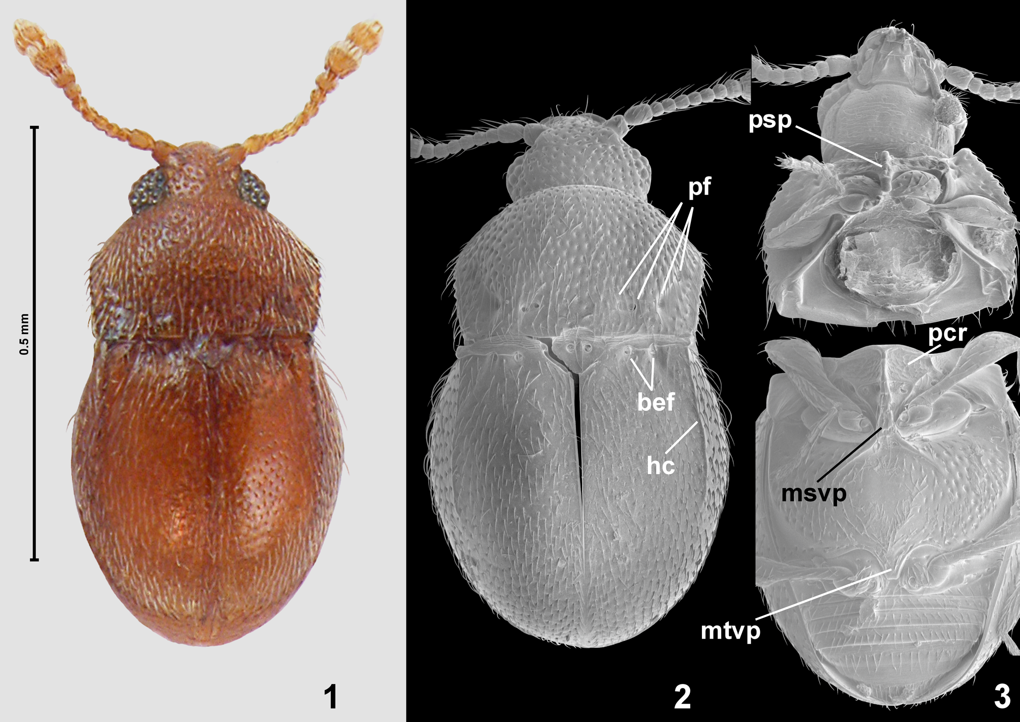
Description. Body ( Figs 1–2 View FIGURES 1–3 ) suboval, strongly convex, densely setose, pronotum and elytra lacking macrosetae.
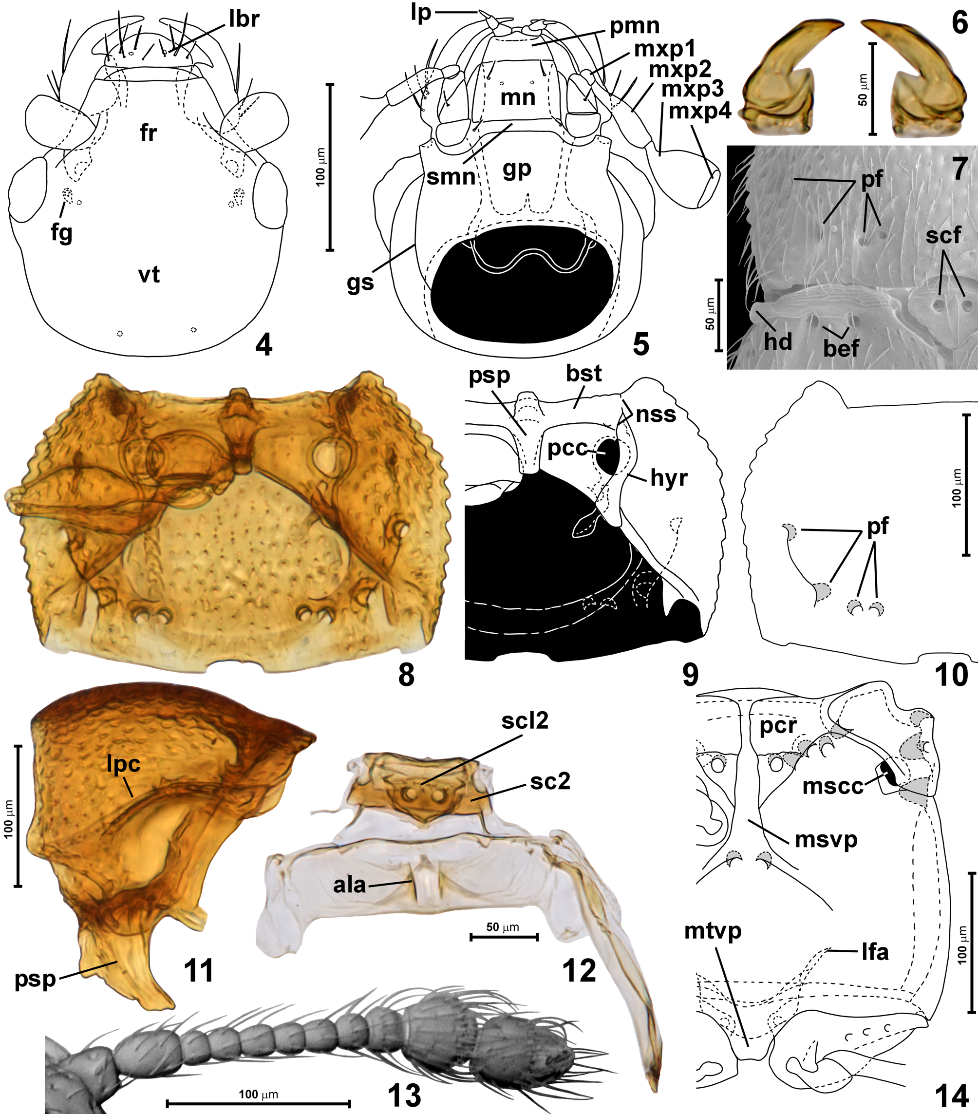
Head capsule ( Figs 1–3 View FIGURES 1–3 , 4–5 View FIGURES 4–14 ) short and broad, in intact beetles tilted so strongly that plane of vertex is nearly perpendicular to the long body axis; frons ( Fig. 4 View FIGURES 4–14 ; fr) and vertex ( Fig. 4 View FIGURES 4–14 ; vt) confluent, together about as long as broad, convex; supraantennal tubercles indistinct; frons with pair of tiny tubercles near posterior margin and small frontal glands ( Fig. 4 View FIGURES 4–14 ; fg) visible only in transparent mounts under compound microscope; eyes large, coarsely faceted, strongly convex; gular plate ( Fig. 5 View FIGURES 4–14 ; gp) large, transverse, with sharply marked gular sutures ( Fig. 5 View FIGURES 4–14 ; gs) and strongly transverse reticulate microsculpture, lacking large, oval punctures; posterior tentorial pits indiscernible; submentum ( Fig. 5 View FIGURES 4–14 ; smn) vestigial, with pair of tiny lateral setae. Mentum ( Fig. 5 View FIGURES 4–14 ; mn) rectangular, weakly transverse and slightly narrowing anterad, with pair of lateral subanterior setae; prementum ( Fig. 5 View FIGURES 4–14 ; pmn) short, largely membranous, with an unknown number of paired suckers, labial palps minute and relatively narrowly separated, inserted at sides of prementum, with palpomere I largest, slightly longer than broad, palpomere II slightly shorter and distinctly narrower than I, slightly elongate, palpomere III much narrower and much longer than II, rod-like with narrowed apex, about 3 × as long as broad. Maxilla ( Fig. 5 View FIGURES 4–14 ) with triangular basistipes bearing one lateral submedian seta, elongate palpifer, elongate lacinia and galea, each with sparse and thick mesal setae; maxillary palp with minute palpomere I ( Fig. 5 View FIGURES 4–14 ; mxp1), strongly elongate, slightly clavate palpomere II ( Fig. 5 View FIGURES 4–14 ; mxp2), strongly broadened palpomere III which is less than twice as long as broad and has truncate apex ( Fig. 5 View FIGURES 4–14 ; mxp3), palpomere IV broad and very short, button-like ( Fig. 5 View FIGURES 4–14 ; mxp4). Mandibles ( Fig. 6 View FIGURES 4–14 ) symmetrical, relatively long, each with subquadrate basal portion and long, slender, only slightly curved distal tooth, setose prostheca absent. Labrum ( Fig. 4 View FIGURES 4–14 ; lbr) short and strongly transverse, with strongly rounded anterior margin, lacking marginal velum, with symmetrically distributed dorsal setae.
Antennae ( Figs 1 View FIGURES 1–3 , 13 View FIGURES 4–14 ) long and slender, composed of 11 antennomeres, club symmetrical and trimerous, but antennomere IX only slightly larger than VIII, so that club may appear as dimerous.
Pronotum ( Figs 1–2 View FIGURES 1–3 , 7, 10 View FIGURES 4–14 ) in dorsal view of whole specimen appears semioval ( Figs 1–2 View FIGURES 1–3 ), but its shape is rectangular with rounded sides; anterior margin shallowly but distinctly concave; anterior pronotal corners broadly subtriangular, blunt and distinctly projecting anterad; lateral margins rounded, forming complete, microserrated lateral carinae ( Fig. 11 View FIGURES 4–14 ; lpc); posterior corners well-defined, obtuse-angled, blunt; posterior margin arcuate, with broad and shallow step-wise emargination and additionally emarginated in front of mesoscutellar shield. Pronotum with two pairs of small foveae ( Figs 2 View FIGURES 1–3 , 7, 10 View FIGURES 4–14 ; pf), lateral pair directed mesad and situated on ends of oblique ridge that demarcates flattened sides of pronotum from more convex median area, inner pits directed anterolaterad and anteromesad, situated in shallow impression extending mesad to connect inner pits as diffuse transverse antebasal groove.
Prosternum ( Figs 3 View FIGURES 1–3 , 8–9 View FIGURES 4–14 ) with basisternal portion ( Fig. 9 View FIGURES 4–14 ; bst) much shorter than coxal part; prosternal process ( Figs 9, 11 View FIGURES 4–14 ; psp) in ventral view subrectangular with well-defined anterior tip, in lateral view ( Fig. 11 View FIGURES 4–14 ) subtriangular and strongly elongate, elevated ventrad beyond procoxae, strongly bent posterad; notosternal sutures ( Fig. 9 View FIGURES 4–14 ; nss) obliterated in front of procoxae but again visible just at anterior sternal margin; procoxal cavities ( Fig. 9 View FIGURES 4–14 ; pcc) broadly closed by posterolateral lobes of prosternum that are fused with postcoxal portions of hypomera; hypomeral ridges ( Fig. 9 View FIGURES 4–14 ; hyr) complete but relatively diffuse on SEMs ( Fig. 3 View FIGURES 1–3 ); hypomera outside hypomeral ridges broad and concave; inner portion of each hypomeron behind procoxal cavity covers profurcal fovea and forms small lobe which in lateral view is visible as projection directed posteroventrad behind prosternal process ( Fig. 11 View FIGURES 4–14 ).
Mesonotum ( Figs 7, 12 View FIGURES 4–14 ) subrectangular, with subtriangular, transverse mesoscutellar shield exposed between elytral bases ( Fig. 7 View FIGURES 4–14 ) and bearing two dorsal scutellar foveae ( Fig. 10 View FIGURES 4–14 ; scf), scutoscutellar suture not visible on dorsal surface, but discernible in transparent mounts; mesoscutum ( Fig. 12 View FIGURES 4–14 ; sc2) strongly transverse.
Elytra ( Figs 1–2 View FIGURES 1–3 , 7 View FIGURES 4–14 ) oval, each with humeral denticle ( Fig. 7 View FIGURES 4–14 ; hd) and two asetose basal elytral foveae ( Fig. 7 View FIGURES 4–14 ; bef); apices of elytra rounded together.
Mesoventrite ( Figs 3 View FIGURES 1–3 , 14 View FIGURES 4–14 ) with mesoventral intercoxal process ( Fig. 14 View FIGURES 4–14 ; msvp) moderately narrow and weakly elevated, broadened distad and anteriorly connected to anterior ridge of ventrite, posteriorly with distinct posterolateral arms. Lateral impressions functioning as procoxal rests ( Fig. 14 View FIGURES 4–14 ; pcr) present, asetose, subtriangular, separated at middle and with all margins well-defined. Mesoventrite with one submedian pair of foveae situated just behind procoxal rests and directed dorsad, with three pairs of foveae situated along anteromesal margin of mesocoxal rest and directed mesad and anteromesad, one pair of foveae situated on border between procoxal rest and mesanepisternum + mesepimeron and directed posteromesad, and three pairs of lateral foveae, with openings situated on lateral surface of mesanepisternum + mesepimeron and directed mesad. Additionally, a large lateral fovea is situated on lateral surface of pterothorax, behind mesocoxal cavity, between lateral meso- and metathoracic structures, this fovea is also directed mesad.
Metanotum ( Fig. 12 View FIGURES 4–14 ) largely membranous, short, but with well-defined alacristae posteriorly reaching behind middle of metascutum.
Hind wings absent.
Metaventrite ( Figs 3 View FIGURES 1–3 , 14 View FIGURES 4–14 ) short, slightly transverse; posterior margins of mesocoxal rests not carinate; sides weakly rounded, posterior margin weakly concave at each metacoxa, with relatively narrow and short metaventral intercoxal process ( Fig. 14 View FIGURES 4–14 ; mtvp), which is subtrapezoidal and indistinctly concave posteriorly. Admetacoxal region at each side with an irregular row of foveate punctures along anterior metacoxal margins. Metanepisterna and metepimera narrow.
Metendosternite (= metafurca) ( Fig. 14 View FIGURES 4–14 ) with short and broad stalk and divergent lateral furcal arms ( Fig. 14 View FIGURES 4–14 ; lfa) with adjacent bases (V-shaped).
Legs moderately long and slender; pro- and mesocoxae oval, metacoxae strongly transverse; all trochanters short and subtriangular; all femora distinctly clavate; tibiae broadening distad; tarsi moderately slender.
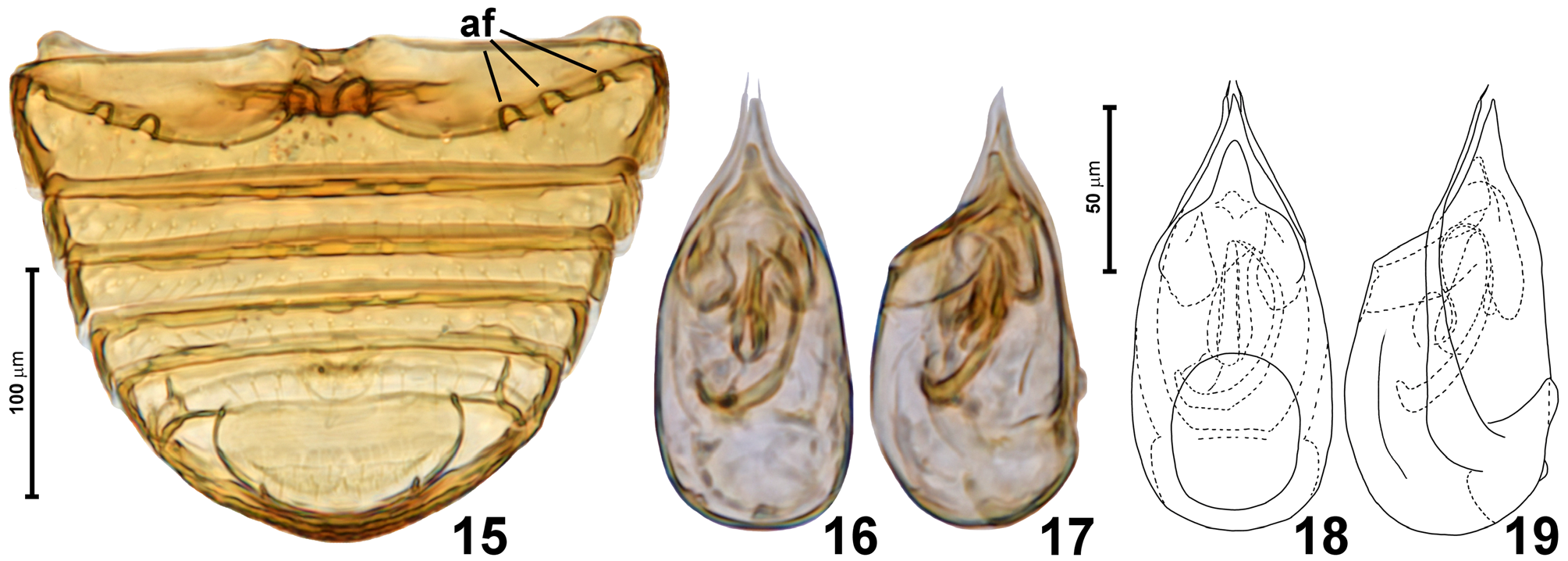
Abdominal sternites ( Fig. 15 View FIGURES 15–19 ) unmodified, except for lateral rows of three abdominal foveae ( Fig. 15 View FIGURES 15–19 ; af) along posterolateral margin of metacoxal rests on sternite III (i.e., first visible), the latter more than twice as long as IV.
Aedeagus ( Figs 16–19 View FIGURES 15–19 ) elongate, with symmetrical median lobe and asymmetrical endophallus, diaphragm present, circular, sub-basal on ventral wall; parameres slender, with apical setae, parameral base with lateral lobes.
Distribution and composition. Foveomicrus is represented by one species known to occur in southern India.
Etymology. The name Foveomicrus combines the prefix foveo - derived from the Latin fovea (a small pit) that refers to the numerous thoracic foveae, with the suffix - micrus, to emphasize the similarity to other members of the ‘ Cephennomicrus group’ of genera. Gender masculine.
Remarks. The button-like maxillary palpomere IV and the presence of frontal glands clearly place Foveomi- crus within the ‘ Cephennomicrus group’ of genera. Indeed, preliminary results of a phylogenetic analysis indicate that this genus is deeply rooted within this clade, and may be a sister group for Cephennococcus + ( Indomicrus gen. n. + ( Trichokrater + Trurlia )) (Jałoszyński, in prep.). The four pairs of pronotal foveae is a unique character, known only in Foveomicrus . Additionally, this genus differs from the remaining members of the ‘ Cephennomicrus group’ in the following characters (among others):
- from Trurlia and Clavomicrus in separated (and not fused) antennomeres X and XI;
- from Pomphopsilla , Indomicrus gen. n. and Cephennococcus in lacking internal prothoracic ‘cavities’;
- from Trichokrater in lacking pronotal setose glandular openings and a symmetrical antennal club;
- from Eutheimorphus in lacking C-shaped pronotal grooves and a different shape of the mesoventral intercoxal process;
- from Cephennomicrus in a vestigial submentum, prosternal process with a well-defined anterior tip and a narrow mesoventral intercoxal process;
- from Lathomicrus and Cephennula in two deep basal elytral and mesoscutellar foveae, and prosternal and mesoventral intercoxal processes not inversely T-shaped in cross-section.
No known copyright restrictions apply. See Agosti, D., Egloff, W., 2009. Taxonomic information exchange and copyright: the Plazi approach. BMC Research Notes 2009, 2:53 for further explanation.