
Hatschekia ostracii Yamaguti, 1953
|
publication ID |
https://doi.org/ 10.5281/zenodo.187869 |
|
DOI |
https://doi.org/10.5281/zenodo.5678630 |
|
persistent identifier |
https://treatment.plazi.org/id/03BC2C7A-010B-FFB8-3DEF-F56AFB5DFD81 |
|
treatment provided by |
Plazi |
|
scientific name |
Hatschekia ostracii Yamaguti, 1953 |
| status |
|
Hatschekia ostracii Yamaguti, 1953
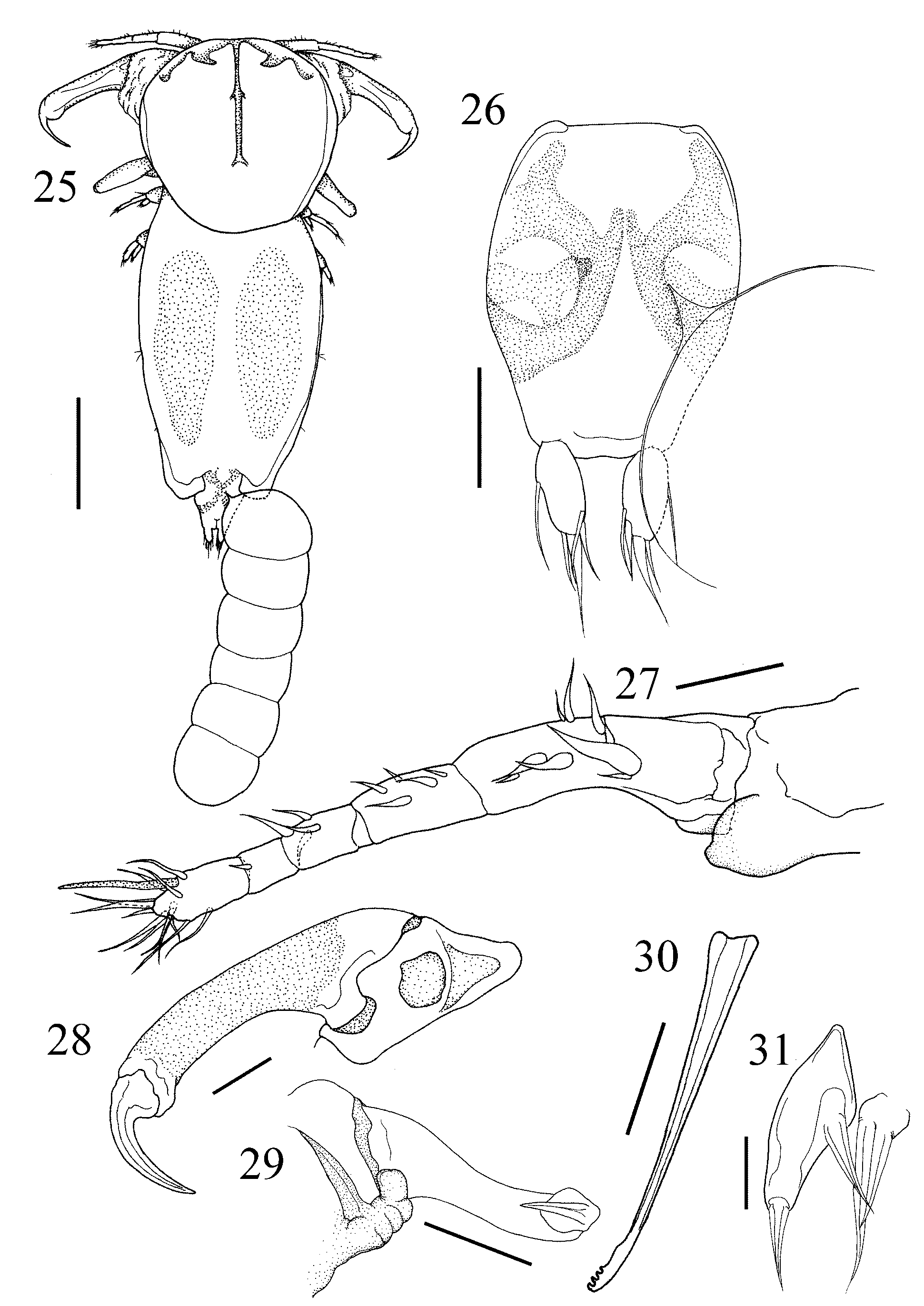
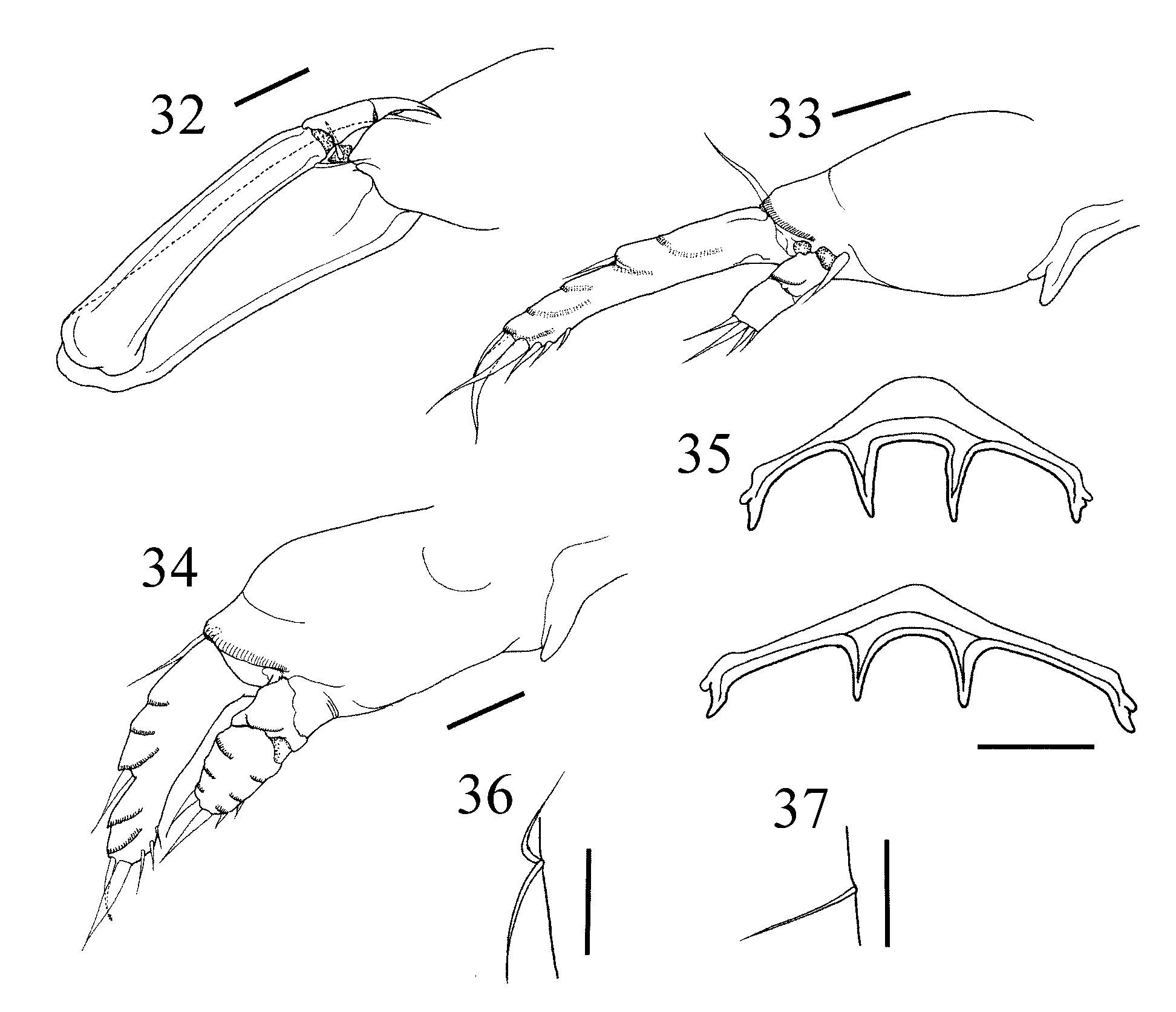
( Figs 25–37 View FIGURES 25 – 31 View FIGURES 32 – 37 )
Hatschekia ostracii: Yamaguti 1953: 228 ; Jones 1985: 256; Kim 1998: 739.
Material examined. 7Ψ (NSMT-Cr 20332), ex Ostracion immaculatus Temminck & Schlegel, Tatsukushi (32°47΄N, 132°51΄E), Kochi, North Pacific Ocean, Japan, 9 May, 2006; 1Ψ (NSMT-Cr 20333), ex Ostracion cubicus L., Ama (26°13΄N, 127°17΄E), Kerama Islands, Okinawa, East China Sea, Japan, 26 May, 2007; 1Ψ (NSMT-Cr 20334), ex O. cubicus L., Sakimotobu (26°38΄N, 127°52΄E), Okinawa, East China Sea, Japan, 25 July, 2007.
Description of female. Body ( Fig. 25 View FIGURES 25 – 31 ) 0.66–1.02 (0.87 ± 0.10) long, excluding caudal rami (n = 9). Cephalothorax suborbicular, longer than wide [0.30–0.46 (0.37 ± 0.04) × 0.28–0.39 (0.35 ± 0.04)], with dorsal, T-shaped chitinous frame. Trunk longer than wide [0.41–0.69 (0.57 ± 0.09) × 0.21–0.39 (0.32 ± 0.06)], with pair of posterior lobes. Abdomen ( Fig. 26 View FIGURES 25 – 31 ) length 0.07–0.11 (0.10 ± 0.01); width 0.06–0.09 (0.08 ± 0.01). Caudal ramus ( Fig. 26 View FIGURES 25 – 31 ) longer than wide [0.03–0.04 (0.03 ± 0) × 0.01–0.02 (0.01 ± 0)] and bears 5 naked setae.
Rostrum with 1 ovoid process on each posterolateral corner ( Fig. 27 View FIGURES 25 – 31 ). Antennule ( Fig. 27 View FIGURES 25 – 31 ) 5-segmented, 0.17–0.25 (0.21 ± 0.02) long; armature formula: 9, 5, 4, 1, 13 + 1 aesthetasc. Antenna ( Fig. 28 View FIGURES 25 – 31 ) 3-segmented; proximal segment (coxa) unarmed; middle segment (basis) covered with surface pits; terminal segment clawlike, unarmed; proximal segment length 0.06–0.11 (0.08 ± 0.02); middle segment length 0.20–0.28 (0.24 ± 0.03); terminal segment length 0.06–0.07 (0.06 ± 0.01); total length 0.32–0.43 (0.38 ± 0.04). Parabasal papilla ( Fig. 29 View FIGURES 25 – 31 ) thumb-shaped, ventrally-directed. Oral cone robust. Mandible ( Fig. 30 View FIGURES 25 – 31 ) slender, with 4 sharp apical teeth. Maxillule ( Fig. 31 View FIGURES 25 – 31 ) bilobate; inner lobe sclerotized, bears 1 proximal and 1 apical elements; outer lobe with 2 attenuate elements. Maxilla ( Fig. 32 View FIGURES 32 – 37 ) 4-segmented; proximal segment unarmed; second segment rodlike, with 1 basal seta; third segment elongate, with 1 distal seta; terminal segment small, with 1 small seta and bifid claw. Maxilliped absent.
Legs 1 and 2 ( Figs 33–34 View FIGURES 32 – 37 ) biramous, with incompletely bimerous exopod and 2-segmented endopod; leg armature formula as follows (armature on terminal segment of rami represented by the mode followed by the range in parentheses):
Intercoxal sclerite of legs 1 and 2 ( Fig. 35 View FIGURES 32 – 37 ) bears 2 short and 2 long processes. Protopods bear row of blunt spinules along distal margin and semicircular wrinkles on anterior surface (it is uncertain whether these wrinkles consist of spinules or not). Rami bear small, spinulate, hyaline sculptures on anterior surface. Leg 1 ( Fig. 33 View FIGURES 32 – 37 ) 0.14–0.16 (0.15 ± 0.01) long; protopod length 0.06–0.08 (0.07 ± 0.01); exopod length [0.07–0.08 (0.08 ± 0.01)] exceeding endopod length [0.02–0.03 (0.02 ± 0)]. Leg 2 ( Fig. 34 View FIGURES 32 – 37 ) length 0.10–0.17 (0.13 ± 0.02); protopod length 0.05–0.09 (0.07 ± 0.01); exopod length 0.05–0.07 (0.06 ± 0.01); endopod length 0.03–0.04 (0.03 ± 0).
Leg 3 ( Figs 25 View FIGURES 25 – 31 , 36 View FIGURES 32 – 37 ) represented by 2 simple setae on mid-lateral surface of trunk. Leg 4 ( Figs 25 View FIGURES 25 – 31 , 37 View FIGURES 32 – 37 ) represented by 1 simple lateral seta on posterior ½ of trunk.
Attachment site. Gill filaments.
Remarks. Hatschekia ostracii was described originally by Yamaguti (1953) based on several gravid female specimens collected from Ostracion cubicus captured in Komatsushima (as Komatusima), Tokushima, Japan. This species was later redescribed by Kim (1998) from female specimens removed from O. immaculatus collected in Korea. Hatschekia ostracii has 4 processes on the intercoxal sclerite of legs 1 and 2 in common with H. balistae Nuñes-Ruivo, 1954 and H. monacanthi Yamaguti, 1939 . However, H. balistae is distinguishable from H. ostracii by the presence of an anteromedian pointed apex on the cephalothorax and absence of posterior lobes on the trunk. Hatschekia ostracii differs from H. monacanthi by lacking a chitinous ring on the cephalothorax and having a significantly longer exopod relative to the endopod in leg 1 [L1ExL/ L1EnL ratio 3.24 ± 0.36 vs. 2.22 ± 0.30 (U-test; p <0.01), Table 1 View TABLE 1 ] and a considerably longer abdomen relative to the body length [AbL/BL ratio 0.12 ± 0.01 vs. 0.03 ± 0.01 (U-test; p <0.001), Table 1 View TABLE 1 ].
No known copyright restrictions apply. See Agosti, D., Egloff, W., 2009. Taxonomic information exchange and copyright: the Plazi approach. BMC Research Notes 2009, 2:53 for further explanation.
|
Kingdom |
|
|
Phylum |
|
|
Class |
|
|
Order |
|
|
Family |
|
|
Genus |
Hatschekia ostracii Yamaguti, 1953
| Uyeno, Daisuke & Nagasawa, Kazuya 2009 |
Hatschekia ostracii:
| Kim 1998: 739 |
| Jones 1985: 256 |
| Yamaguti 1953: 228 |