
Hatschekia iridescens Wilson, 1913
|
publication ID |
https://doi.org/ 10.5281/zenodo.187869 |
|
DOI |
https://doi.org/10.5281/zenodo.5678628 |
|
persistent identifier |
https://treatment.plazi.org/id/03BC2C7A-0103-FFBD-3DEF-F42AFF1CFC38 |
|
treatment provided by |
Plazi |
|
scientific name |
Hatschekia iridescens Wilson, 1913 |
| status |
|
Hatschekia iridescens Wilson, 1913
( Figs 2–13 View FIGURES 2 – 7 View FIGURES 8 – 13 )
Hatschekia iridescens: Wilson 1913: 248 ; Jones 1985: 244; Villalba 1986: 160. Hatschekia diodontis: Yamaguti 1953: 225 .
Material examined. 7Ψ (NSMT-Cr 20327), ex Diodon liturosus Shaw, Aka (26°11΄N, 127°16΄E), Kerama Islands, Okinawa, East China Sea, Japan, 31 July, 2005; 3Ψ (NSMT-Cr 20328), ex Diodon hystrix L., Yonashiro (26°20΄N, 127°57΄E), Kin Bay, Okinawa, North Pacific Ocean, Japan, 14 July, 2007; 2Ψ (NSMT-Cr 20329) ex Diodon holocanthus L., Nishidomari (32°46΄N, 132°43΄E), Otsuki, Kochi, North Pacific Ocean, Japan, 8 November, 2006.
Description of female. Body ( Fig. 2 View FIGURES 2 – 7 ) 0.96–1.27 (1.13 ± 0.12) long, excluding caudal rami (n = 12). Cephalothorax broadly obovate, shorter than wide [0.29–0.44 (0.35 ± 0.05) × 0.36–0.55 (0.47 ± 0.06)], with dorsal, M-shaped chitinous frame. Trunk longer than wide [0.70–0.99 (0.83 ± 0.11) × 0.32–0.44 (0.39 ± 0.04)], bilobed posteriorly. Abdomen ( Fig. 3 View FIGURES 2 – 7 ) shorter than wide [0.04–0.06 (0.05 ± 0.01) × 0.08–0.10 (0.09 ± 0.01)]. Caudal ramus ( Fig. 3 View FIGURES 2 – 7 ) slightly longer than wide [0.02–0.04 (0.03 ± 0.01) × 0.01–0.02 (0.02 ± 0)], bears 5 naked setae.
Rostrum with 1 thumb-shaped process on each posterolateral corner ( Fig. 4 View FIGURES 2 – 7 ). Antennule ( Fig. 4 View FIGURES 2 – 7 ) indistinctly 5-segmented, 0.23–0.34 (0.28 ± 0.04) long, with armature formula: 10, 6, 4, 1, 13 + 1 aesthetasc; base of squat, apically blunt elements fused partially to respective segment. Antenna ( Fig. 5 View FIGURES 2 – 7 ) 3-segmented; proximal segment (coxa) unarmed; middle segment (basis) ornamented with large patch of surface pits (the pits appear as 2 small patches in the figure, but are actually fused into a single patch on the lateral surface); terminal segment claw-like, composed of incompletely fused endopodal segment and claw, armed with 1 minute spine and 1 small seta at midlength; proximal segment length 0.07–0.13 (0.10 ± 0.02); middle segment length 0.13–0.17 (0.15 ± 0.01); terminal segment length 0.09–0.14 (0.12 ± 0.02); total length 0.31–0.43 (0.36 ± 0.04). Parabasal papilla not observed. Oral cone robust. Mandible ( Fig. 6 View FIGURES 2 – 7 ) slender, with 5 sharp teeth apically. Maxillule ( Fig. 7 View FIGURES 2 – 7 ) bilobate; each lobe armed with 2 attenuate elements. Maxilla ( Fig. 8 View FIGURES 8 – 13 ) 4-segmented; proximal segment unarmed; second segment rod-like, with 1 basal seta; third segment elongate, with 1 distal seta; terminal segment small, apically with 1 small seta and bifid claw. Maxilliped absent.
Legs 1 and 2 ( Figs 9–10 View FIGURES 8 – 13 ) biramous, with indistinctly bimerous exopod and 2-segmented endopod; leg armature formula as follows:
Intercoxal sclerite of legs 1 and 2 ( Fig. 11 View FIGURES 8 – 13 ) elongate, unornamented and unmodified. Protopods of legs 1 and 2 with several semicircular surface wrinkles (it is uncertain whether these wrinkles are spinulate or membranous); rami ornamented with semicircles of spinules; some setae unipinnate. Leg 1 ( Fig. 9 View FIGURES 8 – 13 ) 0.12–0.17 (0.15 ± 0.02) long; protopod length 0.06–0.09 (0.08 ± 0.01); exopod length [0.06–0.08 (0.07 ± 0.01)] exceeding endopod length [0.04–0.05 (0.04 ± 0)]. Leg 2 ( Fig. 10 View FIGURES 8 – 13 ) 0.12–0.18 (0.16 ± 0.02) long; protopod length 0.06–0.10 (0.09 ± 0.01); exopod length 0.05–0.09 (0.07 ± 0.01); endopod length 0.04–0.06 (0.05 ± 0.01).
Leg 3 ( Figs 2 View FIGURES 2 – 7 , 12 View FIGURES 8 – 13 ) represented by 2 simple setae on mid-lateral surface of trunk. Leg 4 ( Figs 2 View FIGURES 2 – 7 , 13 View FIGURES 8 – 13 ) represented by 1 simple seta on posterior ¾ of trunk.
Attachment site. Gill filaments.
Remarks. Hatschekia iridescens was described originally by Wilson (1913) based on specimens of both sexes removed from Diodon hystrix collected in Montego Bay, Jamaica. This species was later reported from D. holocanthus in Okinawa, Japan ( Yamaguti 1953). Jones (1985) also redescribed this species using Wilson’s type specimens. Villalba (1986) identified 25 ovigerous females from D. holocanthus in Isla de Pascua, Chile, as H. iridescens . The morphology of his specimens, however, differs from the specimens examined by Wilson (1913), Jones (1985) and the present authors in lacking small posterior lobes on the trunk ( Yamaguti (1953) did not mention the presence or absence of posterior lobes). As such, Villalba’s specimens may not be conspecific with H. iridescens . A re-examination of Villalba’s material is needed to clarify this issue.
Several discrepancies exist between our observations and those of previous authors regarding the fine structural details of H. iridescens . For instance, an M-shaped chitinous frame, consisting of a conspicuous mid-longitudinal part and two somewhat indistinctive lateral parts, was present in our material. Wilson (1913) and Jones (1985), in contrast, reported the mid-longitudinal frame only. These authors perhaps overlooked the lateral parts of the frame. Yamaguti (1953) described a T-shaped chitinous frame on the dorsal surface of the cephalothorax, but his illustration in fact depicted an M-shaped frame.
Wilson (1913, fig. 249) found 15 (3, 4, 0, 8) setae in total on the four distal antennulary segments. Yamaguti (1953, fig. 30) and Jones (1985, fig. 11A) found a total of 26 (6, 4, 3, 1, 12) and 25 (7, 4, 4, 0, 10) setae, respectively, on the antennule, while our specimens have 35 (10, 6, 4, 1, 13 + 1 aesthetasc) elements. Jones (1985, fig. 11A) did not illustrate the relatively smaller setae on the antennule, which suggests that he overlooked them.
The antennal claw in our specimens bears one minute spine and one simple seta. Further, the claw is incompletely 2-segmented, with the proximal and distal segments separated by a thin transverse line. Wilson (1913) found only a swelling, Jones (1985) illustrated only a small medial process, and Yamaguti (1953) described only a spiniform barb on the antennal claw. The elements observed by us are very small and delicate, and the suture line is extremely weak. It is thus plausible that these fine structures of the antennal claw were overlooked by the past authors.
Wilson (1913) noted that the first two leg pairs had bimerous exopods and unimerous endopods. Yamaguti (1953) stated that the exopods of both legs were 2-segmented and their endopods 1- and 2-segmented, respectively. Jones (1985) also reported that the exopods were 2-segmented but the endopods were obscurely 2-segmented in legs 1 and 2. In our specimens, however, legs 1 and 2 had indistinctly 2-segmented exopods and distinctly 2-segmented endopods. This discrepancy may be due to insufficient observation by the previous authors. We observed wrinkles on the anterior surface of the protopods of legs 1 and 2; however, no such information was provided by the previous authors. As these wrinkles are highly transparent, the previous authors may have overlooked these structures.
Hatschekia iridescens resembles H. legouli Nuñes-Ruivo, 1954 in having an M-shaped chitinous frame on the cephalothorax, similar antennulary and swimming leg armature formulae, and squat, apically blunt antennulary elements that lack a complete basal articulation. However, H. iridescens differs from H. legouli by having a considerably smaller antennule length/antenna length ratio [0.78 ± 0.03 vs. 1.16 ± 0.16 (U-test; p <0.01), Table 1 View TABLE 1 ], a less planiform cephalothoracic shield, a slightly higher cephalothorax length/body length ratio [0.31 ± 0.03 vs. 0.22 ± 0.02 (U-test; p <0.001) Table 1 View TABLE 1 ], and relatively smaller posterior lobes on the trunk. A redescription of the male of H. iridescens is needed, as it was poorly described by Wilson (1913). Our finding of H. iridescens on D. liturosus View in CoL represents a new host record.
Copepods
H. iridescens H. legouli H. monacanthi H. ostracii
(n = 12) (n = 7) (n = 7) (n = 9)
Hatschekia legouli: Nuñes-Ruivo 1954: 500 ; Jones 1985: 248.
Material examined. 5Ψ (NSMT-Cr 20330), ex Chilomycterus reticulatus L., Nishidomari (32°46΄N, 132°43΄E), Ohtsuki, Kochi, North Pacific Ocean, Japan, 8 November, 2005; 2Ψ (NSMT-Cr 20331), ex C. reticulatus, Nishinoomote (30°49΄N, 131°2΄E), Tanegashima Island, Kagoshima, East China Sea, Japan, 22 May, 2007.
Description of female. Body ( Fig. 14 View FIGURES 14 – 20 ) 1.05–1.43 (1.23 ± 0.12) long, excluding caudal rami (n = 7). Cephalothorax trapezoid, shorter than wide [0.23–0.34 (0.27 ± 0.04) × 0.41–0.51 (0.47 ± 0.04)], with dorsal, M-shaped chitinous frame. Trunk longer than wide [0.79–1.15 (0.96 ± 0.11) × 0.33–0.48 (0.40 ± 0.05)], with pair of posterior lobes. Abdomen ( Fig. 15 View FIGURES 14 – 20 ) shorter than wide [0.04–0.06 (0.05 ± 0.01) × 0.07–0.09 (0.07 ± 0.01)]. Caudal ramus ( Fig. 15 View FIGURES 14 – 20 ) longer than wide [0.04–0.05 (0.04 ± 0) × 0.02–0.03 (0.02 ± 0)], bears 5 naked setae.
Rostrum with rounded process on each posterolateral corner ( Fig. 16 View FIGURES 14 – 20 ). Antennule ( Fig. 16 View FIGURES 14 – 20 ) incompletely 5-segmented, 0.26–0.36 (0.30 ± 0.03) long; with armature formula: 10, 6, 4, 1 (only 1 specimen has 2 setae on this segment), 13 + 1 aesthetasc; base of squat, apically blunt elements fused partially to respective segment. Antenna ( Fig. 17 View FIGURES 14 – 20 ) 3-segmented; proximal segment (coxa) unarmed; middle segment (basis) ornamented with surface pits; terminal segment claw-like, comprised of incompletely fused endopodal segment and claw, armed with 1 minute inner spine; proximal segment length 0.04–0.07 (0.06 ± 0.01); middle segment length 0.11–0.15 (0.13 ± 0.01); terminal segment length 0.06–0.09 (0.07 ± 0.01); total length 0.22–0.28 (0.26 ± 0.02). Parabasal papilla not observed. Oral cone robust. Mandible ( Fig. 18 View FIGURES 14 – 20 ) slender, with 5 sharp apical teeth. Maxillule ( Fig. 19 View FIGURES 14 – 20 ) bilobate; both lobes bears 2 acuminate elements. Maxilla ( Fig. 20 View FIGURES 14 – 20 ) 4-segmented; proximal segment unarmed; second segment rod-like, with 1 basal seta; third segment elongate, with 1 distal seta; terminal segment small, apically with 1 small seta and bifid claw. Maxilliped absent.
Legs 1 and 2 ( Figs 21–22 View FIGURES 21 – 24 ) biramous, with indistinctly bimerous exopod and 2-segmented endopod; leg armature formula as follows:
Intercoxal sclerite of legs 1 and 2 (not figured) unornamented and unmodified. Protopods with semicircular surface wrinkles (it is uncertain whether these wrinkles are spinulate or membranous); rami ornamented with semicircles of spinules. Leg 1 ( Fig. 21 View FIGURES 21 – 24 ) 0.13–0.15 (0.13 ± 0.01) long; protopod length 0.06–0.08 (0.07 ± 0.01); exopod length [0.06–0.07 (0.06 ± 0)] exceeding endopod length [0.03–0.04 (0.04 ± 0)]. Leg 2 ( Fig. 22 View FIGURES 21 – 24 ) 0.13–0.15 (0.13 ± 0.01) long; protopod length 0.06–0.10 (0.08 ± 0.01); exopod length 0.06–0.08 (0.07 ± 0.01); endopod length 0.04–0.06 (0.04 ± 0.01).
Leg 3 ( Figs 14 View FIGURES 14 – 20 , 23 View FIGURES 21 – 24 ) represented by 2 simple setae on mid-lateral surface of trunk. Leg 4 ( Figs 14 View FIGURES 14 – 20 , 24 View FIGURES 21 – 24 ) represented by 1 simple lateral seta on posterior ¾ of trunk.
Attachment site. Gill filaments.
Remarks. Hatschekia legouli was described originally by Nuñes-Ruivo (1954) based on 49 female specimens from Chilomycterus reticulatus caught in Goree, Senegal. Jones (1985) noted, however, that a redescription of H. legouli was needed because details of leg 1 were not given in the original description. It is worth nothing here that the other appendages of the female also were incompletely described, and the male of H. legouli has not been described yet. We attribute our specimens to H. legouli as they share: a) a trapezoid cephalothorax that is twice as wide as long; b) an M–shaped dorsal chitinous frame on the cephalothorax; c) a trunk with relatively well developed posterior lobes; and d) the antennulary bases protruding beyond the anterior margin of the cephalothorax. In addition, we collected our material from the same host species as Nuñes-Ruivo (1954). As discussed in the Remarks section of H. iridescens above, H. legouli closely resembles H. iridescens , as both species share a similar M-shaped chitinous frame on the cephalothorax, armature on the antennule and swimming legs, and squat, apically blunt antennulary elements that lack a complete basal articulation. Hatschekia legouli , however, differs from H. iridescens in having a trapezoid cephalothorax and the abdomen and caudal rami extending beyond the distal end of the posterior lobes of the trunk. Our finding of H. legouli represents the first record of this species from Japan and the North Pacific Ocean.
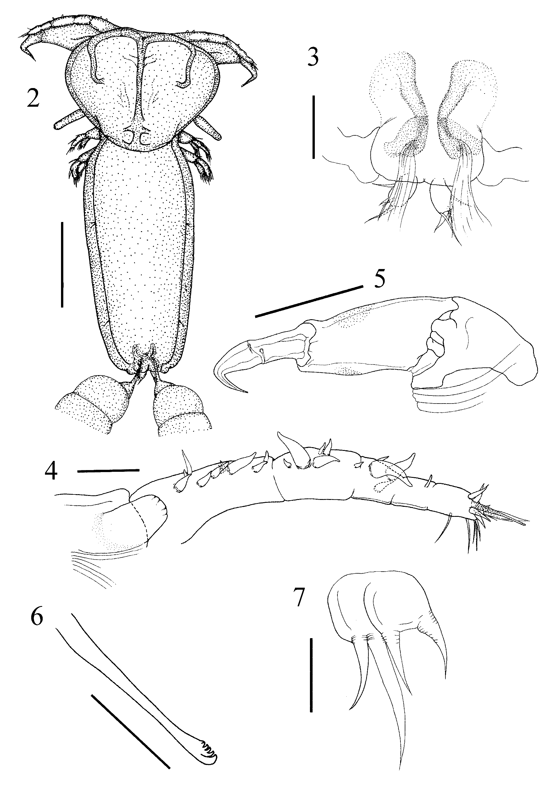
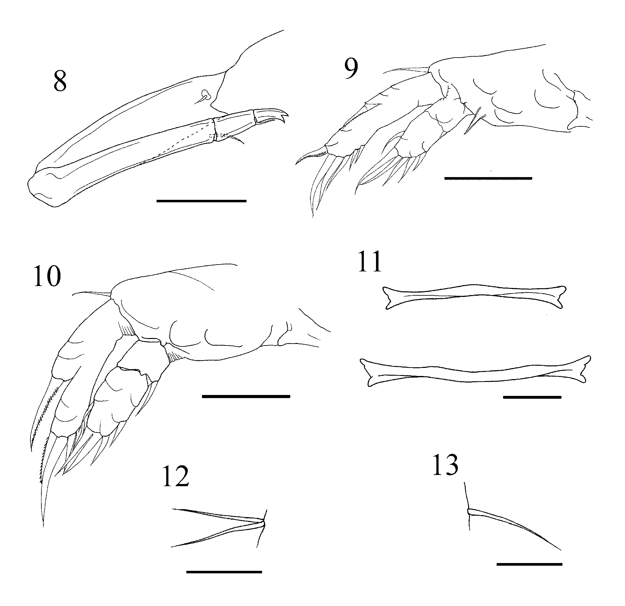

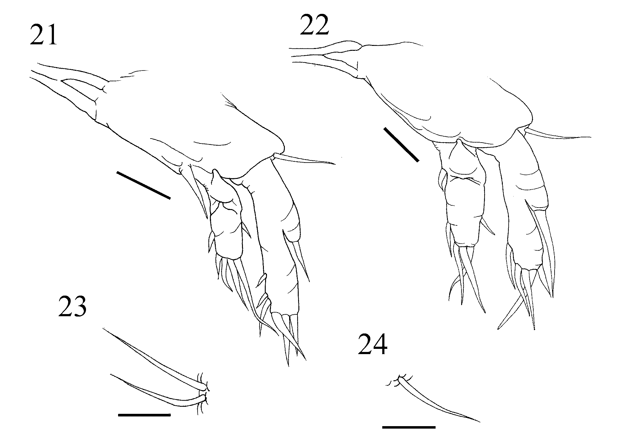
TABLE 1. Ratios of body parts and appendages of females of Hatschekia iridescens, H. legouli, H. monacanthi, and H. ostracii. Data are shown in millimeters as mean ± standard deviation. Abbreviations are as follows: body length (BL), cephalothorax length (CeL), cephalothorax width (CeW), trunk length (TL), trunk width (TW), abdomen length (AbL), abdomen width (AbW), caudal ramus length (CaL), caudal ramus width (CaW), antennule length (A 1 L), antenna length (A 2 L), middle segment length of antenna (A 2 ML), terminal segment length of antenna (A 2 TL), leg 1 length (L 1 L), leg 1 exopod length (L 1 ExL), leg 1 endopod length (L 1 EnL), leg 2 length (L 2 L), leg 2 exopod length (L 2 ExL), and leg 2 endopod length (L 2 EnL).
| CeL/BL | 0.31 ± 0.03 | 0.22 ± 0.02 | 0.27 ± 0.03 | 0.42 ± 0.03 |
|---|---|---|---|---|
| CeW/BL | 0.42 ± 0.05 | 0.39 ± 0.03 | 0.33 ± 0.01 | 0.40 ± 0.02 |
| TL/BL | 0.73 ± 0.03 | 0.78 ± 0.02 | 0.75 ± 0.02 | 0.65 ± 0.03 |
| TW/BL | 0.34 ± 0.03 | 0.32 ± 0.01 | 0.33 ± 0.04 | 0.37 ± 0.03 |
| AbL/BL | 0.05 ± 0.01 | 0.04 ± 0.01 | 0.03 ± 0.01 | 0.12 ± 0.01 |
| AbW/BL | 0.08 ± 0.01 | 0.06 ± 0.01 | 0.09 ± 0.01 | 0.09 ± 0.00 |
| CaL/BL | 0.03 ± 0.00 | 0.04 ± 0.01 | 0.02 ± 0.00 | 0.04 ± 0.01 |
| CaW/BL | 0.01 ± 0.00 | 0.02 ± 0.00 | 0.01 ± 0.00 | 0.02 ± 0.00 |
| CeW/CeL | 1.35 ± 0.31 | 1.74 ± 0.15 | 1.22 ± 0.08 | 0.95 ± 0.06 |
| AbW/AbL | 1.82 ± 0.23 | 1.62 ± 0.25 | 2.96 ± 0.60 | 0.78 ± 0.05 |
| A1L/BL | 0.25 ± 0.03 | 0.24 ± 0.02 | 0.15 ± 0.02 | 0.23 ± 0.01 |
| A2L/BL | 0.33 ± 0.05 | 0.21 ± 0.03 | 0.27 ± 0.03 | 0.44 ± 0.04 |
| A2TL/A2ML | 0.78 ± 0.12 | 0.59 ± 0.11 | 0.20 ± 0.04 | 0.26 ± 0.04 |
| L1L/BL | 0.13 ± 0.01 | 0.11 ± 0.02 | 0.08 ± 0.01 | 0.17 ± 0.02 |
No known copyright restrictions apply. See Agosti, D., Egloff, W., 2009. Taxonomic information exchange and copyright: the Plazi approach. BMC Research Notes 2009, 2:53 for further explanation.
|
Kingdom |
|
|
Phylum |
|
|
Class |
|
|
Order |
|
|
Family |
|
|
Genus |
Hatschekia iridescens Wilson, 1913
| Uyeno, Daisuke & Nagasawa, Kazuya 2009 |
Hatschekia legouli: Nuñes-Ruivo 1954 : 500
| Jones 1985: 248 |
| Nunes-Ruivo 1954: 500 |
Hatschekia iridescens:
| Villalba 1986: 160 |
| Jones 1985: 244 |
| Yamaguti 1953: 225 |
| Wilson 1913: 248 |