
Pachymeniopsis lanceolata
|
publication ID |
https://doi.org/ 10.1515/bot-2021-0013 |
|
DOI |
https://doi.org/10.5281/zenodo.11094332 |
|
persistent identifier |
https://treatment.plazi.org/id/03B97F3C-D42F-D772-5EFA-DA04FACD1DC6 |
|
treatment provided by |
Felipe |
|
scientific name |
Pachymeniopsis lanceolata |
| status |
|
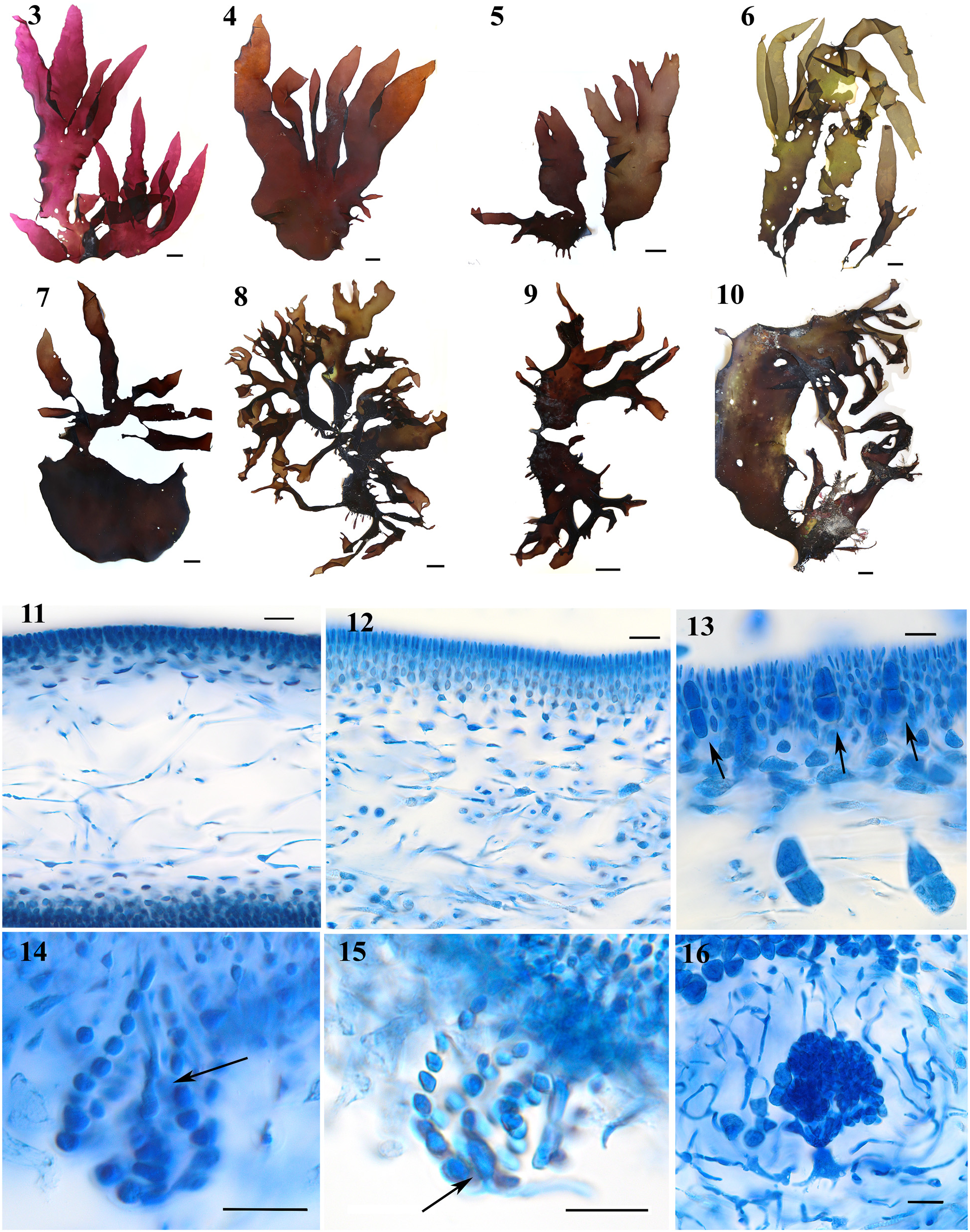
3.2.1 Pachymeniopsis lanceolata ( Figures 3–16 View Figures 3–16 )
Thalli ( Figures 3–10 View Figures 3–16 ) were flattened, 14–35 (60) cm high and 6–15 cm wide, and attached by a discoid holdfast, from which a short ( Figures 5–6, 9–10 View Figures 3–16 ) or nearly non-existent stipe arose ( Figures 3 and 4 View Figures 3–16 ). Thalli were solitary ( Figures 4 and 5 View Figures 3–16 ) or clustered ( Figure 8 View Figures 3–16 ), the blades were broadly lanceolate ( Figures 3, 4 View Figures 3–16 , and 6) or irregularly divided ( Figures 6–8 View Figures 3–16 ). Old thalli became proliferous ( Figures 8 and 10 View Figures 3–16 ). Thalli were purplish-red to brownish, with a membranous texture that became leathery in old plants. Large thalli had a strong chlorine smell. Blades were 200– 600 µm thick ( Figures 11 and 12 View Figures 3–16 ), reaching up to 1 mm in thickness in old specimens. The cortex consisted of anticlinal filaments (6) 8–12 cells long, the cells becoming progressively smaller toward the surface layer of elongate cells ( Figure 12 View Figures 3–16 ). The medulla was composed of sparsely to densely compacted filaments, 2–7 µm in diameter ( Figures 11 and 12 View Figures 3–16 ). Male gametophytes were found in winter, whereas cystocarps and tetrasporophytes were seen in summer. Tetrasporangia ( Figure 13 View Figures 3–16 ) were scattered over the blade (30–53 × 15–21 µm) and were cruciately divided. The carpogonial branch ampullae were monocarpogonial ( Figure 14 View Figures 3–16 ), 45–57 × 29–40 µm. The auxiliary cell ampullae ( Figure 15 View Figures 3–16 ) were 40–46 µm in length and 30–36 µm in width. The auxiliary cell was intercalary in one of the ampullar filaments and had 12 × 7 µm dimensions. Cystocarps ( Figure 16 View Figures 3–16 ) were 100–220 µm in diameter, immersed in the blades and surrounded by a rudimentary pericarp of lax ampullar filaments. The carposporophytes consisted of three synchronously maturing lobes and contained irregularly angular carpospores (15–22 × 10–13 µm).
No known copyright restrictions apply. See Agosti, D., Egloff, W., 2009. Taxonomic information exchange and copyright: the Plazi approach. BMC Research Notes 2009, 2:53 for further explanation.