
Licea testudinacea Nann.
|
publication ID |
https://doi.org/ 10.11646/phytotaxa.629.2.1 |
|
DOI |
https://doi.org/10.5281/zenodo.10390952 |
|
persistent identifier |
https://treatment.plazi.org/id/03B7950C-9B03-FFCC-FF16-04CAFB6FFE13 |
|
treatment provided by |
Plazi |
|
scientific name |
Licea testudinacea Nann. |
| status |
|
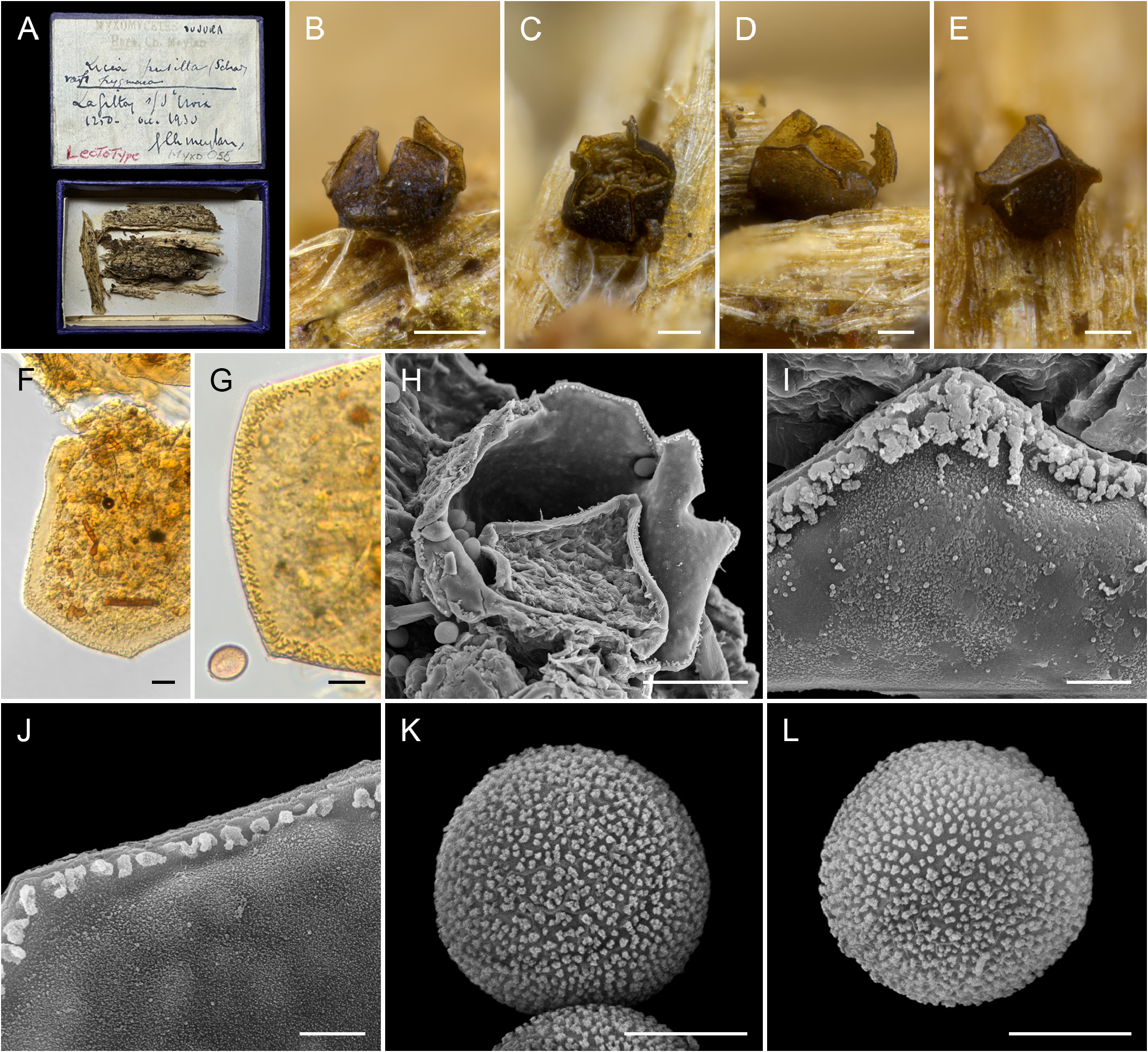
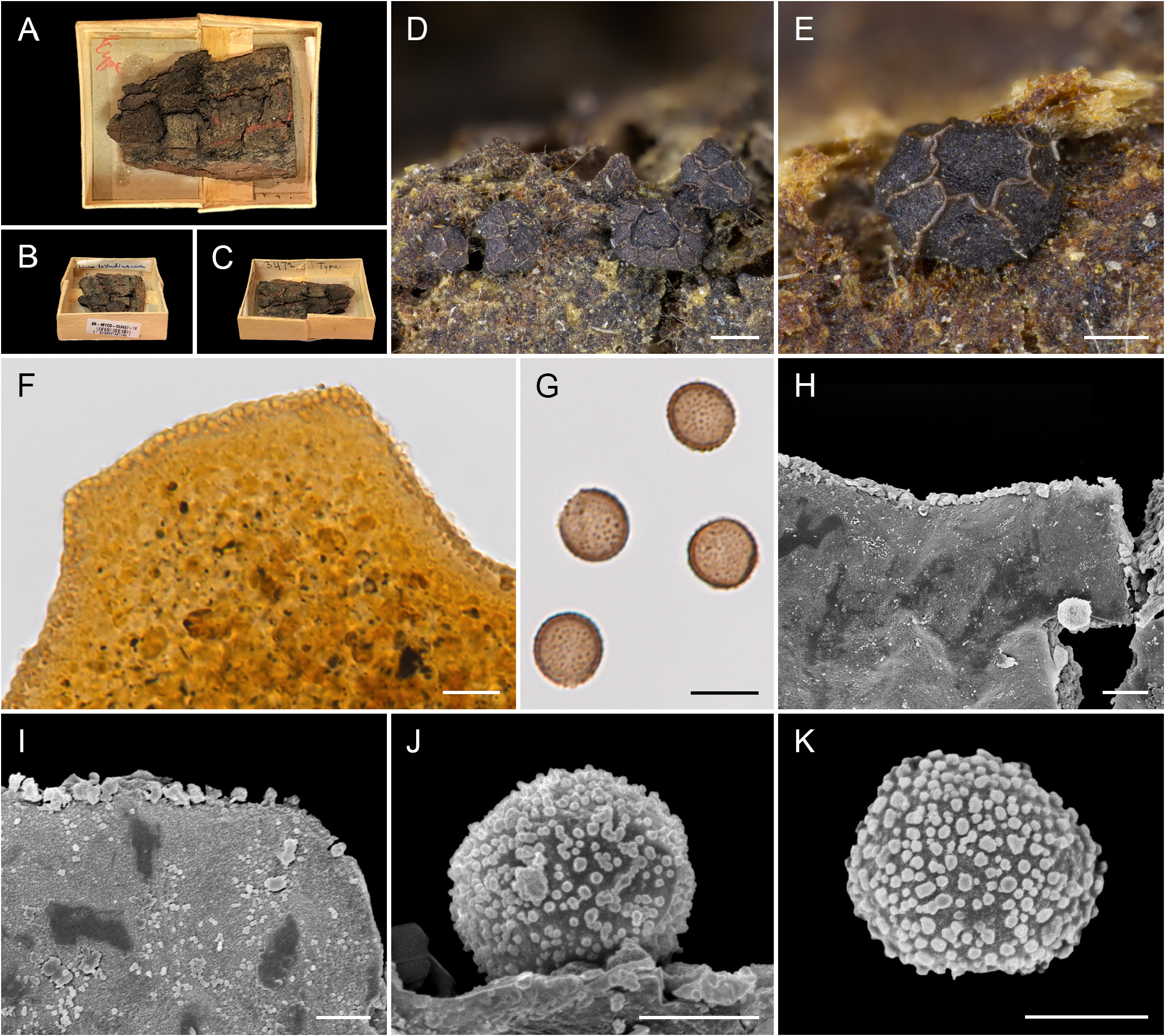
24. Licea testudinacea Nann. -Bremek. Acta Bot. Neerl. 14: 141 (1965) Figs. 20A–K View FIGURE 20
Sporophores sporocarpic, grouped or scattered, sessile. Sporocarps suglobose, 0.1 mm diam., to pulvinate, angular, 0.15 mm high, 0.2–0.8 mm long, dark brown to black, with undulating glossy lighter ridges marking places of dehiscence ( Fig. 19 E View FIGURE 19 ). Peridium double, the outer layer closely appressed and covered with granular refuse material ( Fig. 19 F View FIGURE 19 ), the inner layer inner surface shiny, minutely warted, brownish yellow/orange by TL,; dehiscence along ridges into many small platelets with a band of interlacing outgrowths on platelet margins ( Fig. 19 F View FIGURE 19 ). Spores very dark brown in mass, grey by TL, with a conspicuous paler area, globose, (10–)12–13(–15) µm diam., warted. By SEM the inner peridial surface is densely and minutely warted, with different sized warts ( Fig. 19 I View FIGURE 19 ), the outer layer is covered with granular material; the platelet margins ornamented with a row of prominent different sized outgrowths and pegs, some interlacing ( Figs. 19 H – I View FIGURE 19 ); the epispore is covered by flattened verrucae.
Material examined: Holotypus. NETHERLANDS. Doorwerth, 10-October-1959, NENB 3472 ( BR 5020053457106)! .
Habitat: on bark of living and dead trees, dead trunks of angiosperms and gymnosperms.
Distribution: Norway, Sweden, Lithuania, Russian Federation, United Kingdom, Ireland, Denmark, Netherlands, Belgium, Germany, France, Portugal, India, Japan, USA, Mexico, Guatemala, Costa Rica, Brazil, Australia, New Caledonia.
Icon.: Nannenga-Bremekamp (1965: 141, Figs. 8 A – B View FIGURE 8 ; 1975: 68; 1991: 46), Neubert et al. (1993: 283, Figs. VI: 7–8), Novozhilov et al. (1999: 85, Figs. 4 E – H View FIGURE 4 ), Yamamoto (2006: 39, Figs. 17 A – C View FIGURE 17 ; 2021: 150, Figs. A – C), Johannesen & Vetlesen (2020: 81, Figs. 41 C – E), Barbosa & Cavalcanti (2020: 422, Figs. 3 A – J View FIGURE 3 ).
Notes. This species has dark sporocarps with lighter dehiscence lines, small numerous platelets and dark warted spores with a pale area. It was described with a single peridium and by light microscope it appears single because the two layers are tightly appressed. The author indicates in her later keys ( Nannenga-Bremekamp 2022) that the peridium is in fact double, and this can be seen in Fig. 19H View FIGURE 19 herein. This combination of characters separates it from similar species like L. pygmaea (Meyl.) Ing , L. castanea G. Lister and L. chelonoides Nann. -Bremek.
No known copyright restrictions apply. See Agosti, D., Egloff, W., 2009. Taxonomic information exchange and copyright: the Plazi approach. BMC Research Notes 2009, 2:53 for further explanation.