
Piaroa virichaj, Manzanilla, Osvaldo Villarreal, Giupponi, Alessandro Ponce De Leão & Tourinho, Ana Lúcia, 2008
|
publication ID |
https://doi.org/10.5281/zenodo.183654 |
|
DOI |
https://doi.org/10.5281/zenodo.5680227 |
|
persistent identifier |
https://treatment.plazi.org/id/03B78798-FF8D-4506-FF13-F880FF6DFA22 |
|
treatment provided by |
Plazi |
|
scientific name |
Piaroa virichaj |
| status |
sp. nov. |
Piaroa virichaj View in CoL sp. n.
Figs 1–12 View FIGURES 1 – 2 View FIGURES 3 – 5 View FIGURES 6 – 9 View FIGURES 11 View FIGURE 12 .
Etymology. According to the Piaroa culture Virichaj is the demon that caste spells of fever on natives.
Type material. Male holotype ( MHNLS I 0077). VENEZUELA, AMAZONAS, Tobogán de la Selva, 5º23’10” N 67º36’53” W. 160 m. 2325.xii.2002. Giupponi A., PérezGonzález A. & Villarreal M.O. leg. Paratypes. 1 female and 1 subadult ( MHNLS I 0077). 2 subadults ( MNRJ 04291), the same.
Diagnosis. Total length between 4.92 – 5.56 millimetres. Propeltidium with three pairs of dorsal and anterior setae. With faint, oval eyespots. Movable cheliceral finger with long keel or lamella with three teeth, guard tooth very reduced and curved, unmovable finger with five teeth. Male flagellum flattened dorsoventrally, very elongated, with two dorsal small depressions. Femur IV three times wider than long. Female lateral lobes of spermathecae swelling at base, curving distally, so the bulbs are facing. Apical trifid blunt bulbs with little nodules present.
Description of the male ( holotype). Coloration. Propeltidium, chelicerae, legs I, pedipalpus and basal portion of flagellum yellowishbrown. Mesopeltidium, metapeltidium, abdominal segments and legs lighter. Propeltidium. ( Figs 1–2 View FIGURES 1 – 2 , 6 View FIGURES 6 – 9 ). With two anterior setae (one behind the other), and one pair of medial dorsal setae and other posterior pair. Metapeltidium entire.
Abdomen ( Figs 1–2 View FIGURES 1 – 2 ). Setae: Tergite II with three pairs of microsetae. Tergites I–VIII with a pair of large dorsal; VIII with one pair and one pair of distolateral IX with one pair of large distal and one pair of short and distolateral ones. Segments IX–XII elongated. Segment XII without posterodorsal process. Ventral region. Respiratory spiracles large and oval, slightly sclerotized, darker than sternites.
Flagellum ( Figs. 3–5 View FIGURES 3 – 5 ). Pedicel very long, about 65% of the length of the bulb. Bulb 1.3 time longer than wide. Distal portion 2.6 times the length of the bulb. Bulb flattened dorsoventrally, very elongated, with two dorsal small depressions. Vm2 anterior Dm1; Vm1 at level Dm1; Vm4 at level of proximal border of dorsal depression; Dm4 posteriorly, near of Vl2; Dl3 extremely posterior.
Chelicerae ( Fig. 7 View FIGURES 6 – 9 ). Movable jaw sharp and curving in terminal third, subapical guard tooth present, with 3 acessory teeth, fixed jaw with 5, the proximal larger, three next the same size, distal teeth slightly larger.
Pedipalps ( Fig. 8 View FIGURES 6 – 9 ). Trochanter with a small prolateral spine, with large sharp frontal process, trianglelike shaped, with a row of ventral setae. Femur short and robust, dorsally curved, in shape of clef, thinner at base and wider at apex, dorsal surface three times longer than ventral, dorsal surface with some setae, ventral face armed with apical short and blunt tubercle. Patella surface generally smooth, tubular, three times longer than wide, with short mesoventral spines and a few dorsal and ventral setae. Tibia similar in form with patella, slightly thinner and lesser setae, dorsally curved. Tarsus conic, half of the tibia’s length, with numerous setae, visible sharp spur, and apical inner spine; tarsal claw sharp and curved, slightly larger than half tibial length.
Legs ( Fig. 9 View FIGURES 6 – 9 ). Hubbardiinae pattern, anterodorsal margin of femur IV curved at about a 90° angle.
Measurements. Table 1 View TABLE 1 .
Description of female ( paratype). (Figs 10–11). Medial lobes and gonopod absent, when cleared with peroxide at 3% and examined in glycerin. Chitinized arch with posterior border Ushaped, 2.6 times wider than long; chitinized arch and base lobes joint heartshaped, deep anterior border constriction, reaching 1/3 of structure length.
Natural history. All specimens were collected in a very restricted and small microhabitat, they were found under flat stones, they were upsidedown walking on the stone’s undersurface. The locality ( Fig. 12 View FIGURE 12 ) was a secondary growth forest with a sunless understory. It was very humid and resembled a dry stream bed; with dense litter layer, rotten logs and stones. Presently, this locality has been being used for tourism activities.
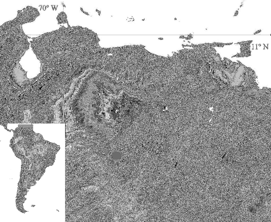
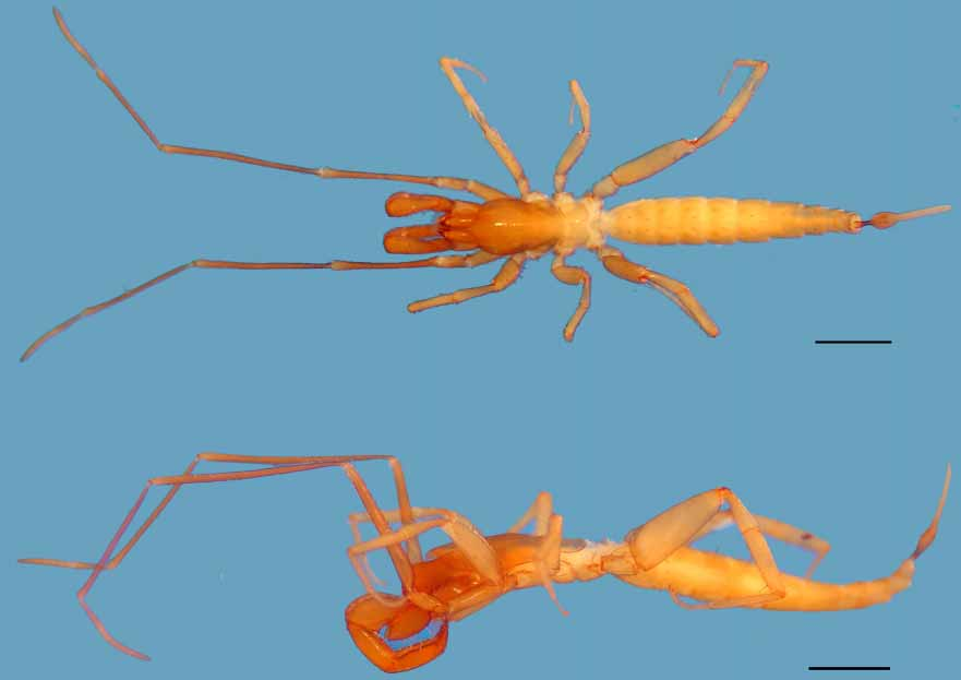
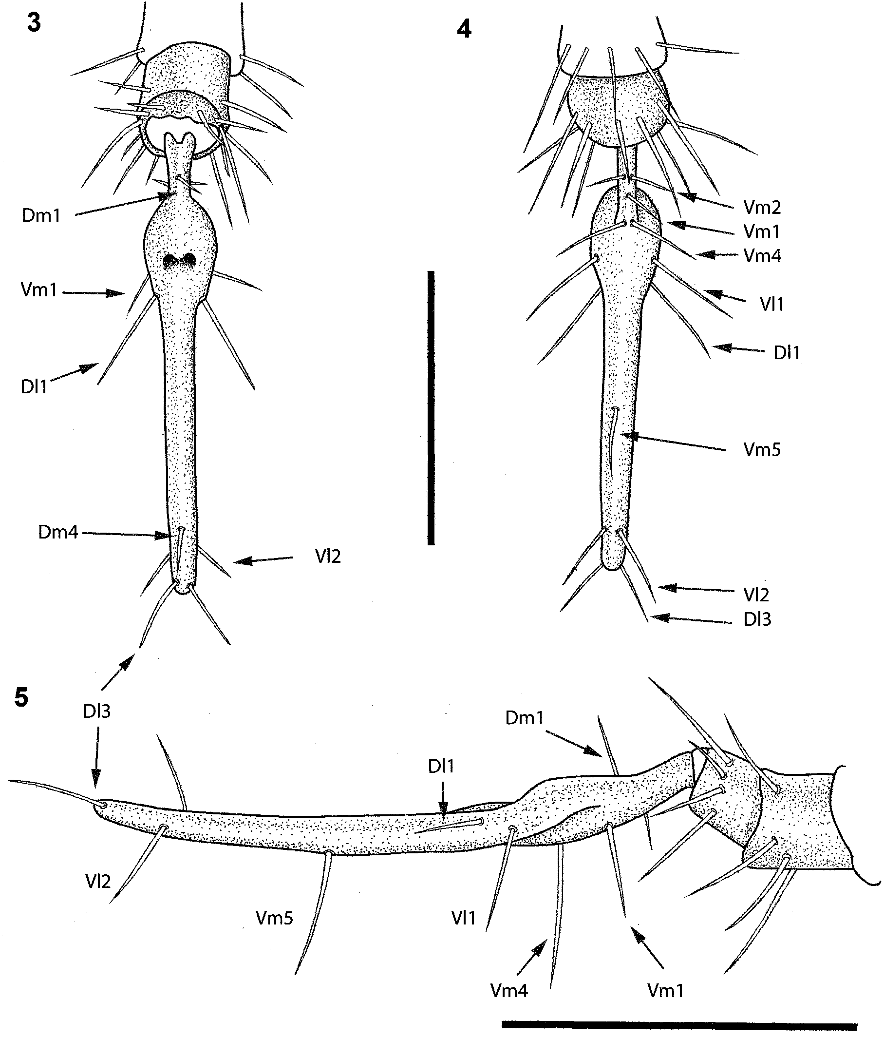
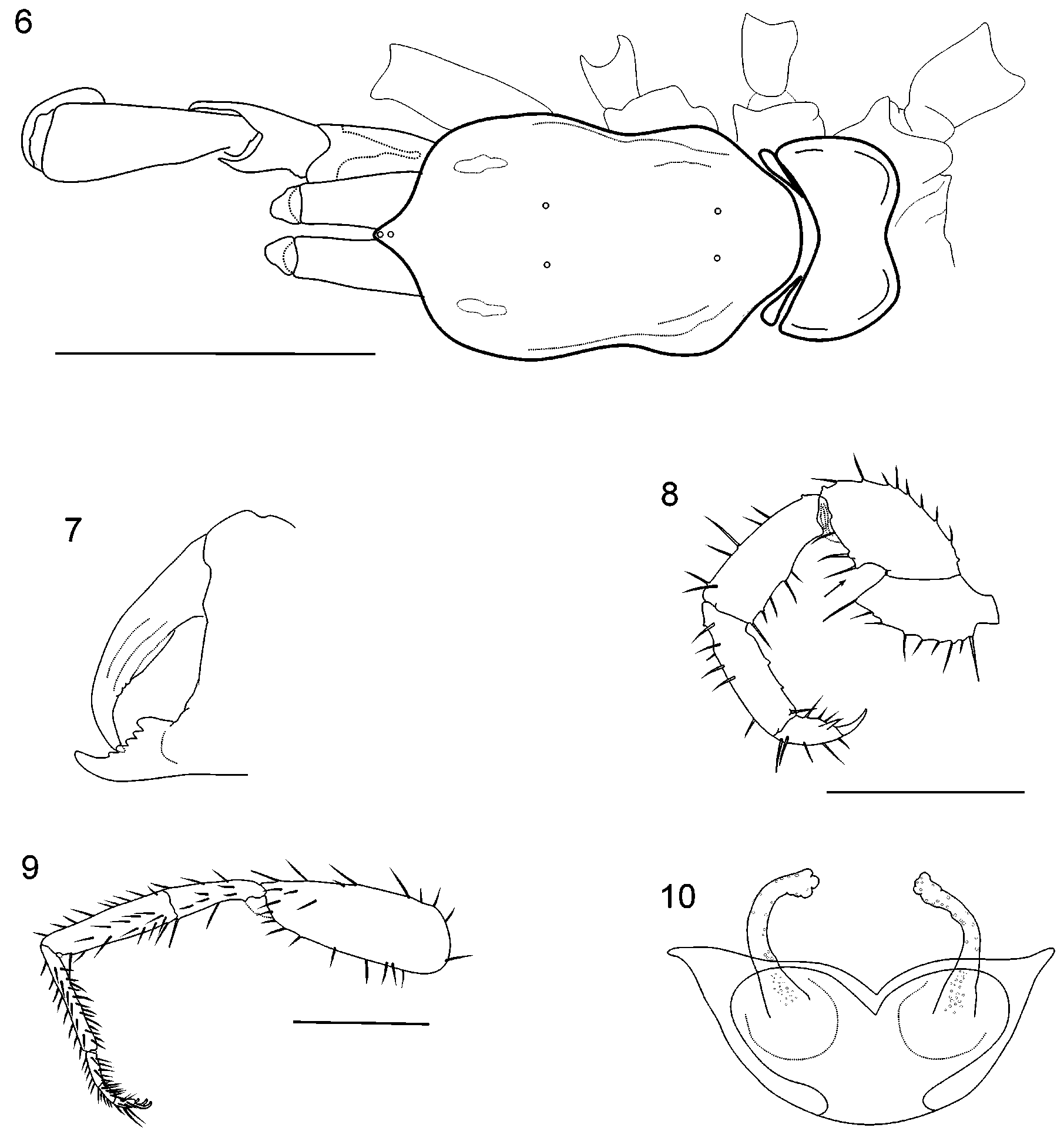
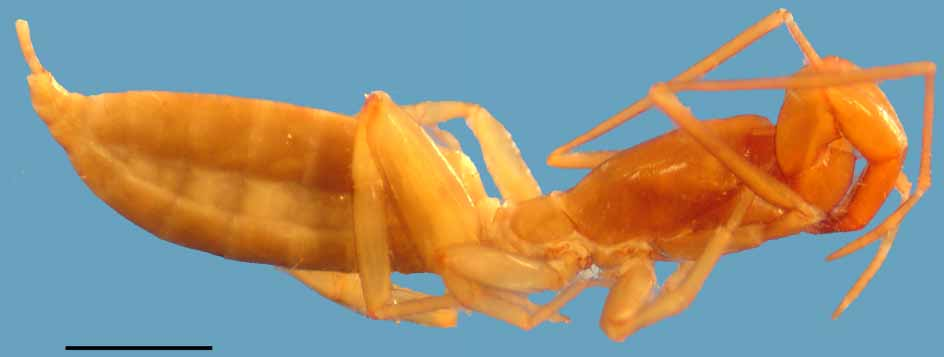
TABLE 1. Piaroa virichaj gen. n. & sp. n. Measurements in millimeters.
| Characters | MALE (MHNLS I0077) | FEMALE (MHNLS I0077) |
|---|---|---|
| Propeltidium: L/W | 1.61/0.87 | 1.53/0.82 |
| Abdomen: L | 3.95 | 3.39 |
| Flagellum: L/W/H | 1.68/0.26/0.21 | broken 0.37/0.10/ 0.8 |
| Pedipalp: L trochanter femur patela tibia tarsus/claw | 0.57 0.79 0.87 0.87 0.39/0.16 | 0.59 0.63 0.68 0.58 0.29/0.16 |
| Leg: I L trochanter femur patela tibia basitarsus telotarsus | 0.57 1.79 2.29 1.71 0.71 0.42 | 0.37 1.58 1.87 1.45 0.42 0.59 |
| Leg: IV L trochanter femur patela tibia basitarsus telotarsus | 0.58 1.76 0.74 0.74 0.81 0.66 | 0.39 1.24 1.34 0.79 0.26 0.32 |
No known copyright restrictions apply. See Agosti, D., Egloff, W., 2009. Taxonomic information exchange and copyright: the Plazi approach. BMC Research Notes 2009, 2:53 for further explanation.
|
Kingdom |
|
|
Phylum |
|
|
Class |
|
|
Order |
|
|
Family |
|
|
SubFamily |
Hubbardiinae |
|
Genus |