
Rhizomyces forcipatus W.Rossi & Feijen, 2018
|
publication ID |
https://doi.org/ 10.5852/ejt.2018.474 |
|
DOI |
https://doi.org/10.5281/zenodo.3845939 |
|
persistent identifier |
https://treatment.plazi.org/id/03B687CF-5124-FF85-EDDD-FCA119507977 |
|
treatment provided by |
Valdenar |
|
scientific name |
Rhizomyces forcipatus W.Rossi & Feijen |
| status |
sp. nov. |
Rhizomyces forcipatus W.Rossi & Feijen sp. nov.
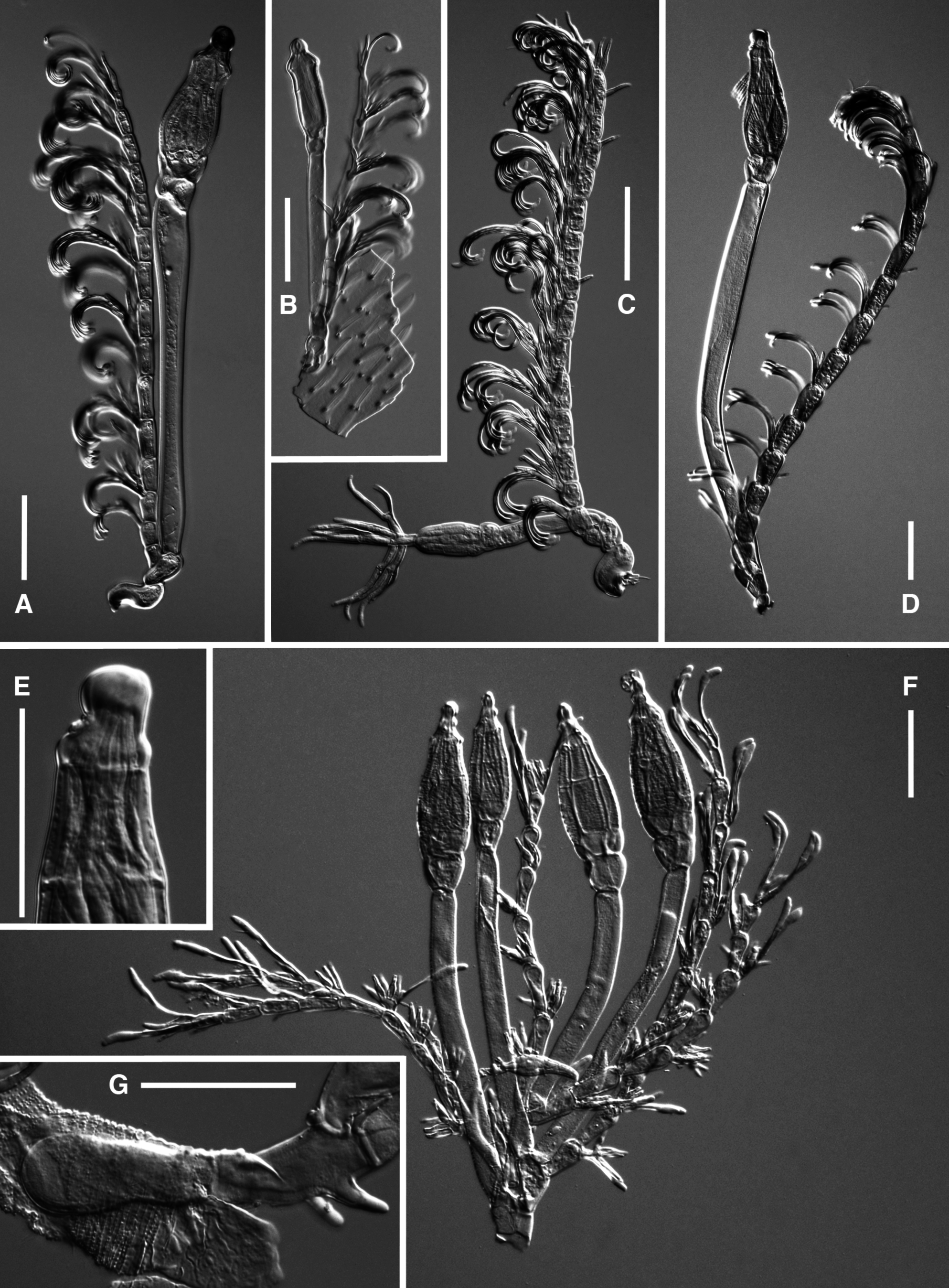
MycoBank No: MB827885 Fig. 1 View Fig A–C
Description
Basal cell hyaline, subsigmoid, more than twice as long as the maximum width, lying flat on the insect, with the black foot placed laterally and flanked by a depression apparently working as a sucker. Suprabasal cell (cell II) variable in size, shape and color, usually smaller and sometimes darker than the basal. Stalk cell of the appendage (cell III) distinctly smaller and darker than cell II, from which it is separated by a slightly oblique septum. Free appendage usually exceeding the perithecial apex, with the axis consisting of 12–16 almost hyaline cells gradually longer and more slender, each bearing a branch arising from the upper, outer angle; these branches consist of an elongate basal cell bearing distally 2–4 bottle-shaped antheridia with darker necks and a one-celled short, hyaline, slender and erect cell which gives rise laterally to a vertical series of 4–8 very slender branchlets; the latter are dark brown, distinctly curved, with hyaline and enlarged tips. Perithecial stalk-cell (cell VI) long, slender, flexuous and hyaline (or almost so). Basal cell region well distinguished, tinged with very pale brown, concolorous with the perithecial venter, consisting of relatively large, subequal cells with their outer margins bulging outwards. Perithecium almost three times longer than maximum width, the venter very slightly inflated, distinctly enlarged below the brownish tip, which tapers rather abruptly to a subspherical apex. Trichogyne consisting of a club-shaped cell from which arise distally 4–5 spreading branchlets which are curved or sigmoid, sometimes bifurcate, gradually tapering towards the apex. Length from foot to perithecial apex 210–345 µm; from foot to the apex of the appendage 250–400 µm; perithecium 70–75 × 25–30 µm; antheridia about 20 µm.
Etymology
From Latin forceps, referring to the forceps-like shape and function of the basal cell.
Types
KENYA: Naro Moru river, on Centrioncus decoronotus Feijen, 1983 , 20 Jul. 1987, H.R. Feijen leg. (holo-: FI 4100a; iso-: FI 4100b); same data as preceding except different host specimens (para-: FI 4099, FI 4101, FI 4102).
MALAWI: Mt. Soche, on sternites of male Centrioncus jacobae Feijen, 1983 , 19 Mar. 1972, H.R. Feijen leg. (para-: FI 4103, RMNH-HF-74); same data as preceding except on wing of the same host specimen (para-: RMNH-HF-90); Mt. Soche, on sternites of male Centrioncus jacobae , 7 Jan. 1973, H.R. Feijen leg. (para-: RMNH-HF-86, RMNH-HF-87); same data as preceding except on wing of female Centrioncus jacobae (para-: RMNH-HF-88); Mt. Ndirande, on wing of female Centrioncus jacobae , 16 Feb. 1974, H.R. Feijen leg. (para-: RMNH-HF-73).
IVORY COAST: Amanikro, on the wing of a female Centrioncus decellei Feijen, 1983 , Sep. 1961 (para-: RMNH-HF-3).
REPUBLIC OF SOUTH AFRICA, Eastern Cape, Hogsback, Tyume Forest, 32.6029° S, 26.9384° E, 1166 m a.s.l., on Teloglabrus sp., 10 Apr. 2010, A.H. Kirk-Spriggs & V. de Swart leg. (para-: FI 4125, FI 4126); Western Cape, Buffelsbos forest, 33.9026° S, 23.6388° E, 400 m a.s.l., on Teloglabrus australis Feijen, 1983 , 27 Mar. 2009, A.H. Kirk-Spriggs leg. (para-: FI 4129).
Remarks
The description is based on the parasites obtained from the sternites of the host insects; thalli growing on the wings are smaller (180–255 µm from foot to perithecial apex: Fig. 1B View Fig ). The thalli observed on Centrioncus jacobae are paler, more slender and longer on average (up to 550 µm from foot to perithecial apex).
Rhizomyces forcipatus sp. nov. is distinguished from all the other species in the same genus by the peculiar shape and function of the basal cell. The latter is turned in, forming a ‘forceps’ which grasps a fold of the exoskeleton of the host insect ( Fig. 1C View Fig ).
Rhizomyces forcipatus sp. nov. bears a superficial resemblance with R. crispatus . However, the latter species has a stockier habitus, a more luxuriant appendage, a distinctly smaller and darker cell III, a darker and subconical tip of the perithecium, and it obviously differs from the new species in the shape of the basal cell.
The presence of this fungus was already reported on various species of Centrioncus Speiser, 1910 and Teloglabrus Feijen, 1983 by Feijen (1983).
Rhizomyces forcipatus sp. nov. is found on several species of the genera Centrioncus and Teloglabrus . This is remarkable, since the species of these genera occur in Afromontane forests and have very limited, allopatric distributions ( Feijen 1983). The rate of parasitism came to 17%; however, this figure was lowered by the large Teloglabrus sanorum Feijen, 1983 sample of young flies. In female flies, R. forcipatus sp. nov. was only found on the wings. In male hosts, the thalli were found on the wings (79%) and on the sterna (30%). Thalli were most often found on both wings together (61%), followed by right wing only (32%). 90% of thalli found on the sterna of males flies were on the left hand side ( Table 2).
No known copyright restrictions apply. See Agosti, D., Egloff, W., 2009. Taxonomic information exchange and copyright: the Plazi approach. BMC Research Notes 2009, 2:53 for further explanation.