
Ischnothyreus fobor, Kranz-Baltensperger, Yvonne, 2011
|
publication ID |
https://doi.org/ 10.5281/zenodo.278111 |
|
DOI |
https://doi.org/10.5281/zenodo.6189536 |
|
persistent identifier |
https://treatment.plazi.org/id/03B587EE-FFC4-FFF6-46A1-5AB6CB180CCF |
|
treatment provided by |
Plazi |
|
scientific name |
Ischnothyreus fobor |
| status |
sp. nov. |
Ischnothyreus fobor View in CoL sp. nov.
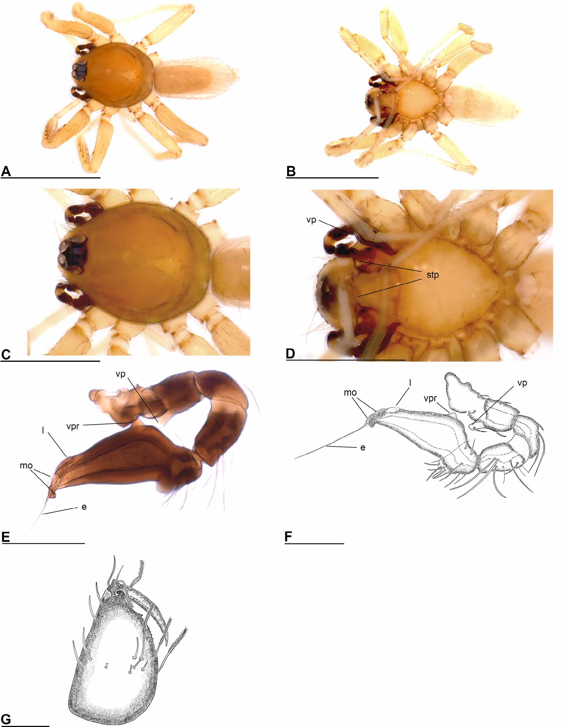
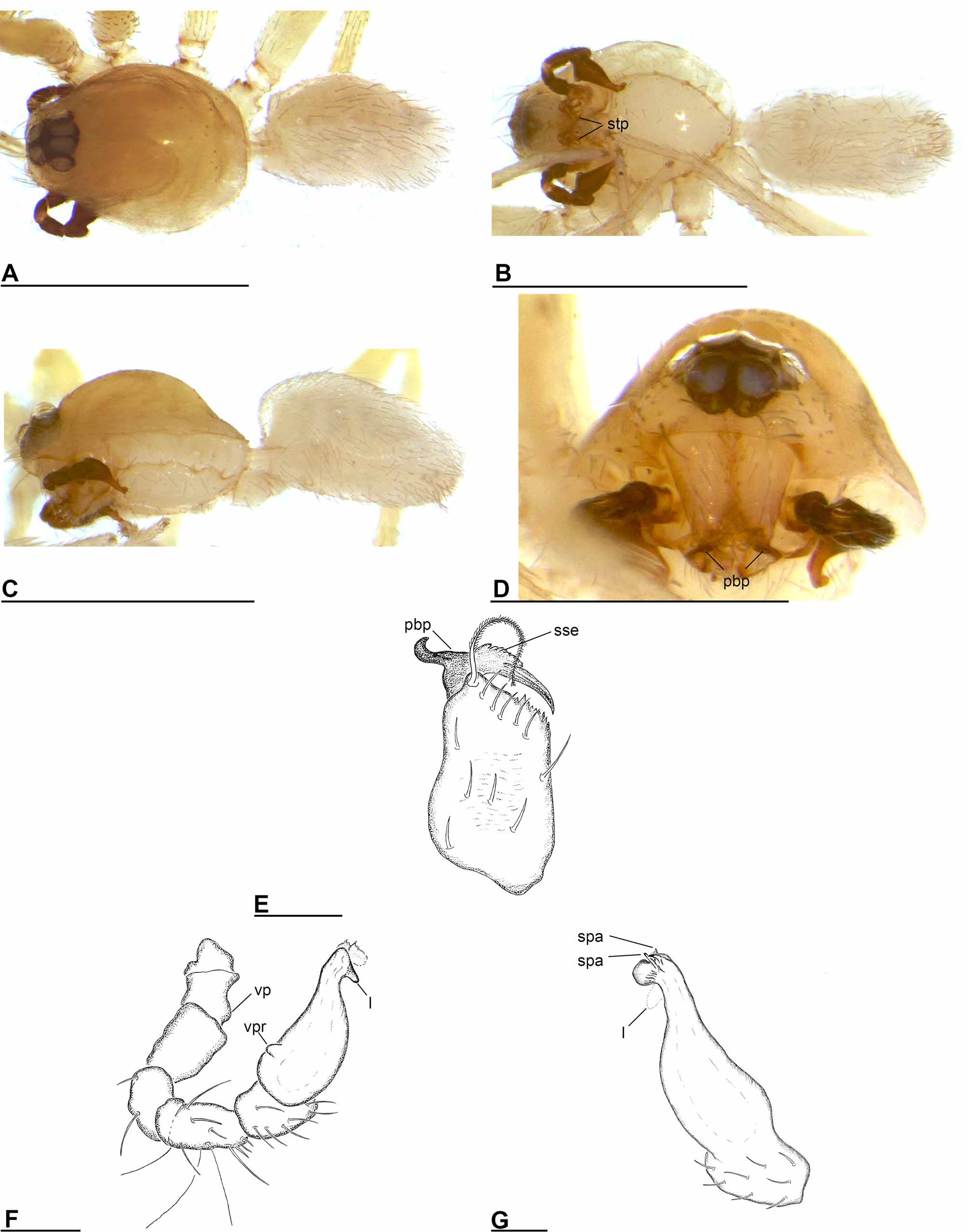
( Figs 20–21 View FIGURE 20 View FIGURE 21 , 35 View FIGURE 35 )
Type material: Holotype: male (PBI_OON 00016082), MALAYSIA: Sabah: Danum Valley Field Centre, 6.– 16.V.1991, leg. C.L. & P.R. Deeleman, RMNH. Paratypes: 2 males & 2 females (PBI_OON 00032229), collected together with holotype, RMNH.
Additional material examined: 2 males (PBI_OON 00031469), MALAYSIA: Sabah: Kinabalu National Park, 1550m, 21.–27.VII.1980, leg. C.L. & P.R. Deeleman ( RMNH). 1 male (PBI_OON 00032231), Bru-88/12. BRU- NEI DARUSSALAM: Brunei-Muara District: près du pont sur le ruisseau Sungai Lubang Barus , route venant de Tutong, 33km de Bandar Seri Begawan, prélèvement de sol au pied de deux grands arbres proches des habitations, env. 20m (translated: close to bridge over Sungai Lubang Barus river, route from Tutong, 33 km from Bandar Seri Begawan, soil extraction at base of two large trees close to habitations, ca. 20m); 16.XI.1988; leg. B. Hauser (Berlese à Seri Begawan) ( MHNG), 1 female (PBI_OON 00015355), Bru-88/33. BRUNEI DARUSSALAM: Belait District: Labi Hills Forest Reserve, Teraja, 42km S de Sungai Liang (12km au Sud de Labi), environs de Rumah Panjang, forêt primaire (translated: Labi Hills Forest Reserve, Teraja, 42km south of Sungai Liang (12km south of Labi), proximity of Rumah Panjang), mixed dipterocarp forest, 40m; 22.XI.1988, leg. C. Lienhard ( MHNG).
Etymology: The specific name, a noun in apposition, is an abbreviation of "found in Borneo".
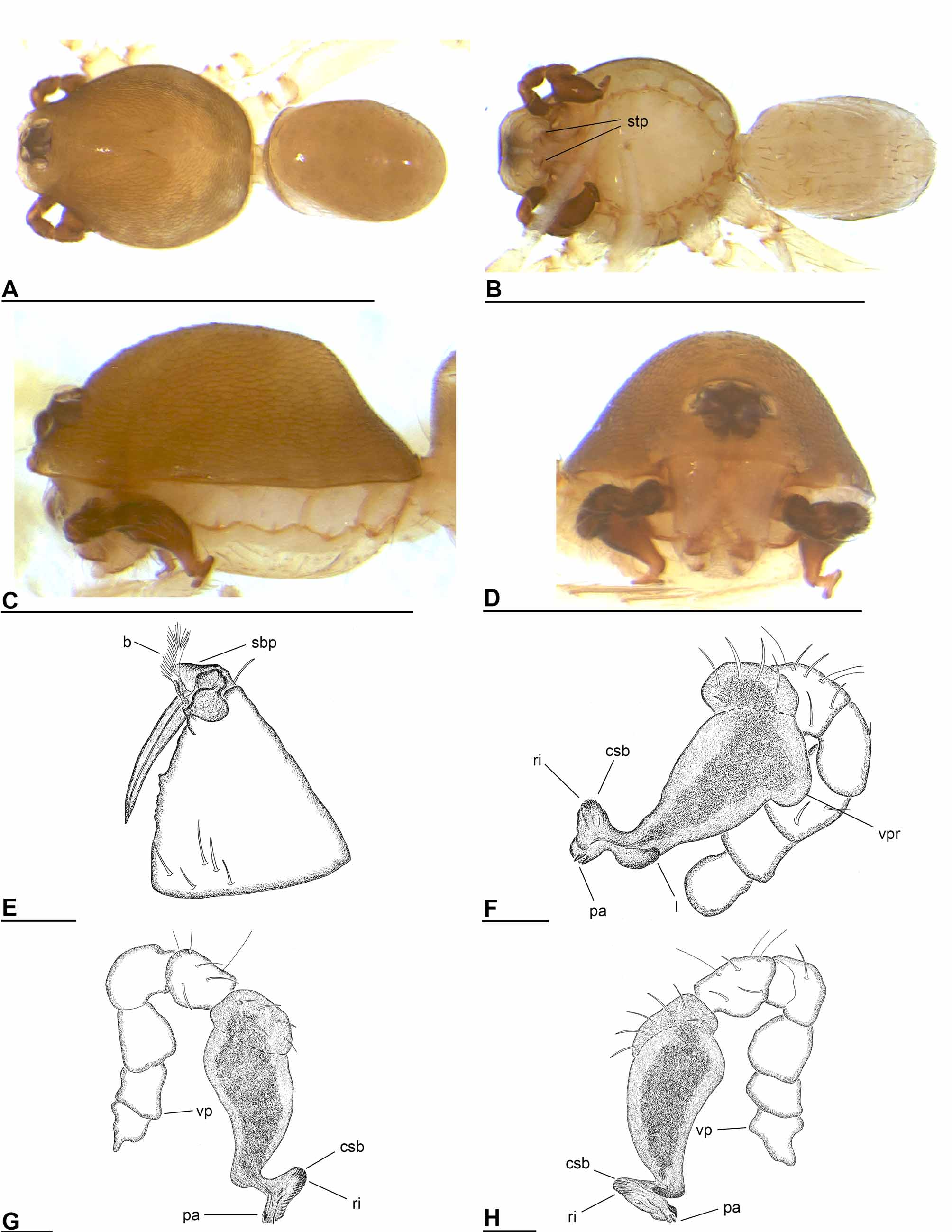
Diagnosis: The male of I. fobor can be recognized by the palpal bulb with its extremely long and thin embolus ( Fig. 20 View FIGURE 20 E–F). The female genital area is characterized by a small strongly winding tube running posteriorly ( Fig. 21 View FIGURE 21 F–G).
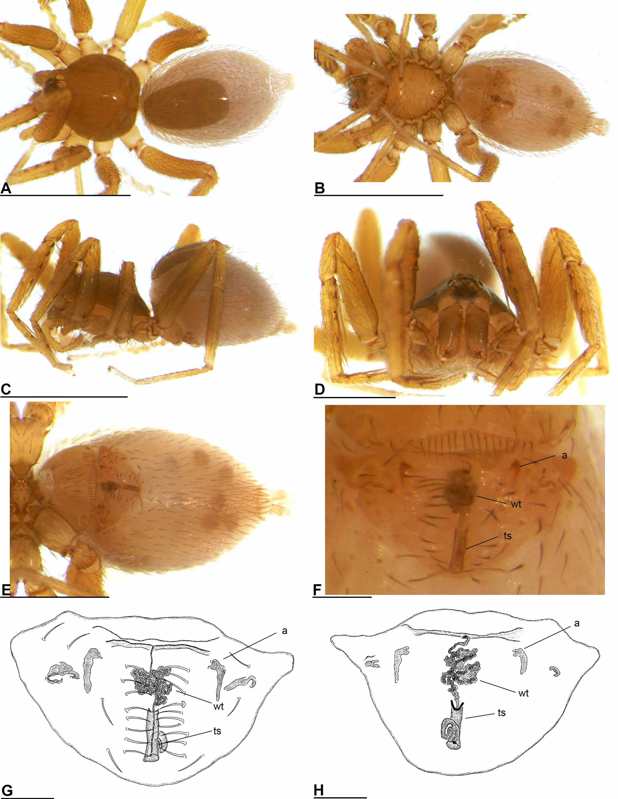
Description: Male (holotype). Total length 1.66 mm. Carapace: domed in lateral view, fovea present, lateral margin slightly rebordered. Clypeus: curved downwards in frontal view. Eyes: ALE touching. Sternum: setae abundant. Mouthparts: chelicerae slightly divergent, without prominent process at base of fangs, fang groove with few small and one larger denticles ( Fig. 20 View FIGURE 20 G). Anterior margin of labium indented at middle. Anteromedian tip of endites with one strong, tooth-like projection (stp) ( Fig. 20 View FIGURE 20 D). Abdomen: scutum extending far dorsal of pedicel. Dorsal scutum covering 1/2 to 3/4 of abdomen, fused to epigastric scutum. Epigastric and postepigastric scutum short, almost rectangular, covering about 2/3 of abdominal length. Spinneret scutum present, incomplete ring, with fringe of needle-like setae. Legs: spine formula: femora: I p0-1-1; II p0-0-1; tibiae: I, II p2-1-1; r2-1-1; metatarsi: I, II p1-1-0; r1-1-0. Genitalia: Palp strongly sclerotized, proximal segments dark brown, trochanter with large ventral projection (vp) ( Fig. 20 View FIGURE 20 D–F), femur normal size, patella shorter than femur, not enlarged, cymbium dark brown, fused with bulb, bulb dark brown, more than two times as long as cymbium, stout, tapering apically, with ventral protuberance (vpr) and lobe (l), embolus (e) very long and filamentous, originating between several membranous outgrowths (mo) ( Fig. 20 View FIGURE 20 E–F).
Female (paratype). Total length 1.68 mm. As in male except as noted. Carapace: without any pattern, broadly oval in dorsal view, pars cephalica strongly elevated in lateral view. Clypeus: straight in frontal view. Eyes: ALE separated by less than their radius. Mouthparts: labium not fused to sternum, anterior margin not indented at middle. Endites unmodified. Femur of female palp with three ventral spines, tibia with three trichobothria. Abdomen: dorsal scutum covering about 1/2 of abdomen, not fused to epigastric scutum. Postepigastric scutum widely hexagonal, covering about 1/3 of the abdominal length, with short posteriorly directed lateral apodemes (a) ( Fig. 21 View FIGURE 21 F). Dense patch of setae anterior to spinnerets present. Legs: spine formula: femora: I p0-1-1; r0-1-1; II p0-0-1; r0-0-1; tibiae: I, II p2-1-1; r2-1-1; metatarsi: I, II p1-1-0; r1-1-0. Genitalia: simple, in the middle of the anterior margin of the postepigastric scutum starts a winding tube (wt), running posteriorly, ending in globlet-like structure (gls) ( Fig. 21 View FIGURE 21 F–G).
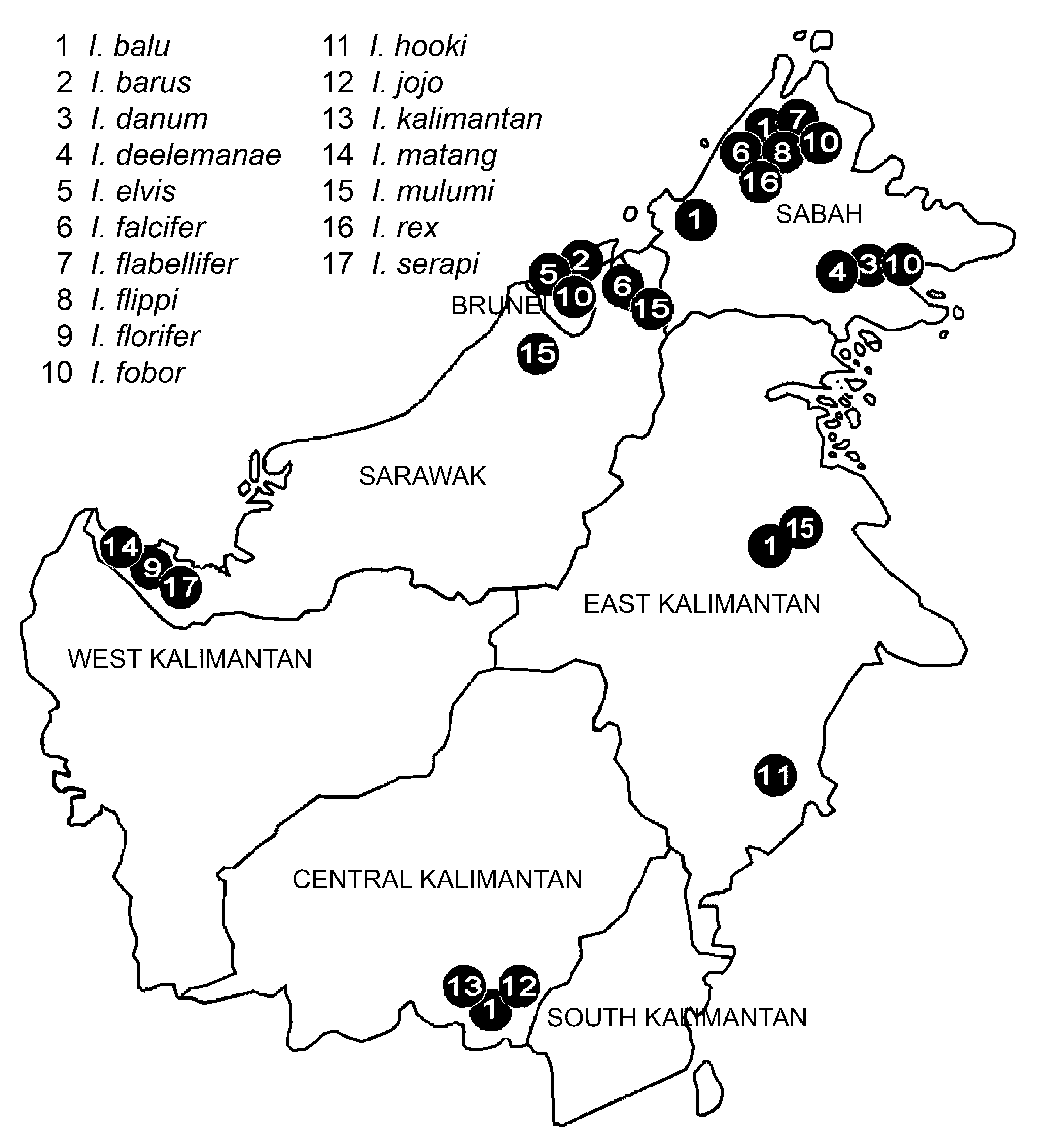
Distribution: Sabah and Brunei ( Fig. 35 View FIGURE 35 ).
Ischnothyreus hooki sp. nov. ( Figs 22–23 View FIGURE 22 View FIGURE 23 , 35 View FIGURE 35 )
Type material: Holotype: male (PBI_OON 00031692), INDONESIA: East Kalimantan : Tenggarong, light forest, leaflitter, 4.VIII.1980, coll. Deeleman, RMNH. Paratypes: 1 male (PBI_OON 00031363), INDONESIA: East Kalimantan : Sepaku, 40km NNW from Balikpapan, degraded primary forest, 2.VIII.1980, coll. Deeleman, RMNH. 2 females (PBI_OON 00031769), INDONESIA: East Kalimantan : Sepaku, 40km NNW of Balikpapan, primary forest, dry period, 21./ 22.VII.1982, coll. Deeleman, RMNH.
Additional material examined: 1 female (PBI_OON 00031777), INDONESIA: East Kalimantan : Sepaku, 40km NNW of Balikpapan, degraded rainforest, litter, 2.–5.VIII.1980, coll. Deeleman ( RMNH). 1 male (PBI_OON 00031628), INDONESIA: East Kalimantan : Sungai Wain Protection Forest, ca. 15 km N of Balikpapan (1°08’36”S, 116°50’59”E), 80m, secondary forest adjacent to primary forest, 5./ 7.X.2008, leg. P. Schwendinger ( MHNG).
Etymology: The specific name is derived from Captain Hook from the novel Peter Pan, written by James M. Barrie, and refers to the similarity of the distal part of the male palp with the left arm of Captain Hook.
Diagnosis: The male of I. hooki can be recognized by the hook-like embolus ( Fig. 22 View FIGURE 22 G–J). With its strongly winding tube, the female genital area of I. hooki is similar to I. fobor , but the winding tube in I. hooki is larger.
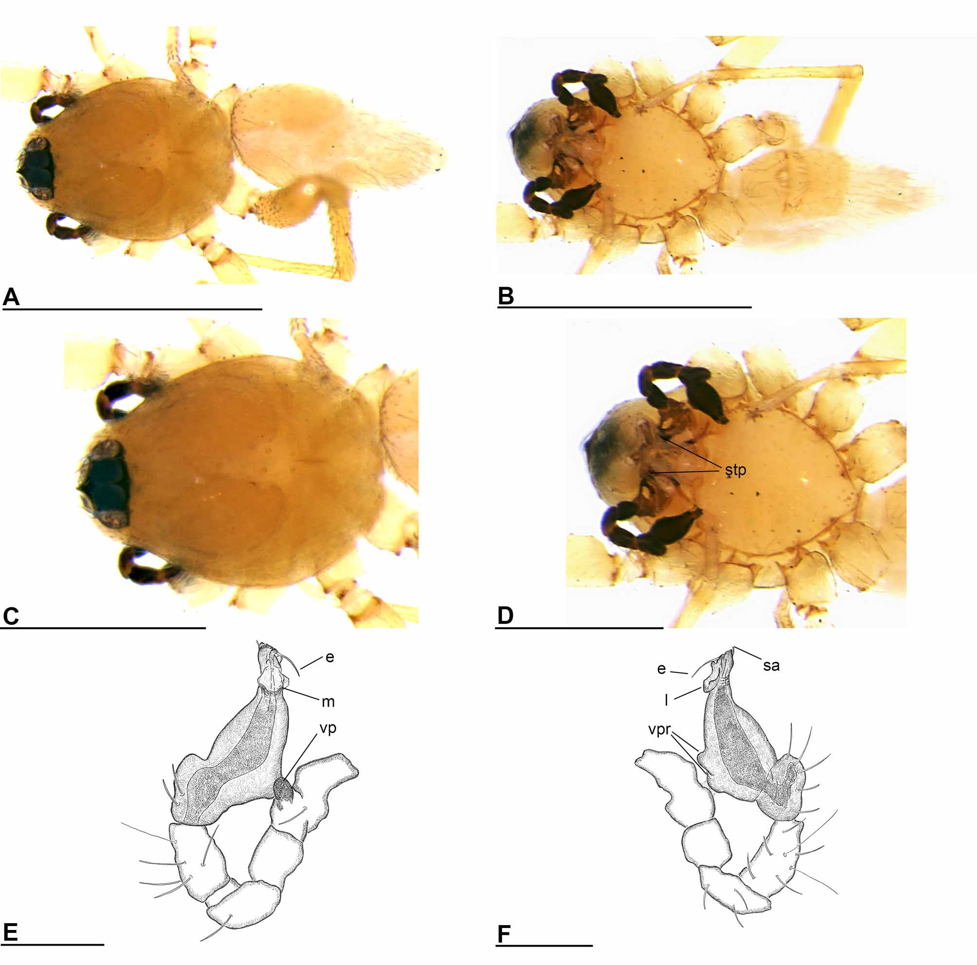
Description: Male (holotype). Total length 1.33 mm. Carapace: broadly oval in dorsal view. Clypeus: curved downwards in frontal view, low, ALE separated from edge of carapace by less than their radius. Eyes: PLE circular, posterior eye row straight from above, procurved from front. Sternum: setae abundant. Mouthparts: chelicerae without prominent process at base of fangs. Labium triangular. Anteromedian tip of endites with one strong, toothlike projection (stp) ( Fig. 22 View FIGURE 22 B, C, F). Abdomen: scutum extending far dorsal of pedicel. Legs: spine formula: femora: I p0-1-1; r0-0-1; II v0-0-2; tibiae: I, II v4-2 -2; metatarsi: I, II v2-2 -0. Genitalia: Sperm pore situated in front of anterior spiracles. Palps strongly sclerotized, proximal segments brown, trochanter with ventral projection, patella about as long as femur, not enlarged, cymbium brown, fused with bulb, bulb brown, more than two times as long as cymbium, stout, tapering apically, with ventral protuberance (vpr) ( Fig. 22 View FIGURE 22 H, J) and lobe (l) ( Fig. 22 View FIGURE 22 . G, I), distal part with ear-like outgrowth (elo) prolaterally ( Fig. 22 View FIGURE 22 H, J), embolus (e) short hook ( Fig. 22 View FIGURE 22 G, I, J).
Female (paratype). Total length 1.80 mm. As in male except as noted. Carapace: without any pattern. Eyes: posterior eye row procurved from above. Mouthparts: endites unmodified. Femur of female palp with four ventral spines, tibia with three trichobothria. Abdomen: scutum not extending far dorsal of pedicel. Dorsal scutum covering less than 1/2 of abdomen, less than 1/4 abdomen width, not fused to epigastric scutum. Postepigastric scutum widely hexagonal, only around epigastric furrow, with short posteriorly directed lateral apodemes (a) ( Fig. 23 View FIGURE 23 E). Dense patch of setae anterior to spinnerets present. Legs: spine formula: femora: I p0-2-1; r0-0-1; II p0-0-2; tibiae: I, II p2-1-1; r2-1-1; metatarsi: I, II p1-1-0; r1-1-0. Genitalia: the posterior margin of the epigastric scutum is lined with numerous needle-like setae. In the middle of the postepigastric scutum is a strongly winding tube (wt) ( Fig. 23 View FIGURE 23 E–H) visible, which runs posteriorly.
Distribution: East Kalimantan ( Fig. 35 View FIGURE 35 ).
Remarks: Male and female of this species were not collected together. However, since the somatic morphology is very similar they are tentatively considered as conspecific.
Ischnothyreus jojo sp. nov. ( Figs 24–25 View FIGURE 24 View FIGURE 25 , 35 View FIGURE 35 )
Type material: Holotype: male (PBI_OON 00032184), INDONESIA: Central Kalimantan : Kaharian, 2°02'S, 113°40'E, 2.–16.IX.1985, swampy primary forest, leaflitter, leg. Suh. Djojosudharmo, coll. Deeleman, RMNH. Paratypes: 3 females (PBI_OON32181), collected together with holotype, RMNH.
Additional material examined: 2 females (PBI_OON 00031468), INDONESIA: Central Kalimantan : Tumbang Tahai, 2°02'S, 113°35'E, primary marshy forest, 2.–13.IX.1985, coll. Deeleman, leg. Suh. Djojosudharmo ( RMNH).
Etymology: The specific name is an arbitrary combination of letters.
Diagnosis: The male of I. jojo can be recognized by the plate-like basal process and the saw-like extension at the base of the cheliceral fang ( Fig. 24 View FIGURE 24 E). The female genital area is characterized by the posteriorly running, strongly winding tube, anteriorly covered by a u-shaped, sclerotized structure ( Fig. 25 View FIGURE 25 E–G).
Description: Male (holotype). Total length 1.76 mm. Clypeus: low, ALE separated from edge of carapace by less than their radius. Eyes: PME largest. Mouthparts: chelicerae slightly divergent, process at base of fangs platelike (pbp), with inwards pointing hook distally, and with a saw-shaped extension (sse), fang groove with few small and one larger denticles ( Fig. 24 View FIGURE 24 D–E). Anterior margin of labium indented at middle. Anteromedian tip of endites with one strong, tooth-like projection (stp) ( Fig. 24 View FIGURE 24 B). Abdomen: epigastric and postepigastric scutum fused. Postepigastric scutum long, almost rectangular, covering about 3/4 of abdominal length. Legs: spine formula: femora: I p0-1-1; r0-0-2; II p0-0-1; r0-0-1; tibiae: I, II p2-1-1; r2-1-1; metatarsi: I, II p1-1-0; r1-1-0. Genitalia: Palp strongly sclerotized, proximal segments brown, trochanter with small ventral projection (vp) ( Fig. 24 View FIGURE 24 F), femur normal size, patella about as long as femur, not enlarged, cymbium brown, fused with bulb, bulb brown, 1 to 1.5 times as long as cymbium, stout, tapering apically, with ventral protuberance (vpr) and lobe (l), apex of bulb roundish with small pointed apophyses (spa), embolus not discernible ( Fig. 24 View FIGURE 24 F–G).
Female (paratype). Total length 2.13 mm. As in male except as noted. Carapace: without any pattern, broadly oval in dorsal view, pars cephalica slightly elevated in lateral view. Clypeus: curved downwards in frontal view. Eyes: posterior eye row straight from above. Sternum: setae abundant. Mouthparts: chelicerae straight; without modification Labium triangular. Endites unmodified. Femur of female palp with four ventral spines, tibia with three trichobothria. Abdomen: dorsal scutum covering less than 1/2 of abdomen, less than 1/4 abdomen width, not fused to epigastric scutum. Epigastric scutum slightly protruding. Postepigastric scutum widely hexagonal, only around epigastric furrow, not fused to epigastric scutum, with short posteriorly directed lateral apodemes (a) ( Fig. 25 View FIGURE 25 E–G). Dense patch of setae anterior to spinnerets present. Legs: spine formula: femora: I p0-1-2; r0-0-2; II p0-0- 1; r0-0-1; tibiae: I, II p2-1-1; r2-1-1; metatarsi: I, II p1-1-0; r1-1-0. Genitalia: the posterior margin of the epigastric scutum is lined with numerous, long, needle-like setae. The epigastric furrow is narrow. The margin of the postepigastric scutum is slightly thickened. In its middle starts a dark thin, winding tube (wt) running posteriorly. The tube is covered ventrally by a sclerotized u-shaped structure (uss). On both sides of the tube are small, curved sclerotized extensions (cse) ( Fig. 25 View FIGURE 25 E–G) visible.
Distribution: Central Kalimantan ( Fig. 35 View FIGURE 35 ).
Ischnothyreus kalimantan sp. nov. ( Figs 26–27 View FIGURE 26 View FIGURE 27 , 35 View FIGURE 35 )
Type material: Holotype: male (PBI_OON 00032180), INDONESIA: Central Kalimantan : Kaharian, 2°02'S, 113°40'E, 2.–16.IX.1985, swampy primary forest, leaflitter, leg. Suh. Djojosudharmo, coll. Deeleman, RMNH. Paratypes: 2 males & 5 females (PBI_OON 00032183), collected with holotype, RMNH.
Etymology: The specific name is a noun in apposition and refers to the Indonesian name of Borneo, the island where this species was collected.
Diagnosis: The male of I. kalimantan can be recognized by the distal part of the palpal bulb, which is blunt and entirely surrounded by a translucent membrane ( Fig. 26 View FIGURE 26 F–G). The female genital area is protruding, the strongly winding tube ends in a rather large sluice, which is folded underneath itself ( Fig. 27 View FIGURE 27 F–H).
Description: Male (holotype). Total length 1.79 mm. Eyes: all subequal. Sternum: as long as wide. Mouthparts: chelicerae straight, fang groove with few small and one larger denticles. Anteriorly directed, slightly sclerotized process (ssp) at base of fangs ( Fig. 26 View FIGURE 26 E). Labium triangular, anterior margin indented at middle. Anteromedian tip of endites with one strong, tooth-like projection (stp) ( Fig. 26 View FIGURE 26 B). Abdomen: scutum extending far dorsal of pedicel. Dorsal scutum orange-brown, covering 1/2 to 3/4 of abdomen width. Epigastric and postepigastric scutum fused, postepigastric scutum short, almost rectangular, covering about 2/3 of abdominal length. Legs: spine formula: femora: I p0-1-1; r0-1-2; II p0-0-1; r0-1-2; tibiae: I, II p2-1-1; r2-1-1; metatarsi: I, II p1-1-0; r1-1-0; Genitalia: Palp strongly sclerotized, proximal segments brown, trochanter with ventral projection (vp) ( Fig. 26 View FIGURE 26 F), femur normal size, patella longer than femur, not enlarged, cymbium brown, fused with bulb, bulb brown, 1 to 1.5 times as long as cymbium, stout, tapering apically, with ventral protuberance (vpr) and lobe (l), distal part of bulb blunt, with several small pointed apophyses (spa), entirely surrounded by translucent membrane (m) ( Fig. 26 View FIGURE 26 F–G).
Female (paratype). Total length 1.88 mm. As in male except as noted. Carapace: orange-brown, without any pattern, broadly oval in dorsal view, pars cephalica slightly elevated in lateral view, fovea present. Endites unmodified. Femur of female palp with five ventral spines, tibia with three trichobothria. Abdomen: scutum not extending far dorsal of pedicel. Dorsal scutum covering about 1/2 of abdomen. Epigastric scutum strongly protruding. Postepigastric scutum widely hexagonal, only around epigastric furrow, not fused to epigastric scutum, with short, posteriorly directed lateral apodemes (a) ( Fig. 27 View FIGURE 27 F–H). Dense patch of setae anterior to spinnerets present. Legs: spine formula: femora: I p0-1-1; r0-1-2; II p0-0-1; r0-1-2; tibiae: I, II p2-1-1; r2-1-1; metatarsi: I, II p1-1-0; r1-1-0. Genitalia: the posterior margin of the epigastric scutum is lined with numerous, long, needle-like setae. The epigastric furrow is wide. The margin of the postepigastric scutum is slightly thickened. In its middle starts a dark, thin, strongly winding tube (wt) running posteriorly, prolonged with a translucent sluice (ts), distally folded underneath itself ( Fig. 27 View FIGURE 27 F–H). The sluice is ventrally lined with numerous, needle-like setae on both sides.
Distribution: Central Kalimantan ( Fig. 35 View FIGURE 35 ).
Ischnothyreus matang sp. nov. ( Figs 28–29 View FIGURE 28 View FIGURE 29 , 35 View FIGURE 35 )
Type material: Holotype: male (PBI_OON 00031470), MALAYSIA: Sarawak: Matang , 122m, leaflitter, 4.IV.1985, coll. Deeleman, leg. C.L. & P.R. Deeleman, RMNH. Paratypes: 1 female (PBI_OON 00032185), collected together with holotype, RMNH. 1 female (PBI_OON 00016081), MALAYSIA: Sarawak: Santubong, 20km N of Kuching, Camp Permai, 1°46'N, 110°19'E, 10m, 5.–10.VIII.2003, leg. A. Schulz (winkler-extraction), AS /03-2, MHNG.
Etymology: The specific name is a noun in apposition taken from the type locality.
Diagnosis: The male of I. matang can be recognized by the large ventral lobe and the collar-like membrane on the palpal bulb ( Fig. 28 View FIGURE 28 E–F). The female genital area shows a strongly winding tube, ending in a goblet-like structure ( Fig. 29 View FIGURE 29 D–E).
Description: Male (holotype). Total length 1.59 mm. Fovea present. Clypeus: low, ALE separated from edge of carapace by less than their radius. Sternum: wider than long. Mouthparts: chelicerae slightly divergent, without prominent process at base of fangs, fang groove with few denticles. Anteromedian tip of endites with one strong, tooth-like projection (stp) ( Fig. 28 View FIGURE 28 D). Abdomen: scutum extending far dorsal of pedicel. Dorsal scutum covering less than 1/2 of abdomen, more than 1/2 to most of abdomen width, fused to epigastric scutum. Epigastric scutum and postepigastric scutum fused. Legs: spine formula: I p0-1-2; r0-0-1; tibiae: I p2-1-1; r2-1-1; metatarsi: I p1-1-0; r1-1-0. Genitalia: Sperm pore situated in front of anterior spiracles. Palp strongly sclerotized, proximal segments brown, trochanter with large ventral projection (vp) ( Fig. 28 View FIGURE 28 E), patella about as long as femur, not enlarged, cymbium brown, fused with bulb, bulb brown, more than two times as long as cymbium, stout, tapering apically, with double ventral protuberance (vpr) and lobe (l), distal part with small, spine-like apophysis (sa) dorsally. Collar-like membrane (m) on retrolateral side, embolus (e) slightly curved ( Fig. 28 View FIGURE 28 E–F).
Female (paratype). Total length 1.90 mm. As in male except as noted. Carapace: without any pattern, broadly oval in dorsal view. Fovea absent. Sternum: longer than wide, setae abundant. Labium triangular, anterior margin indented at middle. Endites unmodified. Abdomen: Dorsal scutum less than 1/4 abdomen width. Epigastric scutum strongly protruding. Postepigastric scutum widely hexagonal, covering about 1/3 of the abdominal length, not fused to epigastric scutum, with short posteriorly directed lateral apodemes (a) ( Fig. 29 View FIGURE 29 D). Spinneret scutum with a fringe of needle-like setae. Dense patch of setae anterior to spinnerets present. Legs: spine formula: femora: I p0- 1-2; r0-0-1; II p0-0-1; r0-0-1; tibiae: I, II p2-1-1; r2-1-1; metatarsi: I, II p1-1-0; r1-1-0. Genitalia: the posterior margin of the epigastric scutum is lined with numerous, long, needle-like setae. The epigastric furrow is rather wide. The margin of the postepigastric scutum is thickened. In its middle starts a thin, strongly winding tube (wt), ending straight in elongated, narrow, goblet-like structure (gls), which is protruding from the scutum ( Fig 29 View FIGURE 29 D–E).
Distribution: Sarawak ( Fig. 35 View FIGURE 35 ).
Remark: Since only one male specimen (the holotype) and two female specimens are currently available, a more accurate examination (preparation of the chelicerae, for example) was not possible; further examinations should be done as soon as more specimens become available.
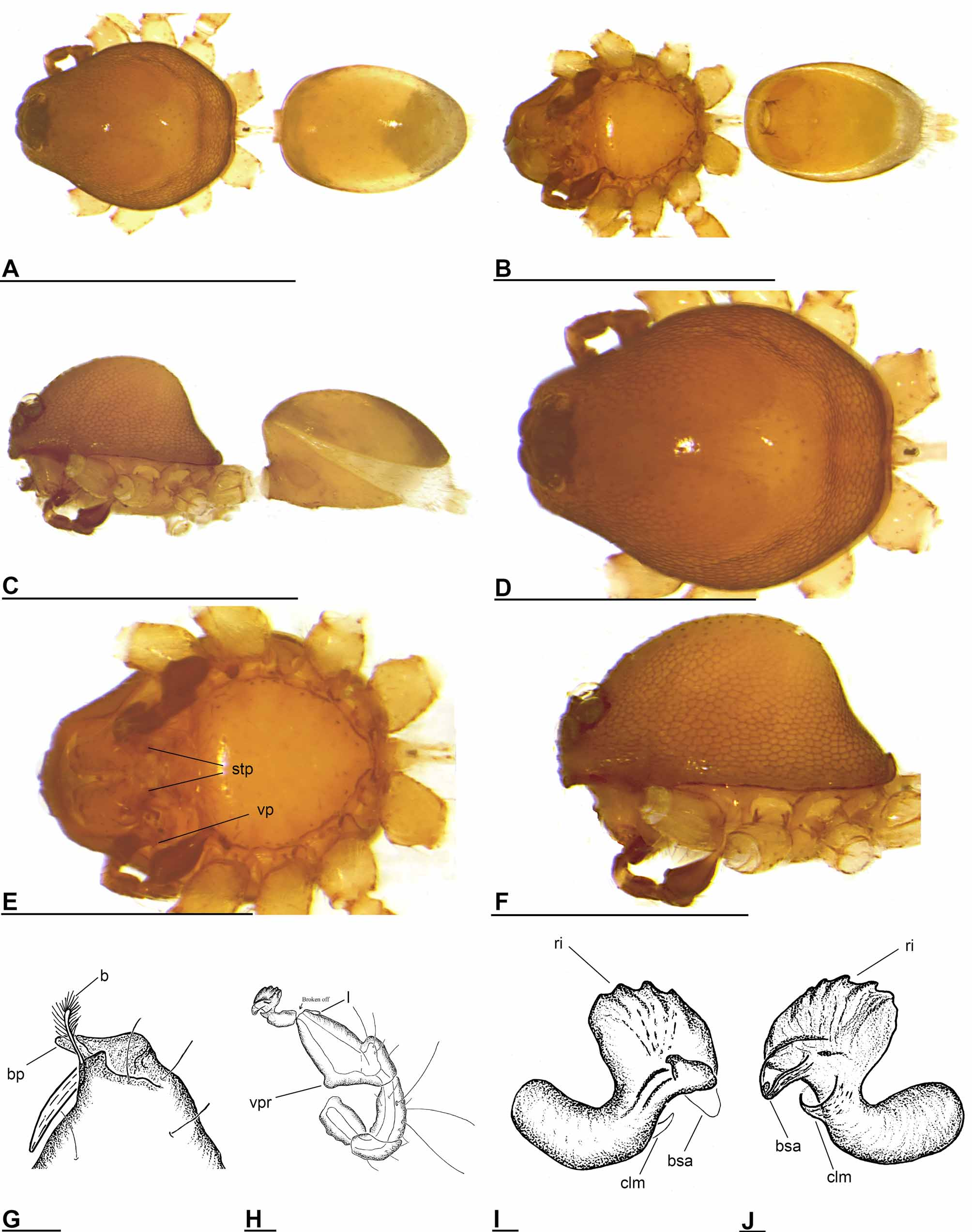
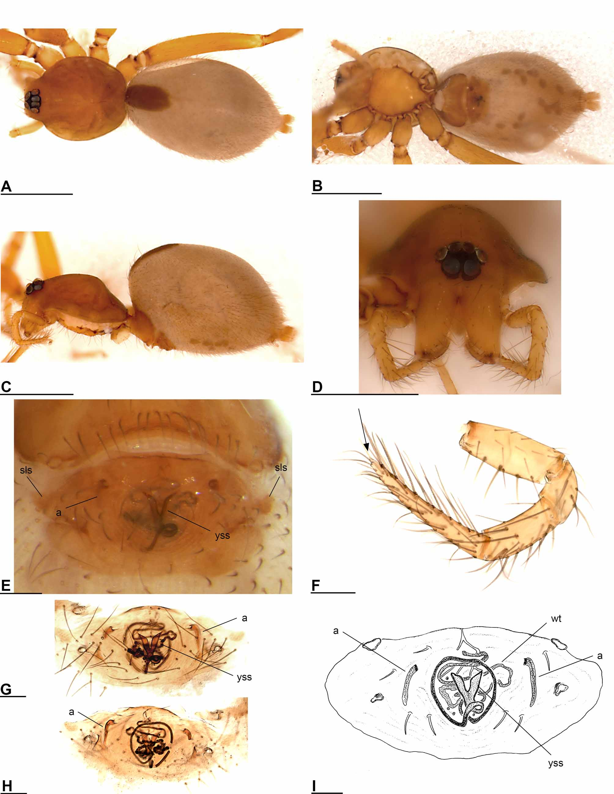
Ischnothyreus mulumi sp. nov. ( Figs 30–31 View FIGURE 30 View FIGURE 31 , 35 View FIGURE 35 )
Type material: Holotype: male (PBI_OON 00012470), AS /03-7. MALAYSIA: Sarawak: Mulu, National Park, 100km SEE of Miri, 4°00'N, 114°49'E, 200m, 19.–24.VIII.2003, leg. A. Schulz (winkler-extraction), MHNG. Paratypes: 4 females (PBI_OON 00032196), same data as holotype, MHNG.
Additional material examined: 1 male & 1 female (PBI_OON 00036304), BRUNEI DARUSSALAM: Temburong District: Ashton Trail near Kuala Belalong Field Studies Centre, 21km SSW Bangar, 04°32.513'N, 115°09.300'E, 100–150m, primary mixed dipterocarp forest, sifting leaf litter, 1.X.2009, C. Griswold & N. Chousou Polydouri UT004, CASENT 9036044 ( CAS). 2 females (PBI_OON 00036326), same data as PBI_OON 0 0 0 36304 except miniwinkler extraction of concentrated leaf litter, 3.X.2009 ( CAS). 1 male (PBI_OON 00036335), same data as PBI_OON 0 0 0 36304 except 4–9.X.2009 ( CAS). 1 female (PBI_OON 00036333), same data as PBI_OON 0 0 0 36304 ( CAS). 1 male (PBI_OON 00036325), same data as PBI_OON 0 0 0 36326 ( CAS). 1 female (PBI_OON 00035260), same data as PBI_OON 0 0 0 36326 ( CAS). 1 female (PBI_OON 00031652), INDO- NESIA: East Kalimantan : Berau Distr., Hutan Wisata Sei Tangap, ca. 8 km W of Tanjungredeb (2°08’04”N, 117°24’39”E), 30m (primary forest), 2.X.2008, leg. P. Schwendinger ( MHNG). 1 female (PBI_OON 00031644), INDONESIA: East Kalimantan : Berau Distr., Hutan Mayang Mangurai, ca. 15 km SW of Tanjungredeb (2°06’13”N, 117°24’05”E), 20m (secondary forest), 30.IX.2008, leg. P. Schwendinger ( MHNG).
Etymology: The specific name is a noun in apposition taken from the type locality.
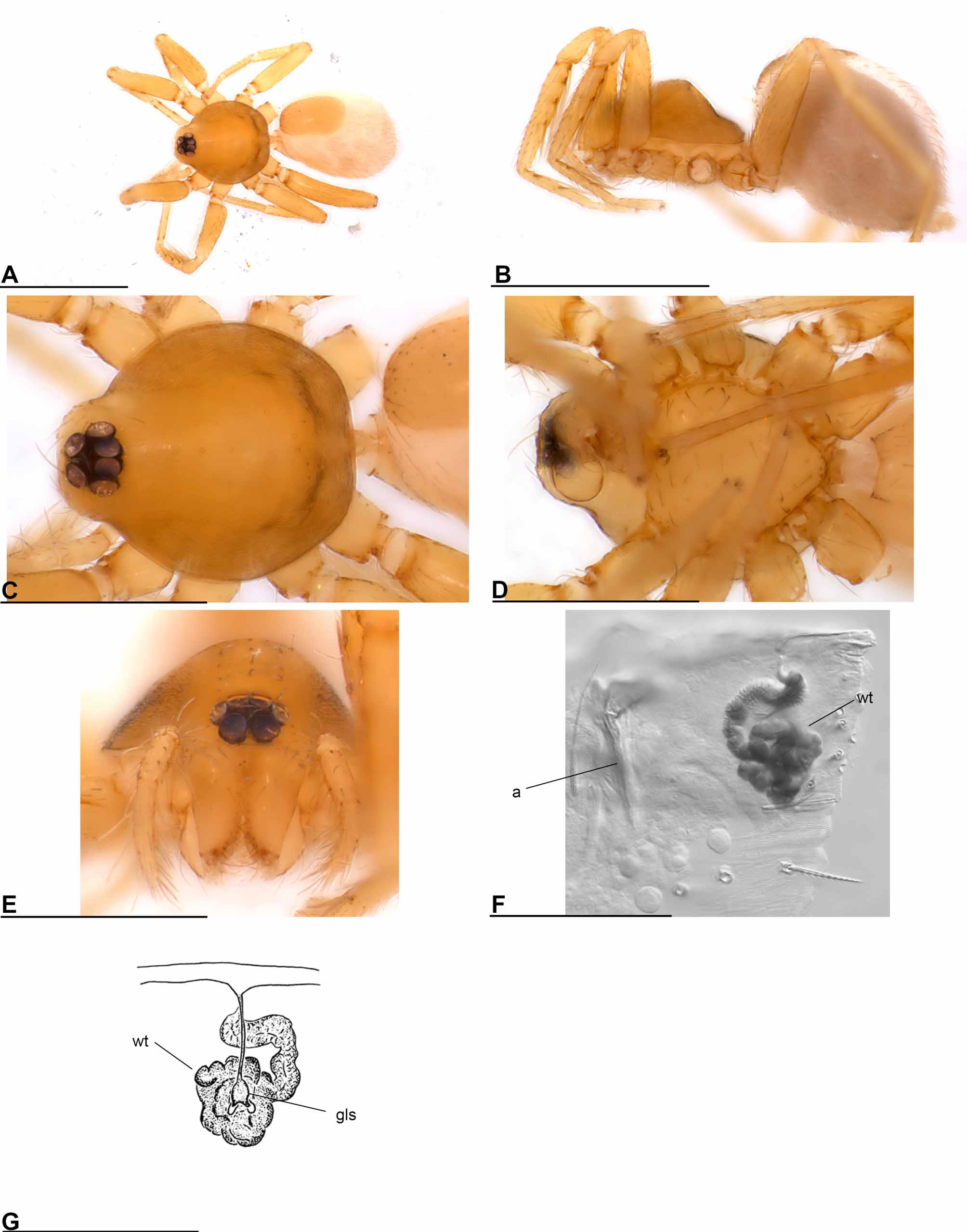
Diagnosis: The male of I. mulumi can easily be recognized by the unusual, crown-shaped terminal part of the bulb, with a beak-like apophysis on the ventral side ( Fig. 30 View FIGURE 30 H–J). The female genital area is characterized by the strongly winding tube, ending posteriorly in a circle, surrounded by numerous short setae ( Fig. 31 View FIGURE 31 G–J).
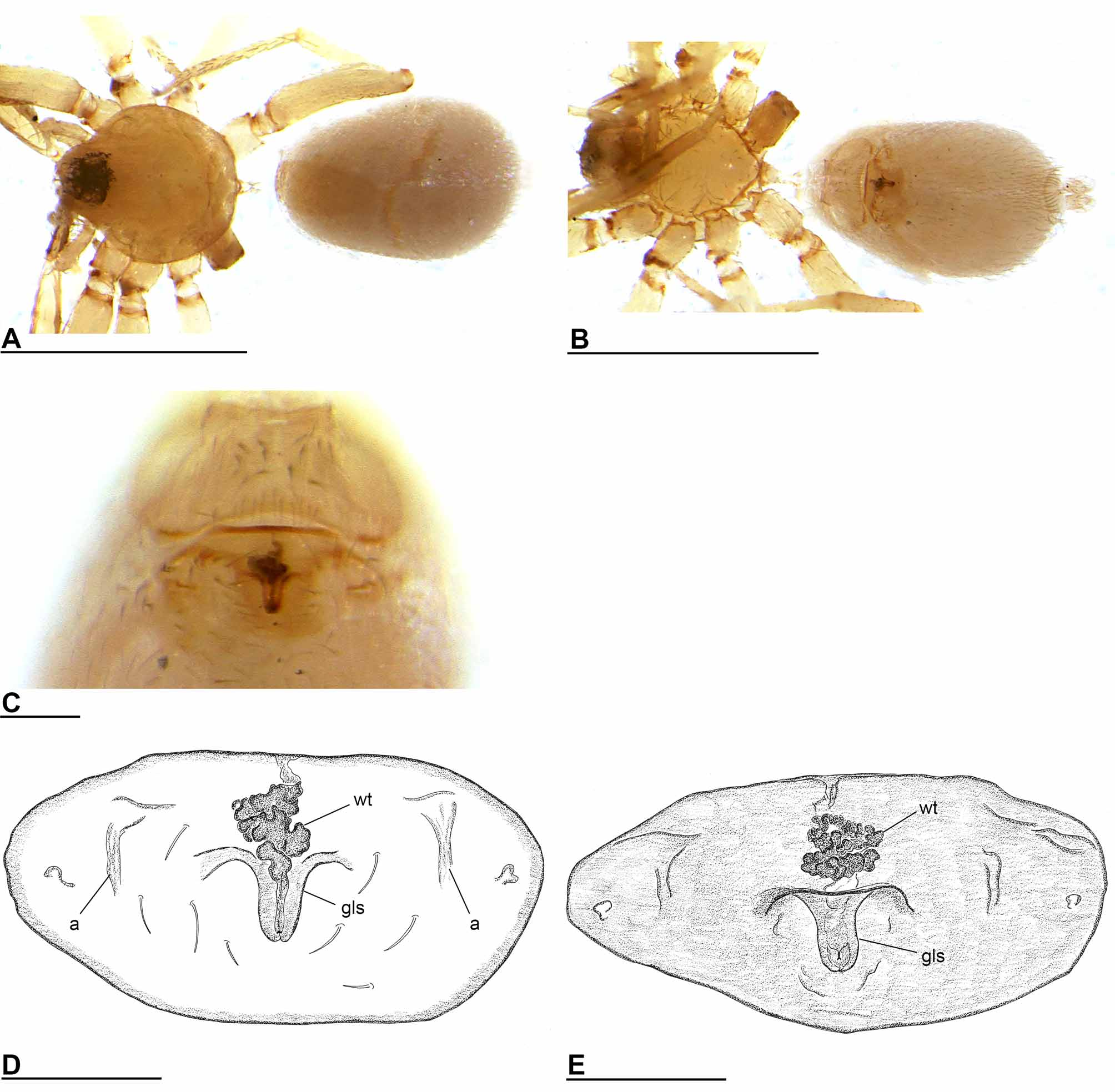
Description: Male (holotype). Total length 1.53 mm. Carapace: orange-brown, broadly oval in dorsal view, domed in lateral view, surface of elevated portion of pars cephalica strongly reticulate, sides strongly reticulate. Sternum: as long as wide, setae abundant. Mouthparts: chelicerae with anteriorly directed process (bp) at base of fangs and brush (b) on retromargin ( Fig. 30 View FIGURE 30 G), fang groove with few denticles. Anterior margin of labium indented at middle. Anteromedian tip of endites with one strong, tooth-like projection (stp) ( Fig. 30 View FIGURE 30 E). Abdomen: book lung covers large, elliptical. Pedicel tube medium, scutum extending far dorsal of pedicel. Dorsal scutum strongly sclerotized, orange, anteriorly and posteriorly darkened, covering full length of abdomen, middle surface and sides striated. Epigastric and postepigastric scutum strongly sclerotized, fused, postepigastric scutum long, almost rectangular, covering about 3/4 of abdominal length. Spinneret scutum with fringe of long setae. Legs: spine formula: femora: I p0-1-1; r0-1-0; II p0-0-1; tibiae: I, II p2-1-1; r2-1-1; metatarsi: I, II p1-1-0; r1-1-0. Genitalia: Palp strongly sclerotized, proximal segments brown, trochanter with ventral projection (vp) ( Fig. 30 View FIGURE 30 E), patella about as long as femur, not enlarged, cymbium brown, fused with bulb, bulb brown, more than twice as long as cymbium, stout, tapering apically, with ventral protuberance (vpr) and lobe (l) ( Fig. 30 View FIGURE 30 H), end of bulb enlarged, ribbed (ri), crown-shaped. Membranous, beak-shaped apophysis (bsa) and collar-like membrane (clm) ventrally ( Fig. 30 View FIGURE 30 I–J).
Female (paratype). Total length 1.66 mm. As in male except as noted. Carapace: without any pattern, pars cephalica strongly elevated in lateral view. Sternum: longer than wide. Mouthparts: without basal process and brush. Labium anterior margin not indented at middle. Endites unmodified. Femur of female palp with three ventral spines. Abdomen: epigastric scutum slightly protruding (arrow) ( Fig. 31 View FIGURE 31 C). Postepigastric scutum widely hexagonal, covering about 1/3 of the abdominal length, not fused to epigastric scutum, with short, curved, posteriorly directed lateral apodemes (a) ( Fig. 31 View FIGURE 31 F). Dense patch of setae anterior to spinnerets present. Legs: spine formula: femora: I p0-1-1; r0-1-0; II p0-0-1; v0-0-1; tibiae: I, II p2-1-1; r2-1-1; metatarsi: I, II p1-1-0; r1-1-0. Genitalia: the posterior margin of the epigastric scutum is lined with numerous needle-like setae. The margin of the postepigastric scutum is thickened. At its middle starts a dark, thin, strongly winding tube running backwards (wt), ending in a dark, half-circle (hc), surrounded by numerous setae ( Fig. 31 View FIGURE 31 G–J).
Distribution: Sarawak, Brunei and East Kalimantan ( Fig. 35 View FIGURE 35 ).
Remark: The material collected in Brunei, 2009, is kept in 97% ethanol. The specimens are very dark.
Ischnothyreus rex sp. nov. ( Figs 32–33 View FIGURE 32 View FIGURE 33 , 35 View FIGURE 35 )
Type material: Holotype: male (PBI_OON 00016064), MALAYSIA: Sabah: Kinabalu National Park, Power Station, 1960m, 4.V.1991, leg. C.L. & P.R. Deeleman, RMNH. Paratype: female (PBI_OON 00016065), collected with holotype, RMNH.
Etymology: The specific epithet is a noun in apposition meaning “king” in Latin. It refers to the crown-shaped process on the base of the male cheliceral fang.
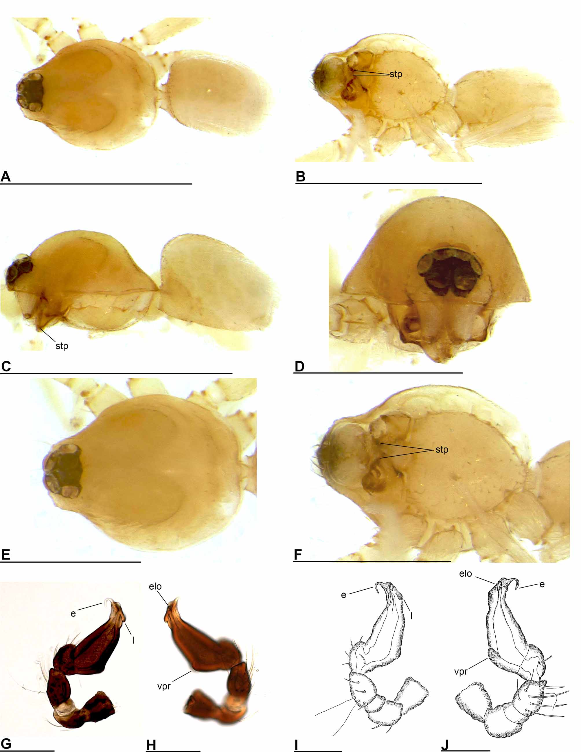
Diagnosis: Very large species. The male of I. rex resembles the male of I. fobor in having a very long and thin embolus ( Fig. 32 View FIGURE 32 I–J), but can easily be distinguished from the latter by the crown-shaped basal process on the cheliceral fang ( Fig. 32 View FIGURE 32 F–H). The female of I. rex resembles the female of I. flippi in having a sclerotized y-shaped structure in the genital area, but can be distinguished from the latter by the presence of a dark circle, which seems to be a prolongation of the y-shaped structure ( Fig. 33 View FIGURE 33 G–I).
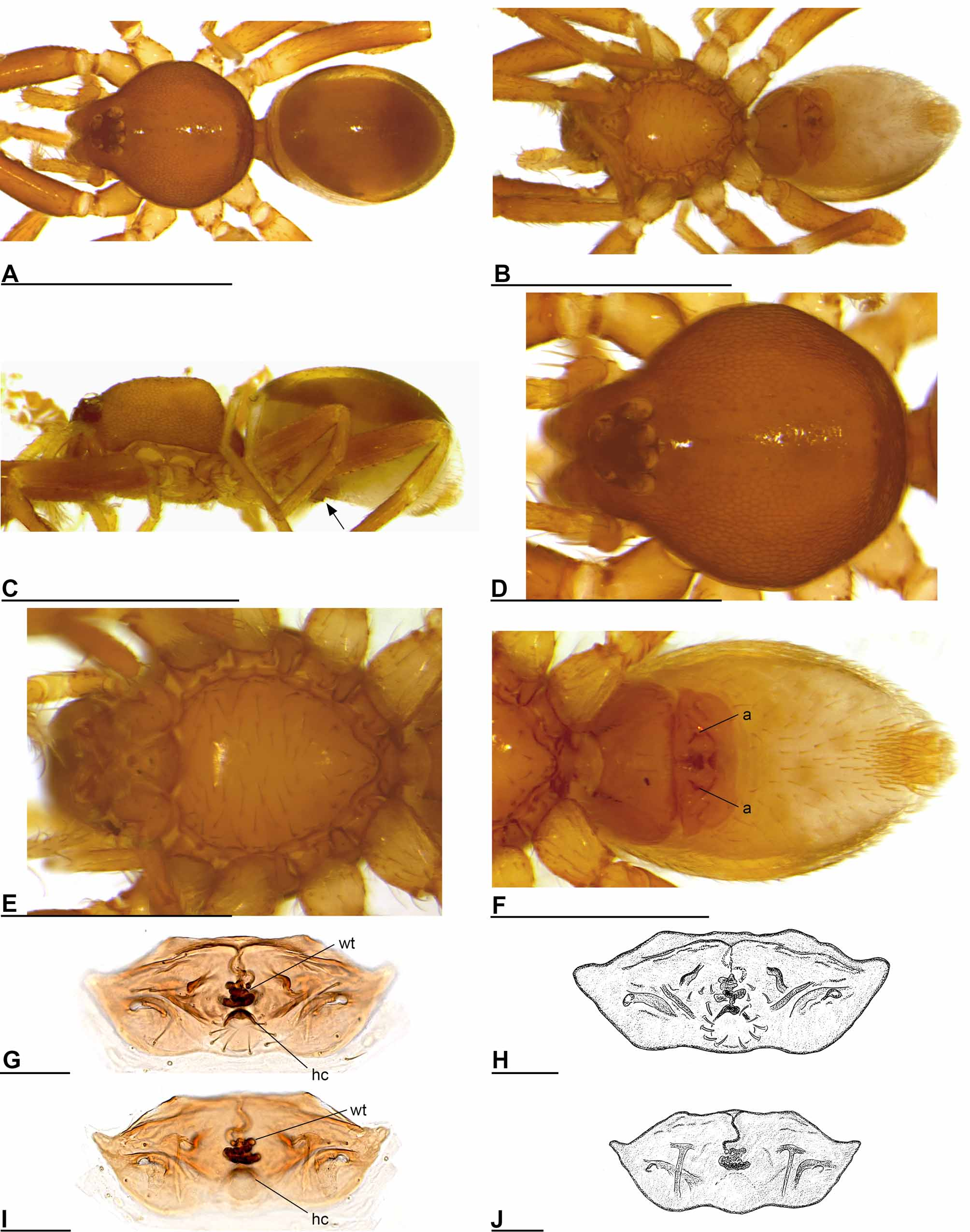
Description: Male (holotype). Total length 2.94 mm. Carapace: orange-brown, slightly elevated in lateral view, surface and sides of elevated portion of pars cephalica finely reticulate, fovea present, lateral margin rebordered. Sternum: longer than wide, pale orange, uniform, not fused to carapace, surface smooth, setae abundant. Mouthparts: chelicerae slightly divergent, with a prominent, crown-shaped process (csp) at the base of the fangs ( Fig. 32 View FIGURE 32 F–H). Anteromedian tip of endites with one strong, tooth-like projection (stp) ( Fig. 32 View FIGURE 32 E). Abdomen: book lung covers large, elliptical (blc) ( Fig. 32 View FIGURE 32 C). Scutum extending far dorsal of pedicel. Dorsal scutum yellow-brown, fused to epigastric scutum. Spinneret scutum present, incomplete ring, with fringe of long setae. Colulus present. Legs: spine formula: femora: I p0-1-1; II p0-1-0; tibiae: I, II p2-1-1; r2-1-1; metatarsi: I, II p1-1-0; r1-1-0. Each tibia with three trichobothria, each metatarsus with one trichobothrium. Genitalia: Palp strongly sclerotized, proximal segments almost black, trochanter with ventral projection (vp) ( Fig. 32 View FIGURE 32 E), patella about as long as femur, not enlarged, cymbium almost black, fused with bulb, bulb almost black, 1 to 1.5 times as long as cymbium, stout, tapering apically, with ventral protuberance (vpr) and lobe (l), embolus (e) extremely long and thin, filamentous, base of embolus with several membranous outgrowths (mo) ( Fig. 32 View FIGURE 32 I–J).
Female (paratype). Total length 3.62 mm. As in male except as noted. Carapace: without any pattern, broadly oval in dorsal view. Mouthparts: chelicerae without prominent basal process. Anteromedian tip of endites unmodified. Femur of female palp with six ventral spines, tibia with three trichobothria, end of tarsus finger-like ( Fig. 33 View FIGURE 33 F, arrow). Abdomen: pedicel tube medium. Dorsal scutum covering less than 1/2 of abdomen, not fused to epigastric scutum. Epigastric scutum slightly protruding, with small lateral sclerites (sls) ( Fig. 33 View FIGURE 33 E). Postepigastric scutum widely hexagonal, only around epigastric furrow, not fused to epigastric scutum, with short, posteriorly directed lateral apodemes (a) ( Fig. 33 View FIGURE 33 E, G–I). Legs: spine formula: femora: I p0-1-1; r0-0-2; II p0-0-1; tibiae: I, II p2-1-1; r2-1-1; metatarsi: I, II p1-1-0; r1-1-0. Genitalia: the posterior margin of the epigastric scutum is lined with numerous needle-like setae. The margin of the postepigastric scutum is thickened. At its middle is a y-shaped, sclerotized structure visible (yss), whose posterior part leads to a tube. After overlapping itself, the tube runs as a circle, surrounds the y-shaped structure and finally leads to the strongly winding tube (wt) ( Fig. 33 View FIGURE 33 E, G–I).
Distribution: Sabah ( Fig. 35 View FIGURE 35 ).
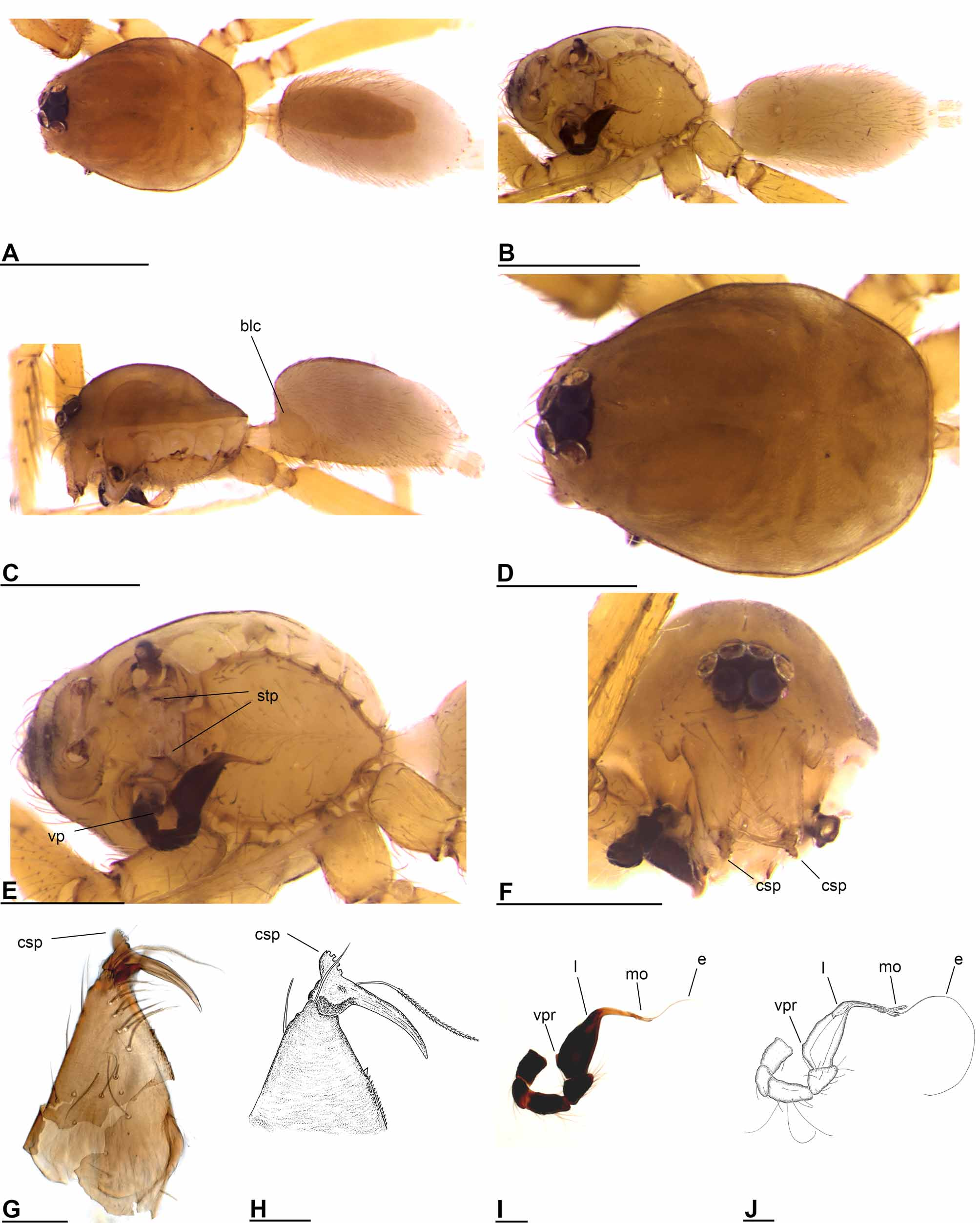
Ischnothyreus serapi sp. nov. ( Figs 34 View FIGURE 34 , 35 View FIGURE 35 )
Type material: Holotype: male (PBI_OON 00032200), MALAYSIA: Sarawak: route Kuching– Matang , Gunung Serapi , prélèvement du sol dans la forêt le long de la route vers la station TV (translated: soil extraction in forest along street in direction of television station), 670m, 9.XII.1987, leg. B. Hauser (Berlese à Kuching), Sar-87/64, MHNG. Paratype: male (PBI_OON 00016218), same data as for holotype, MHNG.
Etymology: The specific name is a noun in apposition taken from the type locality.
Diagnosis: The male of I. serapi can be recognized by the anvil-shaped distal part of the palpal bulb ( Fig. 34 View FIGURE 34 F–H).
Description: Male (holotype). Total length 1.15 mm. Carapace: whole carapace strongly reticulate. Eyes: all subequal, PLE circular. Mouthparts: chelicerae, endites and labium yellow. Chelicerae with small process (sbp) at base of the fangs. Brush (b) on retromargin near the basal process ( Fig. 34 View FIGURE 34 E). Anteromedian tip of endites with one strong, tooth-like projection (stp) ( Fig. 34 View FIGURE 34 B). Abdomen: dorsal scutum strongly sclerotized, covering full length of abdomen, more than 1/2 to most of abdomen width, middle surface striated, sides striated. Epigastric and postepigastric scutum strongly sclerotized, fused. Legs: spine formula: femora: I p0-1-1; r0-0-1; II p0-0-1; tibiae: I, II p2- 1-1; r2-1-1; metatarsi: I, II p1-1-0; r1-1-0. Genitalia: Palp strongly sclerotized, proximal segments brown, trochanter normal size, with ventral projection (vp), femur normal size, patella about as long as femur, not enlarged, cymbium brown, fused with bulb, bulb brown, more than two times as long as cymbium, stout, tapering apically, with ventral protuberance (vpr) and lobe (l), distal part of bulb laterally enlarged, anvil-shaped, ribbed (ri), with crown-shaped basis (csb) and small, pointed apophyses distally (pa) ( Fig. 34 View FIGURE 34 F–H). Embolus not clearly discernible.
Female: Unknown.
Distribution: Sarawak ( Fig. 35 View FIGURE 35 ).


No known copyright restrictions apply. See Agosti, D., Egloff, W., 2009. Taxonomic information exchange and copyright: the Plazi approach. BMC Research Notes 2009, 2:53 for further explanation.