
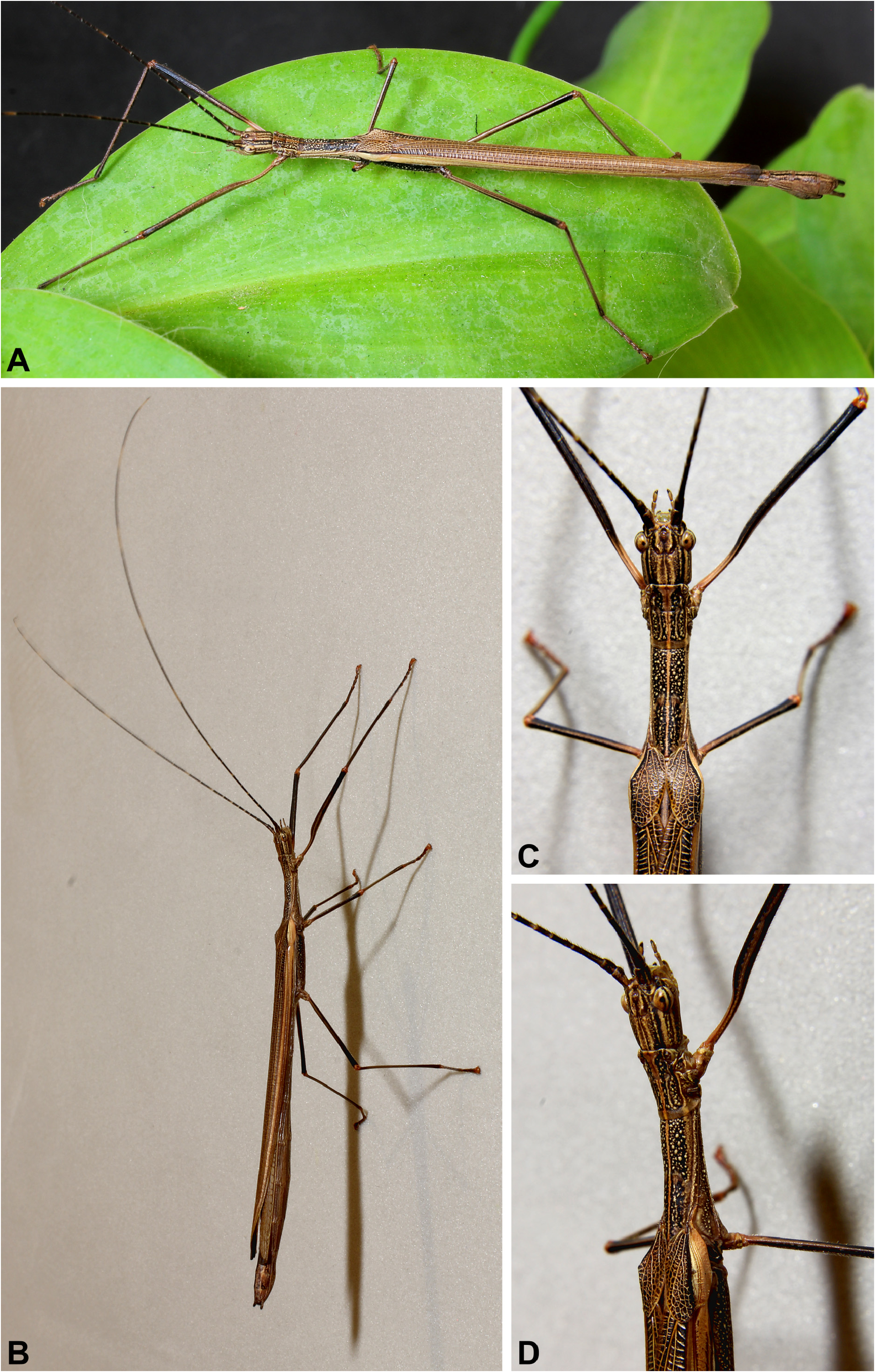
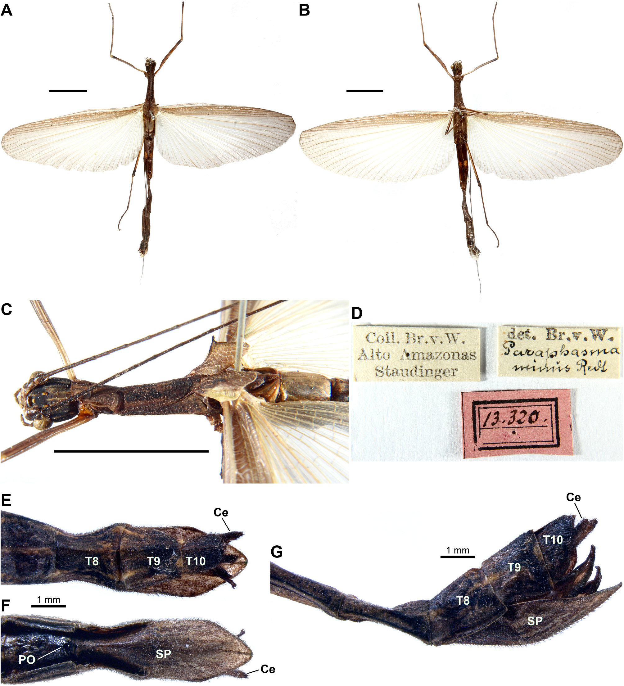
Paraphasma (Chiquetto-Machado & Cancello, 2021)
|
publication ID |
https://doi.org/10.11646/zootaxa.5122.1.1 |
|
publication LSID |
lsid:zoobank.org:pub:EC13A69D-D6FA-4926-AC59-648A5626C9B9 |
|
persistent identifier |
https://treatment.plazi.org/id/03B587AA-FF96-FF8C-FF2A-F993FD2DF001 |
|
treatment provided by |
Plazi |
|
scientific name |
Paraphasma |
| status |
|
Key to the species of Paraphasma View in CoL
Males
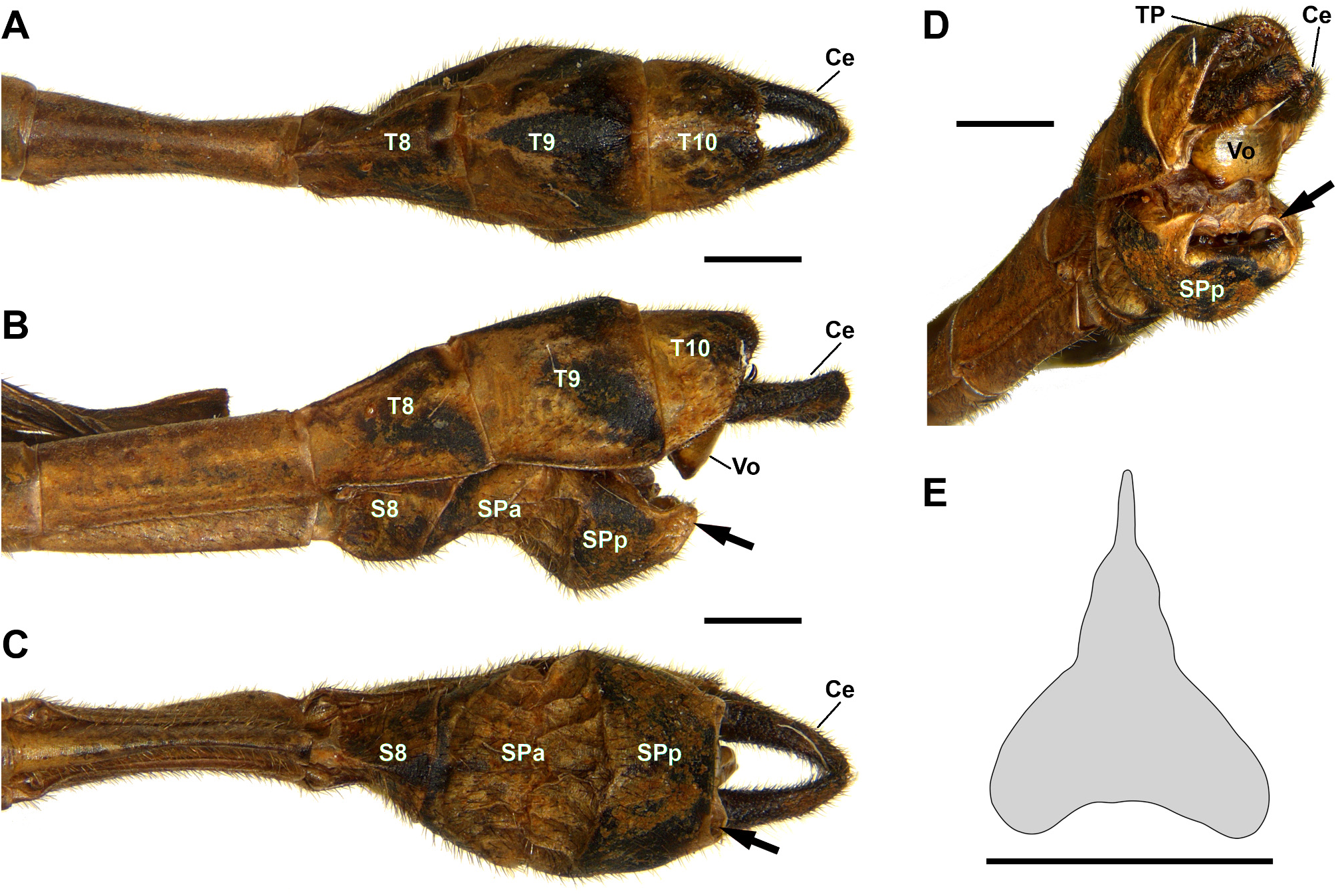
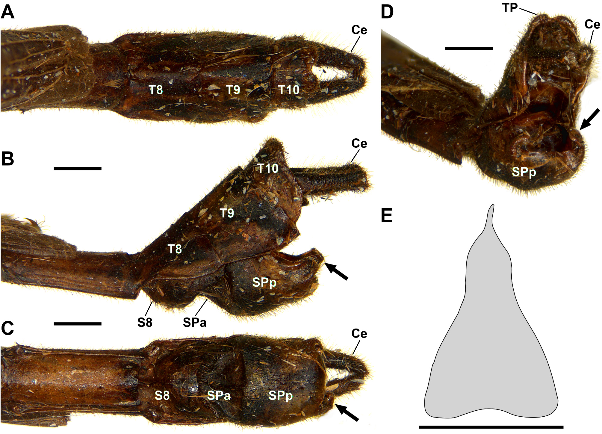
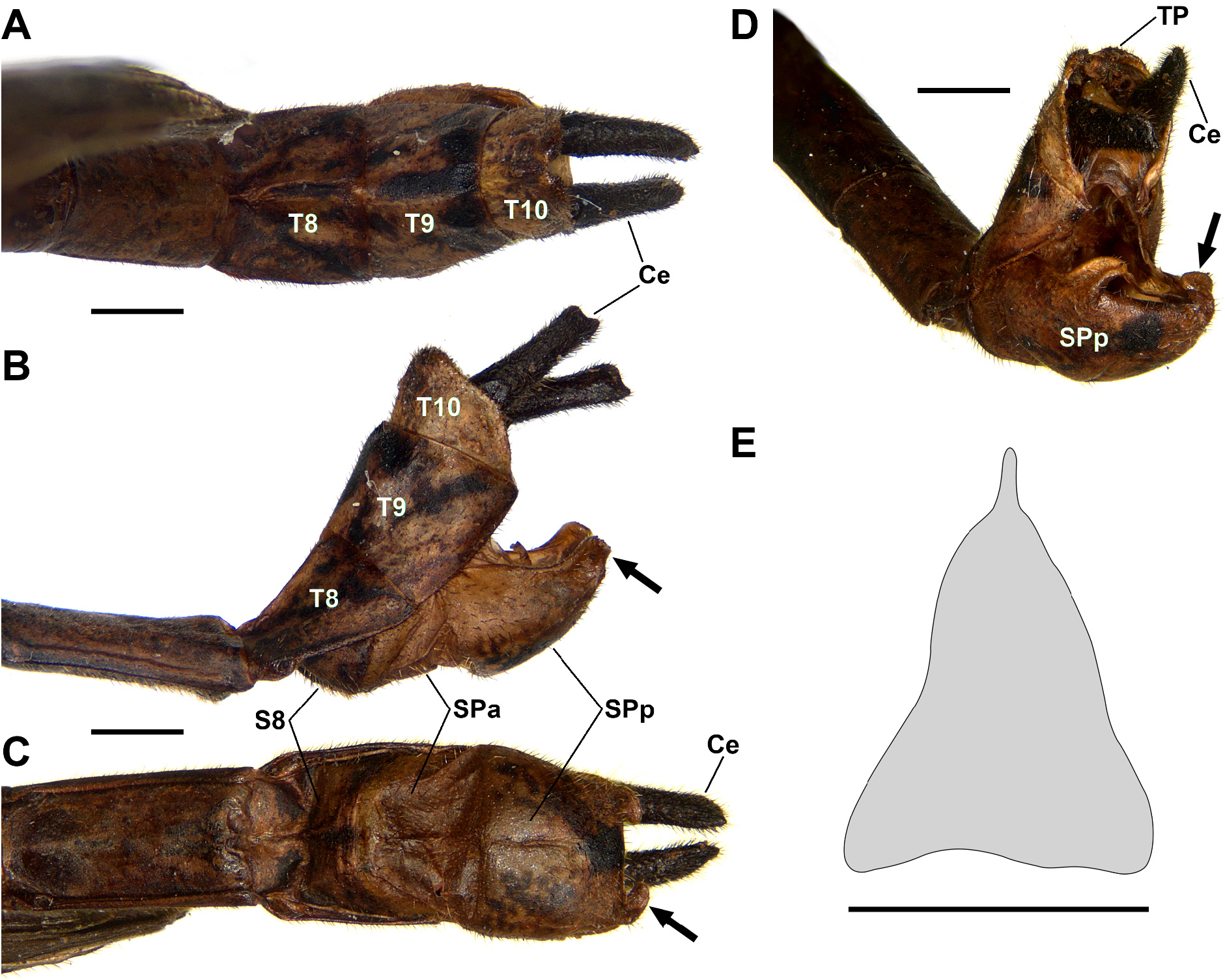
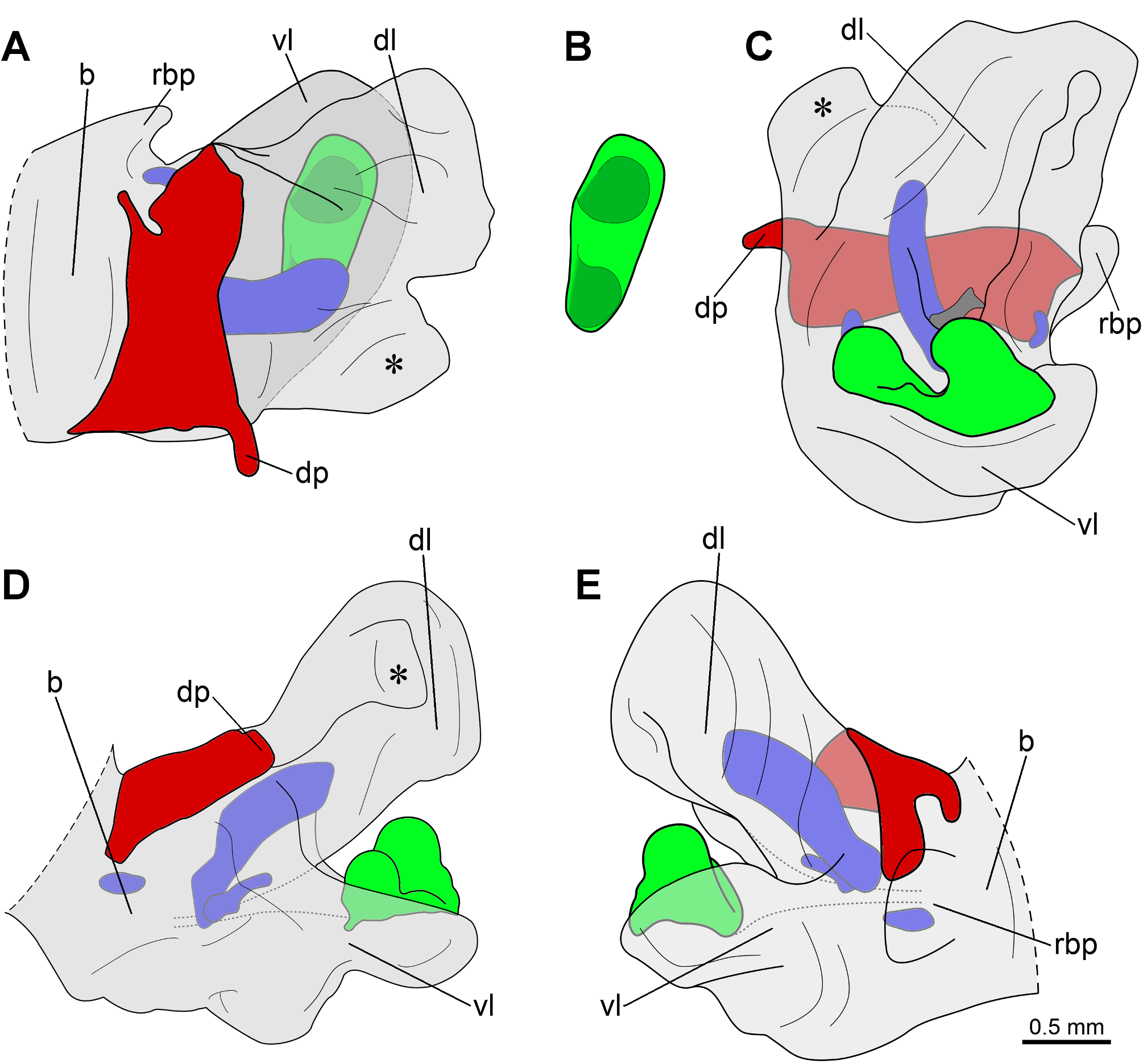
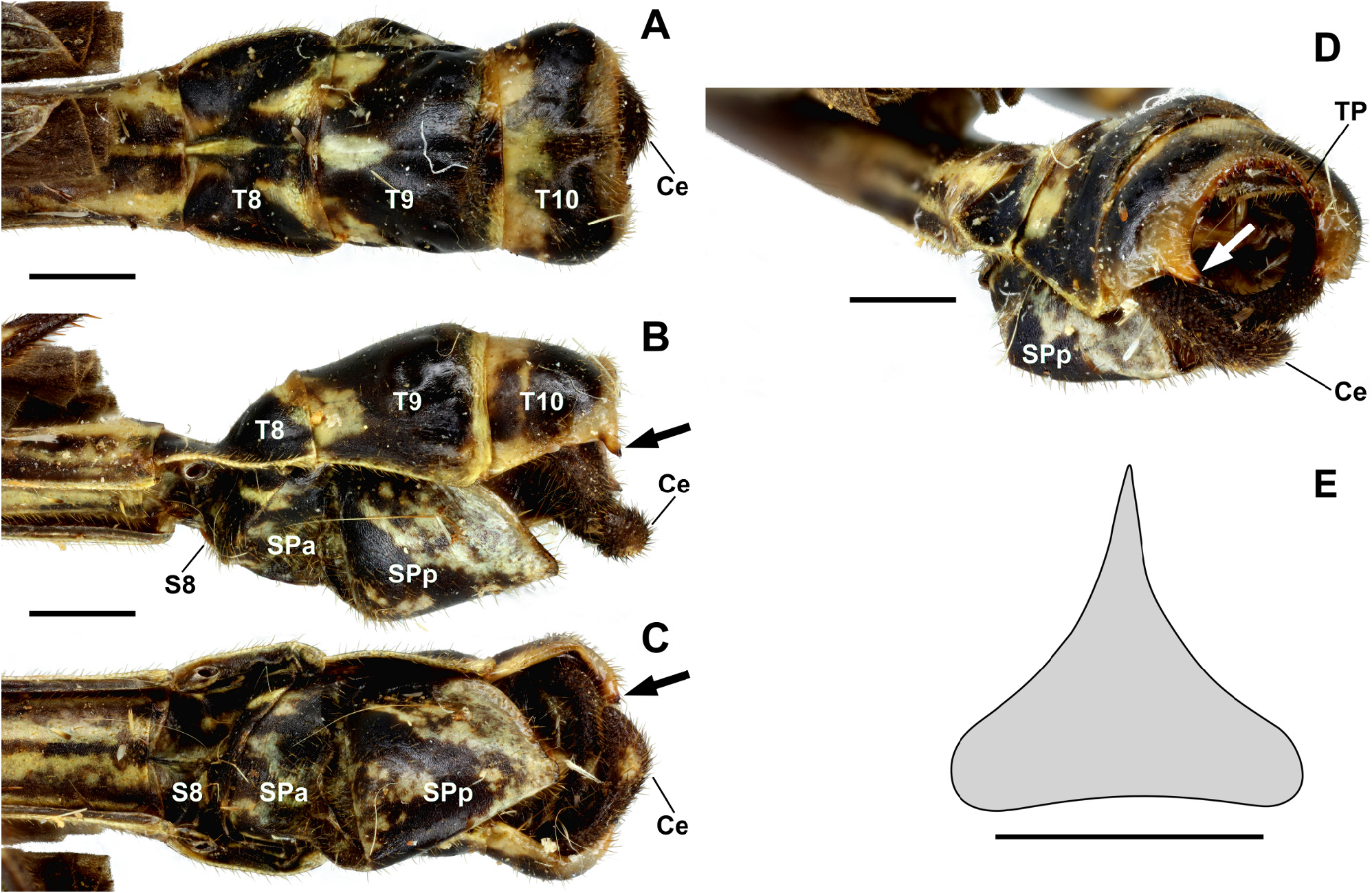
1. Anterior region of subgenital plate with a pair of lateral projections, sometimes covered by tergite IX ( Fig. 41B,D View FIGURE 41 : arrows; Fig. 45E View FIGURE 45 : arrowhead)..................................................................................... 2
- Anterior region of subgenital plate without lateral projections ( Figs 3 View FIGURE 3 , 8 View FIGURE 8 , 12 View FIGURE 12 , 21 View FIGURE 21 , 27 View FIGURE 27 , 31 View FIGURE 31 , 36 View FIGURE 36 ).......................... 3
2. Tegmina with apical margin distinctly acuminate and shoulder pads developed into large, triangular, somewhat dull spines ( Fig. 40 View FIGURE 40 ); anterior region of subgenital plate with a pair of large, digitiform lateral projections surpassing the posterior margin of tergite IX ( Fig. 41B,D View FIGURE 41 : arrows); posterior margin of subgenital plate acuminate ( Fig. 41B,C View FIGURE 41 )... Paraphasma trianguliferum View in CoL
- Tegmina with apical margin rounded and shoulder pads developed into sharp spines ( Fig. 44 View FIGURE 44 ); anterior region of subgenital plate with a pair of small, approximately styliform lateral projections entirely covered by tergite IX ( Fig. 45E View FIGURE 45 : arrowhead); posterior margin of subgenital plate truncate in lateral view and V-shaped in caudal view ( Fig. 45B,D View FIGURE 45 ).................................................................................................... Paraphasma umbretta View in CoL
3. Posterolateral regions of tergite X with a pair of spiniform projections ( Fig. 36B–D View FIGURE 36 : arrows); cerci strongly incurved, with acuminate apex ( Fig. 36A–D View FIGURE 36 ); posterior margin of subgenital plate acuminate ( Fig. 36B,C View FIGURE 36 )..................................................................................................... Paraphasma spinicauda View in CoL sp. nov.
- Tergite X without spiniform projections; cerci straight or slightly incurved, with spatulate apex ( Figs 3 View FIGURE 3 , 8 View FIGURE 8 , 12 View FIGURE 12 , 21 View FIGURE 21 , 27 View FIGURE 27 , 31 View FIGURE 31 ); posterior margin of subgenital plate truncate, laterally forming a pair of approximately triangular expansions with malleable aspect ( Figs 3 View FIGURE 3 , 8 View FIGURE 8 , 12 View FIGURE 12 , 21 View FIGURE 21 , 27 View FIGURE 27 , 31 View FIGURE 31 : arrows)................................................................... 4
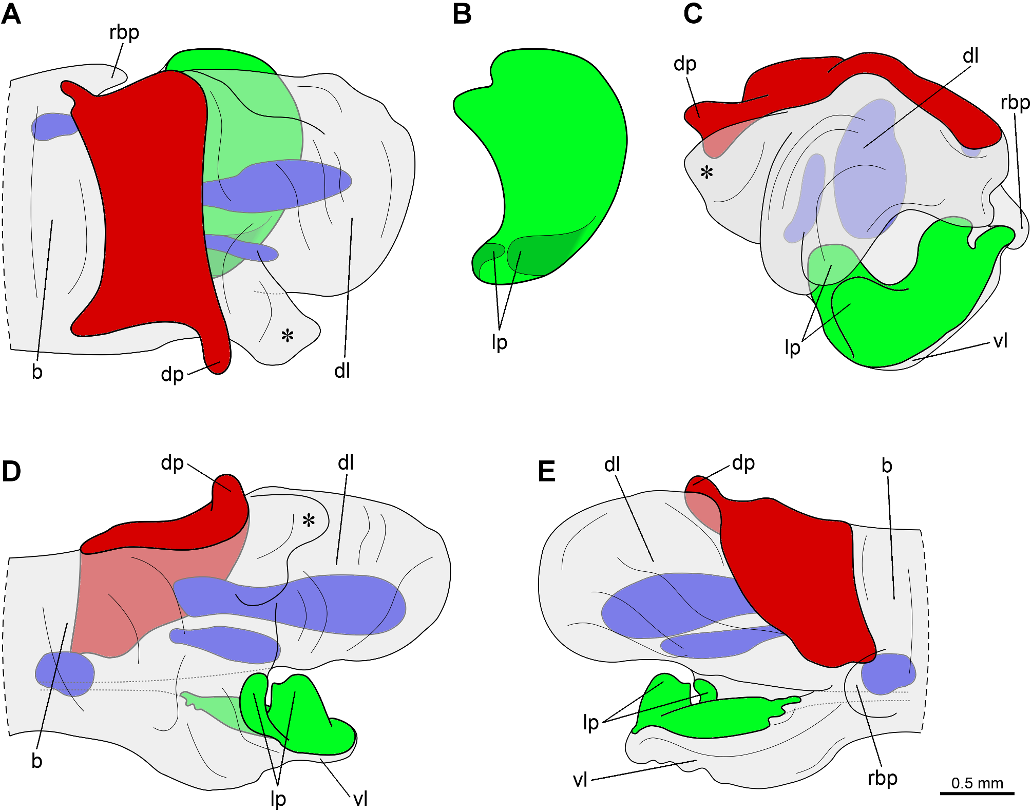
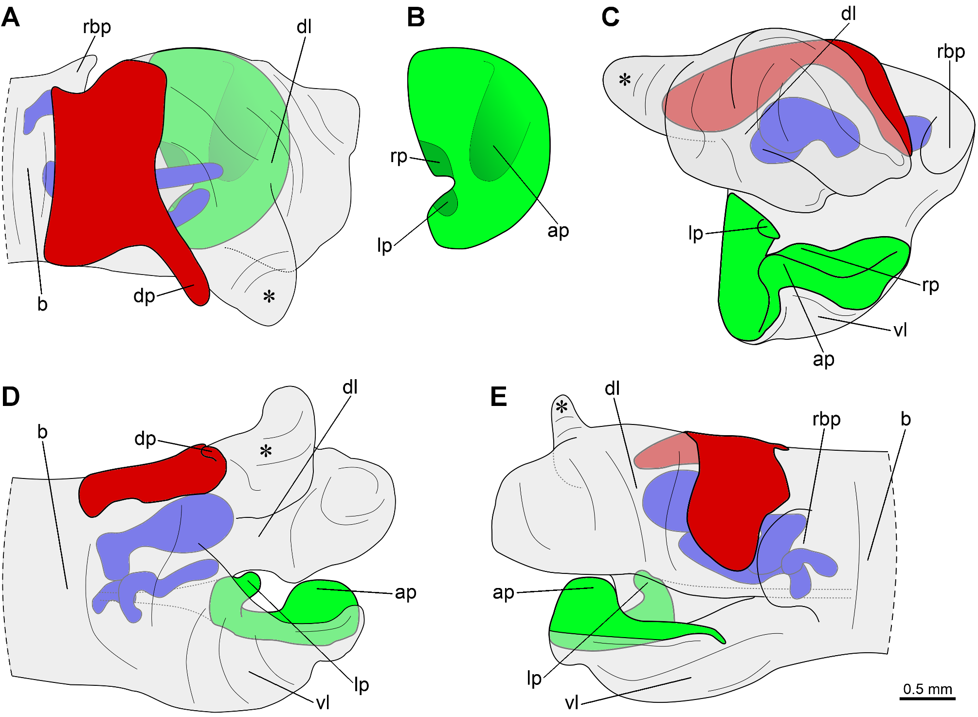
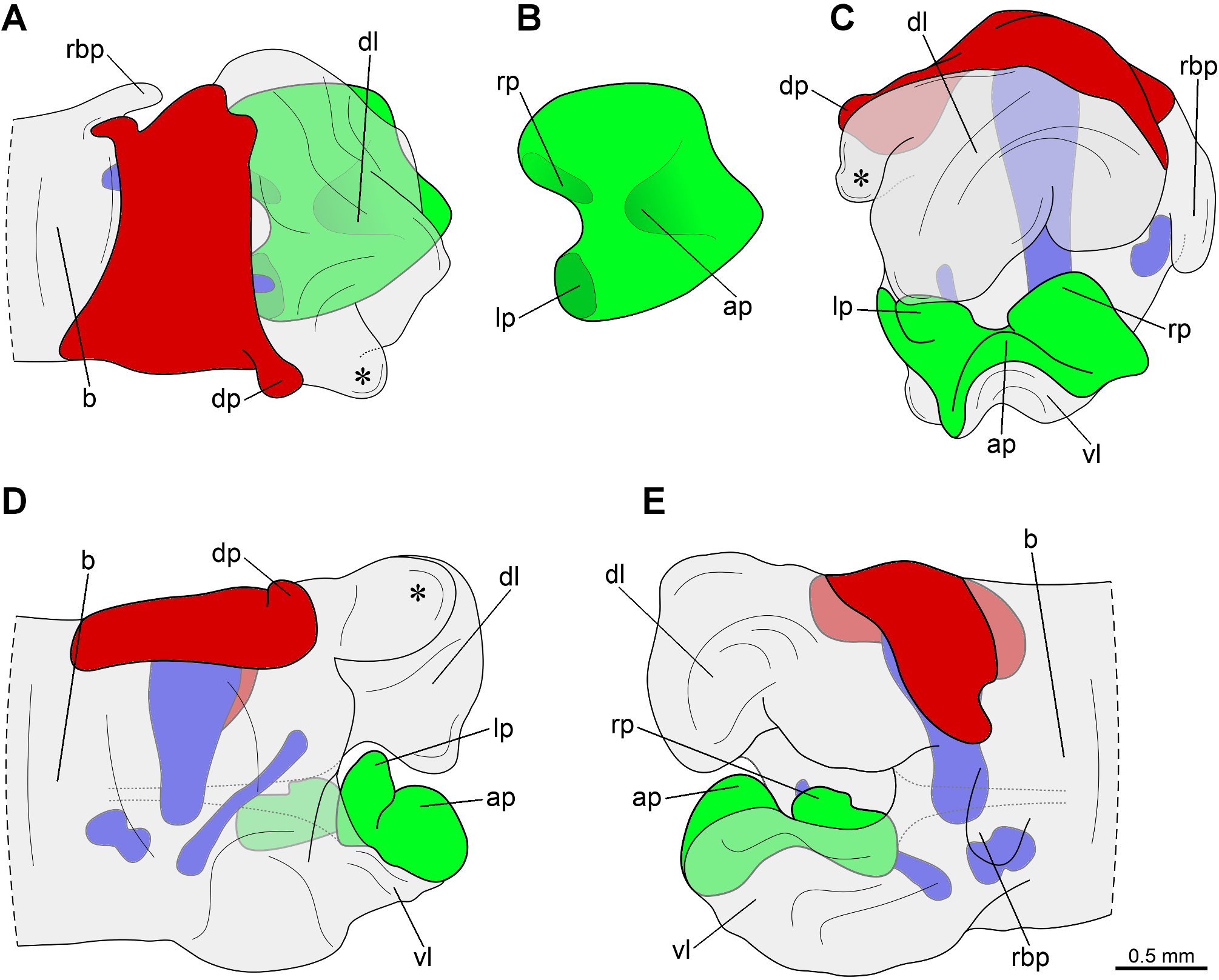
4. Apex of cerci internally concave ( Figs 12 View FIGURE 12 , 31 View FIGURE 31 ); in the phallic organ, sclerite of the ventral lobe restricted to the central region of the inner face of the ventral lobe, bearing two rounded protuberances ( Figs 14 View FIGURE 14 , 32 View FIGURE 32 , in green)......................... 5
- Apex of cerci not concave ( Figs 3 View FIGURE 3 , 8 View FIGURE 8 , 21 View FIGURE 21 , 27 View FIGURE 27 ); in the phallic organ, sclerite of the ventral lobe covering the entire inner face of the ventral lobe and slightly reaching the outer face, bearing one to three protuberances ( Figs 4 View FIGURE 4 , 9 View FIGURE 9 , 22 View FIGURE 22 , 28 View FIGURE 28 , in green)....... 6
5. In the phallic organ, sclerite of the ventral lobe roughly oval in dorsal view, with the right protuberance directed anteriorly and the left one directed caudally ( Fig. 14 View FIGURE 14 , in green)............................................. Paraphasma laterale View in CoL
- In the phallic organ, sclerite of the ventral lobe oblong in dorsal view, with the two protuberances positioned side by side and projected towards each other ( Fig. 32 View FIGURE 32 , in green).................................... Paraphasma sooretama View in CoL sp. nov.
6. Tegmina shoulder pads spiniform or dull; in the phallic organ, sclerite of the ventral lobe bearing three protuberances (i.e., apical, left basal and right basal protuberances), all of them undivided ( Figs 22 View FIGURE 22 , 28 View FIGURE 28 : ap, lp, rp)........................ 7
- Tegmina shoulder pads spiniform; in the phallic organ, sclerite of the ventral lobe bearing one or two protuberances (right basal protuberance absent) ( Fig. 4 View FIGURE 4 : ap, lp; Fig. 9 View FIGURE 9 : lp), with the left basal one partially or completely divided in two............ 8
7. Tegmina shoulder pads spiniform or dull; in the phallic organ, sclerite of the ventral lobe roughly triangular in dorsal view, with somewhat acuminate posterior margin ( Fig. 28B View FIGURE 28 ), and with the three protuberances blunt and conspicuous ( Fig. 28 View FIGURE 28 : ap, lp, rp).................................................................................... Paraphasma minus View in CoL
- Tegmina shoulder pads dull; in the phallic organ, sclerite of the ventral lobe roughly semicircular in dorsal view, with rounded posterior margin ( Fig. 22B View FIGURE 22 ), and with the three protuberances as follows: apical protuberance large and rounded ( Fig. 22 View FIGURE 22 : ap), left basal one strongly protruding ( Fig. 22 View FIGURE 22 : lp), and right basal one inconspicuous, restricted to a gentle bulge ( Fig. 22 View FIGURE 22 : rp)................................................................................... Paraphasma marginale View in CoL
8. In the phallic organ, sclerite of the ventral lobe bearing two protuberances: the apical one, which is narrow and elongate ( Fig. 4 View FIGURE 4 : ap), and the left basal one, which is rounded and partially divided in two (approximately saddle-shaped) ( Fig. 4 View FIGURE 4 : lp).................................................................................... Paraphasma conspersum View in CoL
- In the phallic organ, sclerite of the ventral lobe bearing only the left basal protuberance ( Fig. 9 View FIGURE 9 : lp), which is divided into two regions, the anterior larger and approximately lamellate, and the posterior small and rounded................................................................................................... Paraphasma indistinctum View in CoL sp. nov.
Females
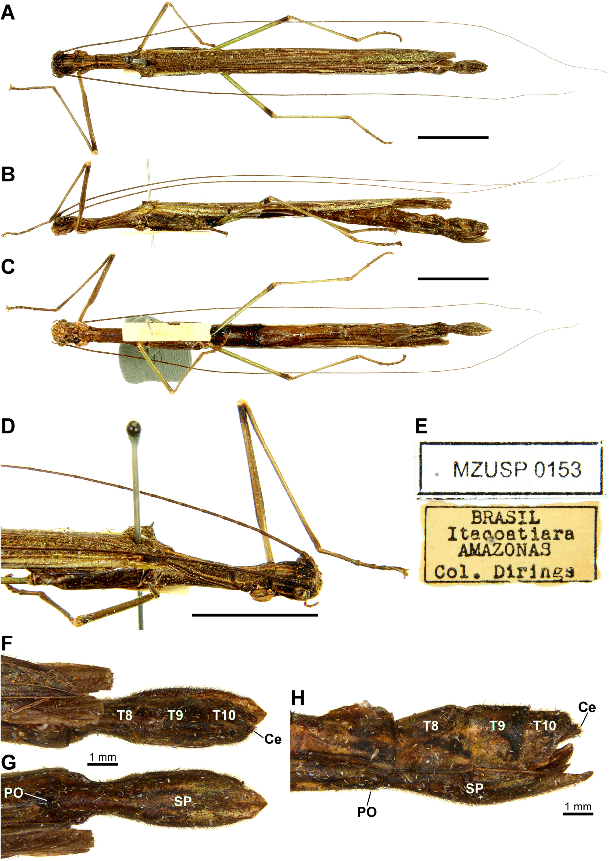
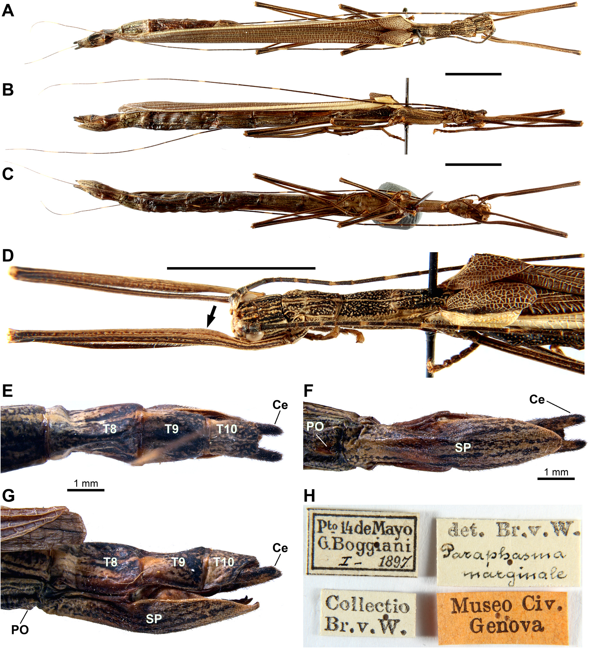
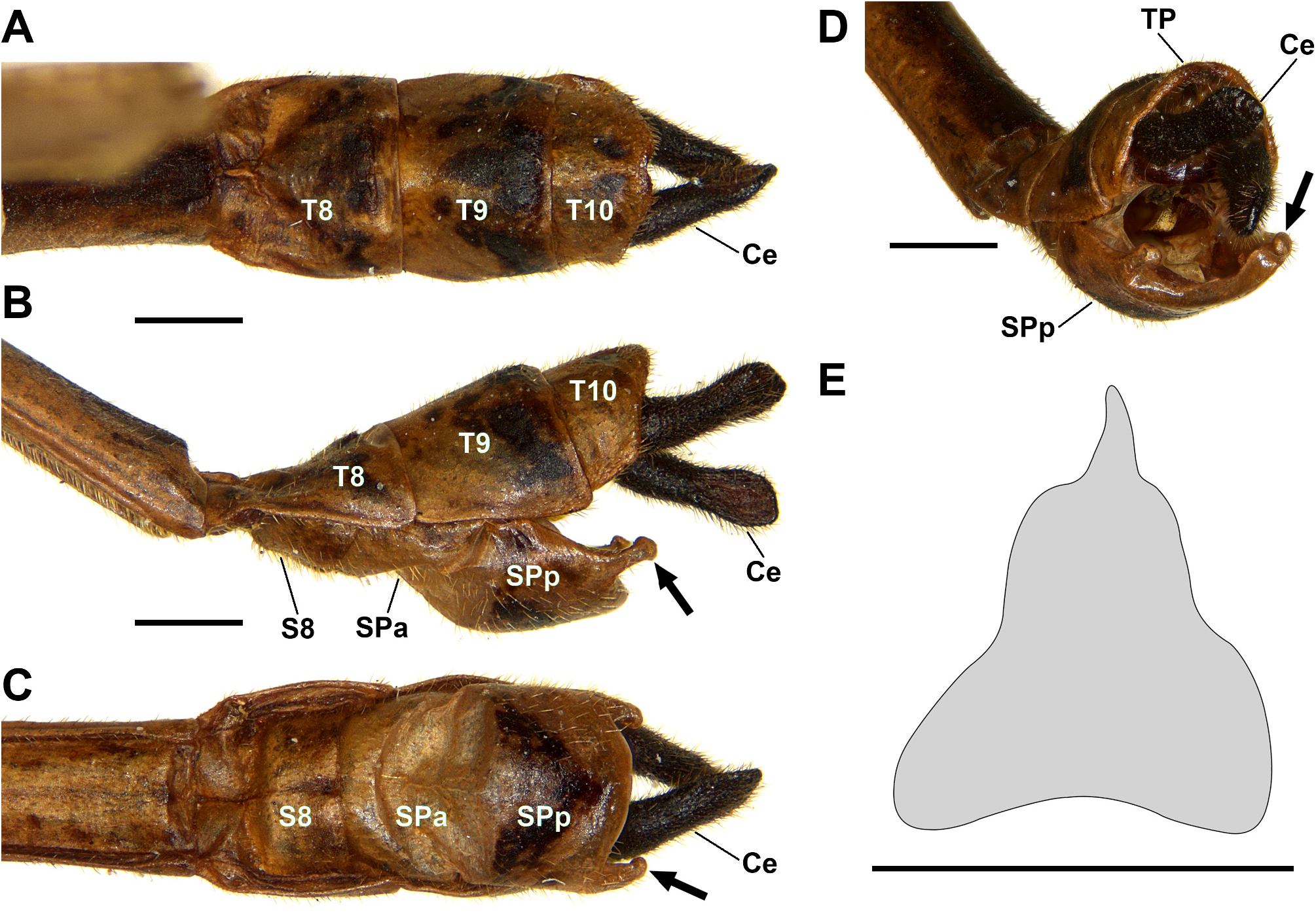
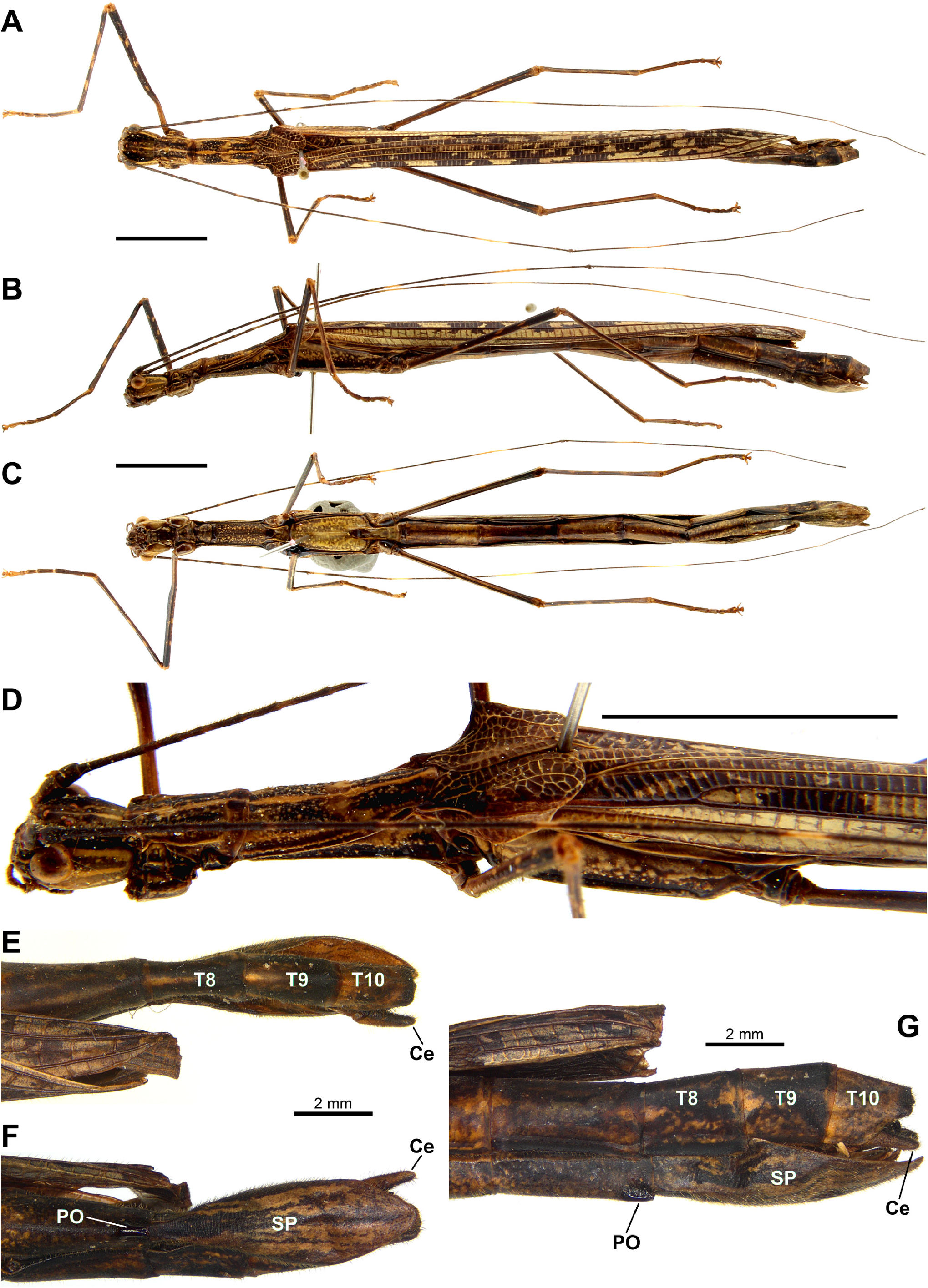
1. Praeopercular organ strongly pronounced, developed into a distinct bulge with blunt apex, sometimes surpassing the posterior margin of sternite VII ( Fig. 38G,H View FIGURE 38 )............................................ Paraphasma spinicauda View in CoL sp. nov.
- Praeopercular organ fairly discreet ( Fig. 5F,G View FIGURE 5 , 23F,G View FIGURE 23 , 33G View FIGURE 33 , 47F,G View FIGURE 47 ), developed into a small, longitudinal protuberance not surpassing the posterior margin of sternite VII.............................................................. 2
2. Tegmina with apical margin distinctly acuminate and shoulder pads developed into large, triangular, somewhat dull spines ( Fig. 42A–C View FIGURE 42 ); combined length of metathorax and median segment about 2.5x the length of mesothorax.................................................................................................. Paraphasma trianguliferum View in CoL
- Tegmina with apical margin rounded or weakly acuminate and shoulder pads developed into sharp spines or dull protuberances ( Figs 5C,D View FIGURE 5 , 10D View FIGURE 10 , 23D View FIGURE 23 , 29C View FIGURE 29 , 33D View FIGURE 33 , 47D View FIGURE 47 ); combined length of metathorax and median segment at most 2x the length of mesothorax......................................................................................... 3
3. Posterior margin of sternite VII straight; tegmina shoulder pads spiniform ( Fig. 47A,D View FIGURE 47 ); body lacking the pair of light lateral stripes extending along head, prothorax, mesothorax and costal region of tegmina and hind wings ( Figs 47A–D View FIGURE 47 , 49 View FIGURE 49 )......................................................................................... Paraphasma umbretta View in CoL
- Posterior margin of sternite VII with a rounded indentation; tegmina shoulder pads spiniform or dull; body with a pair of light lateral stripes extending along head, prothorax, mesothorax and costal region of tegmina and hind wings ( Figs 15C View FIGURE 15 , 19A View FIGURE 19 , 23B View FIGURE 23 , 25 View FIGURE 25 , 33B,D View FIGURE 33 )......................................... Paraphasma conspersum View in CoL , Paraphasma indistinctum View in CoL sp. nov., Paraphasma laterale View in CoL , Paraphasma marginale View in CoL , Paraphasma minus View in CoL , Paraphasma sooretama View in CoL sp. nov. (examination of the male is necessary for distinguishing among these species)


No known copyright restrictions apply. See Agosti, D., Egloff, W., 2009. Taxonomic information exchange and copyright: the Plazi approach. BMC Research Notes 2009, 2:53 for further explanation.