
Stenopsyche griseipennis McLachlan Population Variant
|
publication ID |
https://doi.org/ 10.11646/zootaxa.3717.1.5 |
|
publication LSID |
lsid:zoobank.org:pub:50CD1E66-36B6-428F-80C3-C814F6479D40 |
|
DOI |
https://doi.org/10.5281/zenodo.6151357 |
|
persistent identifier |
https://treatment.plazi.org/id/03B387B6-7E3D-FFF2-EBF9-FA4BFC8BFB56 |
|
treatment provided by |
Plazi |
|
scientific name |
Stenopsyche griseipennis McLachlan Population Variant |
| status |
|
Stenopsyche griseipennis McLachlan Population Variant View in CoL
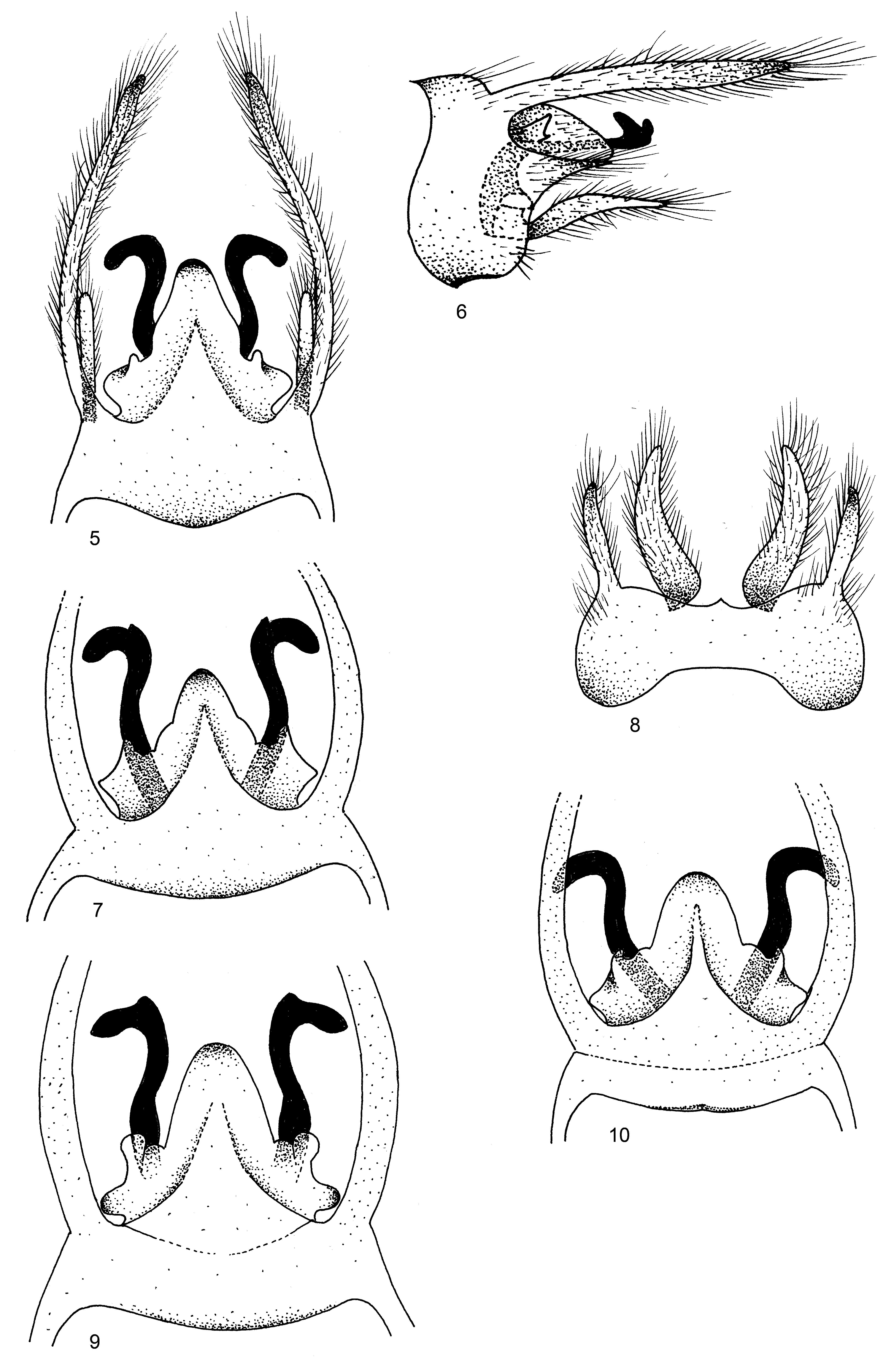
( Figs. 1–4 View FIGURES 1 – 4 )
Material examined. 1♂, INDIA: Uttarakhand; Diwalighal, 1400 m, 10-v-2009, Pandher & Parey (NPC); Diwalighal, 1400 m, 10-v-2009, Pandher & Parey, 2♂, 2♀ (NPC); Reetha Sahib, 1200 m, 12-vi-2008, Pandher & Parey, 1♂, 1♀ (NPC). Himachal Pradesh; Andhretta, 900 m, 10-vi-2010, Pandher, 1♂ (NPC). Sikkim; Mangan, 1700 m, 15-v-2010, Vikram, 1♂ (NPC).
Diagnosis. Stenopsyche griseipennis McLachlan belongs to the S. marmorata Group and is allied to S. haimvatika Schmid and S. marmorata Navás. In general appearance the genitalia of the population variant illustrated here closely resemble those of S. griseipennis McLachlan. However , there is variation in the superior arms of the inferior appendages which do not converge medially, though apically curved outward, each with a long neck and a small head in the holotype described by McLachlan, whereas in the population variant studied here the superior arms of the inferior appendages converge medially, are apically curved outward, with a very small neck and a long pointed beak. Similarly, the structure of phallus also shows marked differences from that of its type species and similar species. The phallotheca of S. griseipennis has spines scattered all over it and the endotheca has a bunch of slender, hair-like spines at its base; the phallotheca of S. haimvatika has dark, thick and backwardly directed spines basally in its ventral half and the phallotheca of S. marmorata has slender, dense spines apically on its lateral side and thick, scattered, backwardly directed spines ventrolaterally (Plate VI, Figs. 4 View FIGURES 1 – 4 , 5, 7 View FIGURES 5 – 10 of Schmid, 1969). However, in the population variant under study the phallotheca bears scattered small spines dorsolaterally, long backwardly directed thick spines basally on its dorsolateral margin, and scattered thick, long spines ventrally; the endotheca is without spines ( Fig. 4 View FIGURES 1 – 4 ).
Description. Adult male: large sized, typical for genus, length from front of head to tip of folded forewings 24 mm; overall body color in alcohol yellow, antennae yellow, dark brownish at joints of flagellar segments and each scape large, conical; head infuscate with golden yellow pubescence, vertex brownish, ocelli large and pale golden; thorax yellowish brown; forelegs each with 2 dark bands on tibia and 1 on tarsus; midlegs each with 4 dark bands, 2 each on tibia and tarsus; hind legs yellow and without any bands on tibiae and tarsi. Antennae 1.5 times length of forewing; interocular distance almost equal to breadth of eye, maxillary palpi 3.8 mm in length, segment 3 more than twice as long as segments 1 and 2 combined, segment 5 flexible and almost equal to length of first 4 segments combined; labial palpi 1.45 mm long, segment 3 almost equal to length of first two segments. Spurs: 3,4,4. Pronotum covered with one pair each of median pronotal setal warts and lateral pronotal setal warts.
Forewings with irregular, brown reticulate pattern anteriorly, lacking in anal areas. Forewing length 17 mm; veins Sc, R1, Cu, Cu1b and 1A thickened; discoidal and median cells closed, discoidal cell smaller, narrow and shorter than median cell. Thyridial cell long and somewhat broader towards apex. Forks I, II, III, IV and V present.
Hind wings clear, broader than forewings; veins Sc, R1 and Cu1 thickened, Sc and R1 separately joining R2+3, forming supra-discoidal cell, and then joining wing margin as common vein Sc+R1+R2+R3, supra-discoidal cell positioned above discoidal cell, very minute; discoidal cell closed, only about 1.5x supra-discoidal cell; median cell absent. Thyridial cell long, narrow and closed. Forks II, III and V present, forks I and IV absent.
Male genitalia ( Figs. 1–4 View FIGURES 1 – 4 ). Segment IX large, produced anterad dorsally and subventrally, bearing numerous bristles, with prominent and sharp apicolateral side piece on each side posteriorly. Body of tergum X membranous, roughly triangular, apically cleft; dorsal projections of tergum X very minute, with long and apically rounded intermediate projections arising at their bases. Preanal appendages long, broad, thick and arising at junction of segments IX and X. Superior arms of inferior appendages almost twice as long as body of tergum X, broader basally, converging medially, curved outward apically, each with narrow neck and head resembling those of bird. Ventral arm of each inferior appendage finger-like laterally and pointed in lateral view, ventrally oval, bearing setae all over. Phallotheca almost cylindrical, longer than endotheca; endotheca lacking spines. Phallotheca bearing scattered small spines dorsolaterally, backwardly directed long and thick spines basally on dorsolateral margins and scattered thick, long spines ventrally.
Distribution. India: Uttarakhand, Himachal Pradesh, Sikkim.

No known copyright restrictions apply. See Agosti, D., Egloff, W., 2009. Taxonomic information exchange and copyright: the Plazi approach. BMC Research Notes 2009, 2:53 for further explanation.