
Biapertura sibirica ( Sinev, Karabanov & Kotov, 2020 )
|
publication ID |
https://doi.org/10.11646/zootaxa.4885.3.1 |
|
publication LSID |
lsid:zoobank.org:pub:784B14D1-7B68-42F1-81A1-9EAB8DFD7E79 |
|
persistent identifier |
https://treatment.plazi.org/id/03B287B6-FFE2-FF8E-BDB5-FE52FEF2AFB1 |
|
treatment provided by |
Plazi (2020-11-29 19:49:34, last updated 2020-12-08 21:43:21) |
|
scientific name |
Biapertura sibirica ( Sinev, Karabanov & Kotov, 2020 ) |
| status |
comb. nov. |
Biapertura sibirica ( Sinev, Karabanov & Kotov, 2020) comb. nov.
Lilljeborg 1901: 459, Pl. 17: fig 12 ( Alona affinis partum); Werestschagin 1911: 555 ( Alona affinis var. dentata ); Behning 1941: 315 ( Alona affinis var. dentata ); Smirnov 1971: 472–474, Fig. 590 ( Alona affinis dentata ); Sinev 1997: 53, Fig. 4C View FIGURE 4 ( Alona affinis var. dentata ); Garibian et al. 2019: 49–52, Fig. 8 View FIGURE 8 ( Alona cf. affinis ); Sinev et al. 2020: Fig. 1–5 View FIGURE 1 View FIGURE 2 View FIGURE 3 View FIGURE 4 View FIGURE 5 , 6 View FIGURE 6 A–F ( Alona ).
Type locality. Lake near road between Amga river and Churapcha, Sakha (Yakutia) Autonomous Republic, Russia, 61.80453° N, 133.2409° E GoogleMaps .
Type material: holotype (parthenogenetic female), allotype (adult male) and paratypes in Zoological Museum of M. V. Lomonosov Moscow State University , access numbers Ml–186, Ml–187 and Ml–188, respectively .
Material studied earlier. See Sinev 1997 ( A. affinis var. dentata ) and Sinev et al. 2020 for the list of material from Eurasia, and for detailed description.
Diagnosis. Female. Length of adult 0.68–0.94 mm. Body oval ( Fig. 17A View FIGURE 17 ), maximum height at the middle of the body; height/length ratio about 0.6 in adults. Postero-dorsal angle with 1–3 denticles and few setulae ( Fig. 17 View FIGURE 17 B–D). Main head pores ( Fig. 17E View FIGURE 17 ) with PP 1.5-2.3 in adults. Lateral head pores located about 0.9-1.3 IP distance from midline, at level before anterior major head pore. Postabdomen ( Fig. 17F View FIGURE 17 ) subrectangular, with parallel margins, weakly narrowing at the end. Length about 2.5 height. Ventral margin straight. Dorsal margin weakly convex to straight in postanal portion and weakly concave in anal one, with distal part about 2.5 times longer than preanal one, with postanal portion 2.5–2.8 times longer than anal one. Postanal margin ( Fig. 17G View FIGURE 17 ) with 11–13 massive denticles, each with 2–6 spinulae along anterior margin; size of denticles increasing distally. Postabdominal claw ( Fig. 17H View FIGURE 17 ) with basal spine about 0.35 length of the claw. Antenna ( Fig. 17I View FIGURE 17 ) with basal segment of both branches longer than two others, middle segments in both branches shorter than apical segments. Spine on basal segment of exopodite longer than middle segment. Spines on apical segments shorter than apical segments. Thoracic limb I with IDL claw-like seta 1 ( Fig. 17J View FIGURE 17 ). Seta e of endite 2 only slightly longer than seta f.
Male. Length of adult was 0.60–0.67 mm. Body low oval ( Fig. 17K View FIGURE 17 ), with maximum height at the middle or in the third quarter of the body, height/length ratio about 0.54. Postabdomen ( Fig. 17 View FIGURE 17 L–M) with maximum height at postanal angle, evenly narrowing in postanal portion. Length about 2.5x height. Postabdominal claw situated on small protrusion in ventral portion of distal margin. The sperm ducts open above the protrusion. Posteroventral and posterodorsal angles rounded. Dorsal margin weakly convex in postanal portion and weakly concave in anal one. Preanal and postanal angles weakly defined. Distal portion of postabdomen 2–2.5 times longer than preanal one, postanal portion 2.2–2.4 times longer than anal one. Postabdominal claw weakly curved, shorter than in female, significantly shorter than preanal margin. Basal spine about 0.25–0.3 length of claw. Thoracic limb I ( Fig. 17N View FIGURE 17 ) with IDL seta 1 large, about 2/3 length of IDL seta 2, located at distal margin of IDL. Ventral face of the limb under the copulatory brush with a row of about 5 stiff setulae of moderate length; outer face of endite 3 with a row of about 15 shorter setulae, there is a narrow gap between these two rows.
Differential diagnosis. B. sibirica clearly differs from all other species of the group by the presence of denticles on the posteroventral angle of valves in both males and females. It also clearly differs from all species with known male ( B. affinis , B. kendallensis and B. ossiani ) in morphology of male postabdomen.
Distribution and ecology. B. sibirica is common in Asian Russia, and also recorded in Mongolia and in Kyrgyzstan. In Europe this species is rare; it was recorded in a few water bodies of Sweden, Finland, Ukraine, and European Russia. Found in lakes and ponds, oxbow lakes in river valleys, and pools in Sphagnum bogs. Frequently co-exists with B. affinis . Ecology of the species is not studied, but indirect data suggest it is a eurybiotic, acid-tolerant species.
Behning, A. L. (1941) The Cladocerans of the Caucasus. Gruzmedgiz Publishing, Tbilisi, 384 pp. [in Russian]
Garibian, P. G., Chertoprud, E. S., Sinev, A. Y., Korovchinsky, N. M. & Kotov, A. A. (2019) Cladocera and Copepoda (Crustacea: Branchiopoda) of the Lake Bolon and its basin (Far East of Russia). Arthropoda Selecta, 28 (1), 37 - 63. https: // doi. org / 10.15298 / arthsel. 28.1.05
Lilljeborg, W. (1901) Cladocera Sueciae. Nova acta regiae societatis scientatis scientiarum upsaliensis, Seriei Tertiae, 19, 1 - 701.
Sinev, A. Y. (1997) Review of the affinis- group of Alona Baird, 1843, with the description of a new species from Australia (Anomopoda: Chydoridae). Arthropoda Selecta, 6 (3 - 4), 47 - 58.
Sinev, A. Y., Karabanov, D. P. & Kotov, A. A. (2020) A new North Eurasian species of the Alona affinis complex (Cladocera: Chydoridae). Zootaxa, 4767 (1), 115 - 137. https: // doi. org / 10.11646 / zootaxa. 4767.1.5
Smirnov, N. N. (1971) Chydoridae of the world fauna. Fauna SSSR. Rakoobraznie, 1 (2), 1 - 531. [in Russian]

FIGURE 1. Biapertura affinis (Leydig, 1860) from Glubokoe Lake, Moscow Area, Russia.A–K, adult parthenogenetic female. A, lateral view. B–C, posteroventral angle of valves. D, head shield. E, head pores. F, labrum. G, postabdomen. H, postanal margin of postabdomen. I, postabdominal claw. J, antennule. K, antenna. L, ephippial female. M–O, adult male. M, lateral view. N, postabdomen. O, postabdominal claw. (A–L, orig.; M–O, after Sinev et al. 2020)

FIGURE 2. Biapertura affinis (Leydig, 1860) from Glubokoe Lake, Moscow Area, Russia. A–B, lateral view. C, dorsolateral view. D, dorsal view. E, ventral view. F, frontal view. G, ventral part of valve. H, postabdomen. I, postabdominal claw. J, antenna.
FIGURE 3. Biapertura affinis (Leydig, 1860) from Glubokoe Lake, Moscow Area, Russia, parthenogenetic female.A–B, head pores. C–E, posterior dorsal pores.
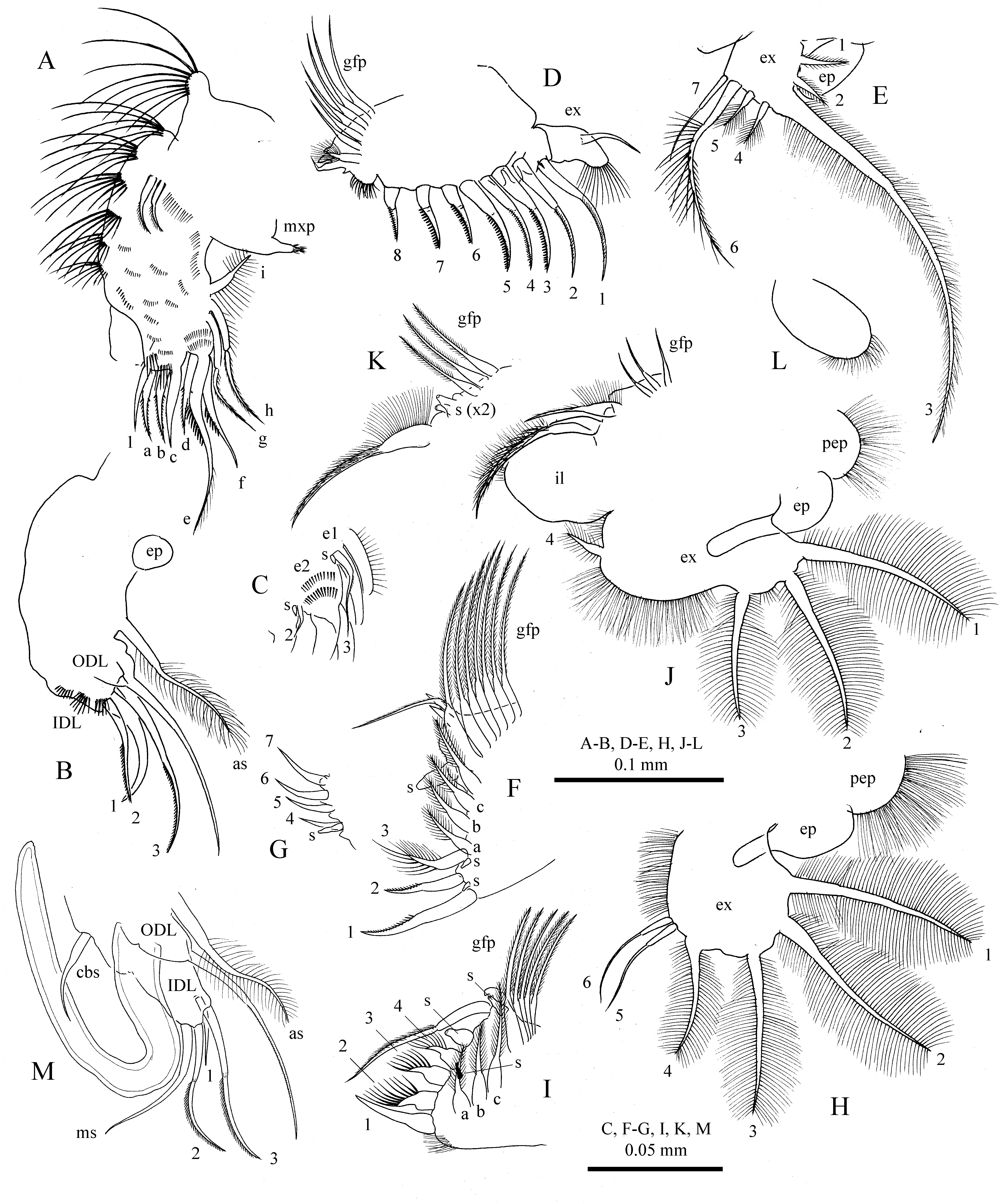
FIGURE 4. Biapertura affinis (Leydig, 1860) from Glubokoe Lake, Moscow Area, Russia. A–L, thoracic limbs of adult parthenogenetic female. A, limb I. B, IDL and ODL of limb I. C. inner setae of endites 1–2 of limb I. D, limb II. E, exopodite of limb III. F–G, inner and outer parts of inner portion of limb III. H, exopodite of limb IV. I, inner portion of limb IV. J–K, limb V and its gnathobase filter plate. L, limb VI. M, copulatory hook and IDL of thoracic limb I of adult male. (A–L, orig.; M, after Sinev et al., 2020)
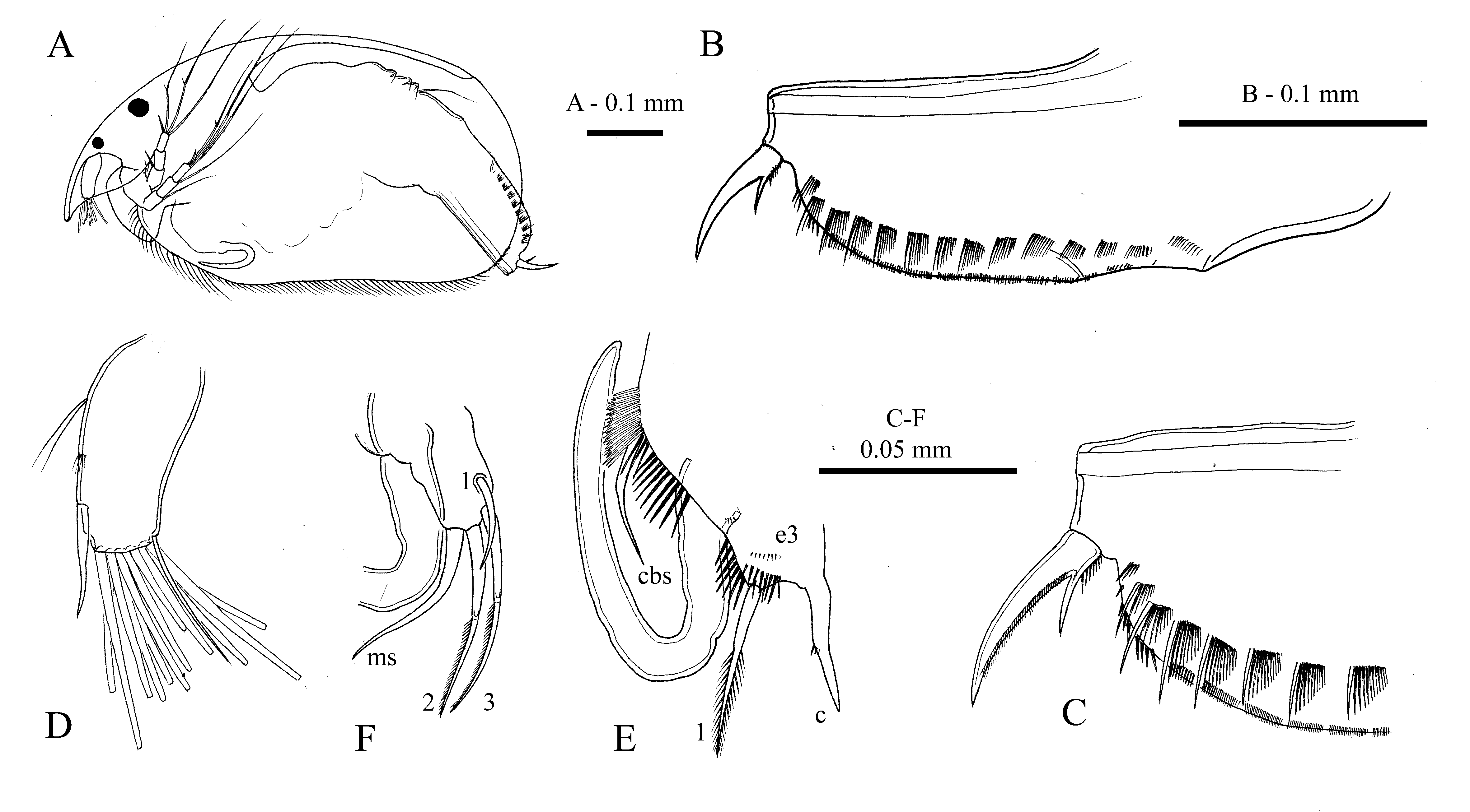
FIGURE 5. Biapertura affinis (Leydig, 1860) from Klein Vaal River, Transvaal Province, Republic of South Africa, adult male. A, lateral view. B, postabdomen. C, distal portion of postabdomen. D, antennule. E, copulatory hook of thoracic limb I. F, IDL of thoracic limb I.

FIGURE 6. Biapertura kendallensis (Henry, 1919) from Ryan’s II Billabong, Bonegilla, east of Wodonga, Victoria, Australia. A–B, juvenile females of instar I and II. C–K, adult parthenogenetic female. C, lateral view. D, ventral margin of valves. E, setae of anterior group. F, setae of posterior group. G, posterior margin of valves. H, posteroventral angle of valves. I–J, head shields. K, head pores. L, ephippial female. M–N, juvenile males of instar I and II. O, adult male.

FIGURE 8. Biapertura kendallensis (Henry, 1919) from near Ryan’s II Billabong, Bonegilla, east of Wodonga, Victoria, Australia.A–B, antenna of adult female. C–F, ephippial female. C–D, same specimen in lateral and dorsolateral view. E, ephippium. F, head pores.
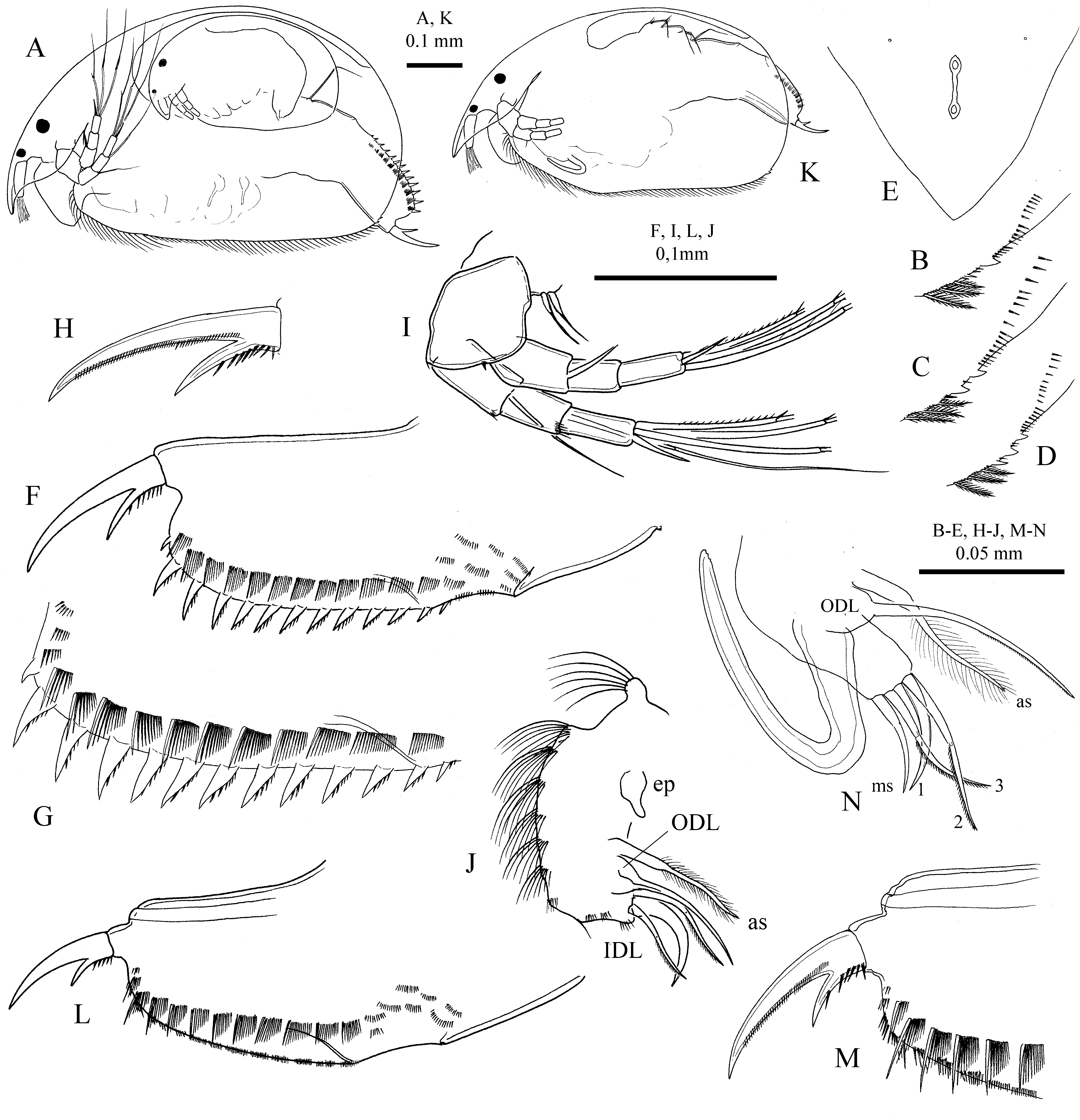
FIGURE 17. Biapertura sibirica (Sinev, Karabanov & Kotov, 2020). A–J, adult parthenogenetic female. A, lateral view. B–D, posteroventral angle of valves. E, head pores. F, postabdomen. G, postanal margin of postabdomen. H, postabdominal claw. I, antenna. J, IDL of thoracic limb I. K–N, adult male. K, lateral view. L, postabdomen. M, distal portion of postabdomen. N, copulatory hook and IDL of thoiracic limb I. (After Sinev et al. 2020.)
| V |
Royal British Columbia Museum - Herbarium |
No known copyright restrictions apply. See Agosti, D., Egloff, W., 2009. Taxonomic information exchange and copyright: the Plazi approach. BMC Research Notes 2009, 2:53 for further explanation.
|
Kingdom |
|
|
Phylum |
|
|
Class |
|
|
Order |
|
|
Family |
|
|
Genus |
1 (by plazi, 2020-11-29 19:49:34)
2 (by ExternalLinkService, 2020-11-29 19:57:24)
3 (by tatiana, 2020-12-04 13:22:27)
4 (by ExternalLinkService, 2020-12-08 21:43:21)
5 (by tatiana, 2020-12-09 12:59:00)
6 (by ExternalLinkService, 2020-12-17 03:22:32)
7 (by ExternalLinkService, 2021-09-19 03:50:43)
8 (by plazi, 2023-10-31 23:38:18)