
Drusus medianus, Marinkovic-Gospodnetic, 1976
|
publication ID |
https://doi.org/ 10.5281/zenodo.1051908 |
|
DOI |
https://doi.org/10.5281/zenodo.6201445 |
|
persistent identifier |
https://treatment.plazi.org/id/03AFA402-FFF9-FFCD-8F82-57347B812FAA |
|
treatment provided by |
Plazi |
|
scientific name |
Drusus medianus |
| status |
|
Description of fifth instar larva of Drusus medianus View in CoL
Case and Larva
Case constructed completely of mineral particles ( Fig. 16 View FIGURES 16 – 21 ), slightly curving, total length 9.98–12.55 mm, width of anterior part 2.42–3.16 mm, width of posterior part 1.72–2.16 mm (n=20). Overall body shape eruciform ( Fig. 17 View FIGURES 16 – 21 ), total length without case 10.17–11.06 mm (n=20).
Head
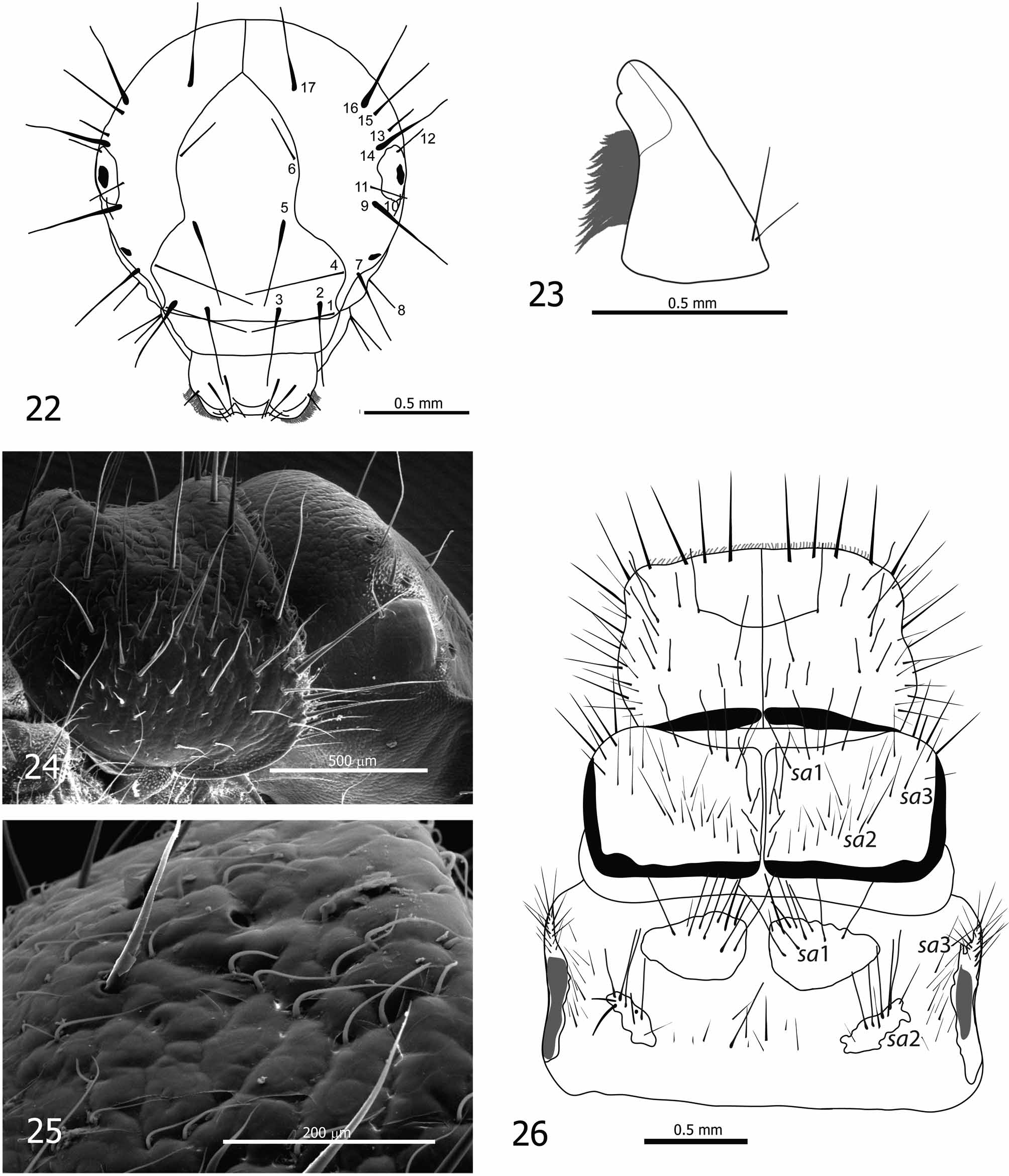
Head capsule hypognathous, ellipsoidal, with width 1.37–1.48 mm (n=20) ( Figs 17–18 View FIGURES 16 – 21 , 22 View FIGURES 22 – 26 ). Head brown, dorsally darker and laterally lighter ( Figs 16–17 View FIGURES 16 – 21 ), with granular surface sculpturing surface ( Fig. 19 View FIGURES 16 – 21 ). Genae of the parietals reddish-brown to yellow with lighter ring around each eye ( Fig. 18 View FIGURES 16 – 21 ). Posterior part of dorsum with numerous, dark muscle attachment spots. Distinct area of spinules (small spines, size approximately 0.015–0.025 mm) positioned laterally on each side of head capsule next to the eye ( Figs 20–21 View FIGURES 16 – 21 ). Spinules also in some specimens on posterior part of each antennal prominence. Frontoclypeal apotome bell-shaped with narrow central region ( Fig. 22 View FIGURES 22 – 26 ). Antennae short, brown to dark brown, each positioned on small, noticeable prominences accompanied by 2 prominent lateral setae (setae no. 7 and no. 9) ( Figs. 18–19 View FIGURES 16 – 21 ). Other primary setae positioned as shown in Fig. 22 View FIGURES 22 – 26 and light or dark and short or long as described for D. ramae .
Labrum symmetrical, brown posteriorly to yellowish anteriorly, with setal brush at anterolateral margins and 5 pairs of thin primary setae on dorsal surface.
Mandibles black, mesal part reddish. Typical for grazers, mesal margin with setal brush and without teeth except 1 small tooth usually present subapically ( Fig. 23 View FIGURES 22 – 26 ). Two setae present laterobasally on each mandible ( Fig. 23 View FIGURES 22 – 26 ).
Labium and maxillae light-brown (yellowish). Maxillary palps 5-segmented.
Thorax
Pronotum dark brown to black ( Figs 16–18 View FIGURES 16 – 21 ) with granular surface sculpturing ( Figs 24–25 View FIGURES 22 – 26 ). Posterior margin rounded, both posterior and lateral margins thick and darkly sclerotized ( Fig. 18 View FIGURES 16 – 21 ). Anterior part (i.e. 50–60 %) of pronotum slightly concave, posterior part with a prominent median hump ( Figs 18 View FIGURES 16 – 21 , 24 View FIGURES 22 – 26 ). Pronotum bearing dark setae, especially laterally and on anterior margin, some of them long and conspicuous ( Figs 18 View FIGURES 16 – 21 , 24 View FIGURES 22 – 26 ). Dorsal and lateral regions of pronotum covered with numerous white recumbent setae ( Figs 24- 25 View FIGURES 22 – 26 ). Prosternal horn present.
Mesonotum sclerites brown, lighter than pronotum, with dark muscle attachment spots and uneven (rugged) surface. Long, dark setae at positions sa 1, sa 2 and sa 3 ( Fig. 26 View FIGURES 22 – 26 ). Posterior and lateral margins thick and darkly sclerotized.
Metanotum with 3 pairs of dorsal sclerites ( Fig. 26 View FIGURES 22 – 26 ). Anteromedian (sa 1) sclerites ellipsoid, with distance between them smaller than their length ( Figs 26 View FIGURES 22 – 26 , 48–49 View FIGURES 46 – 53 ), covered by setae, mainly anteromedially, color similar to mesonotum. Posteromedian (sa 2) sclerites smaller and lighter than sa 1 sclerites ( Figs 26 View FIGURES 22 – 26 , 48–49 View FIGURES 46 – 53 ), diagonally and irregularly ellipsoid and with many dark setae. Group of setae present on membranes between sa 2 sclerites and between sa 2 and sa 3 sclerites. Lateral (sa 3) sclerites longitudinally prolonged, sickleshaped, lighter brown with dark median region, and group of setae anteriorly ( Fig. 26 View FIGURES 22 – 26 ).
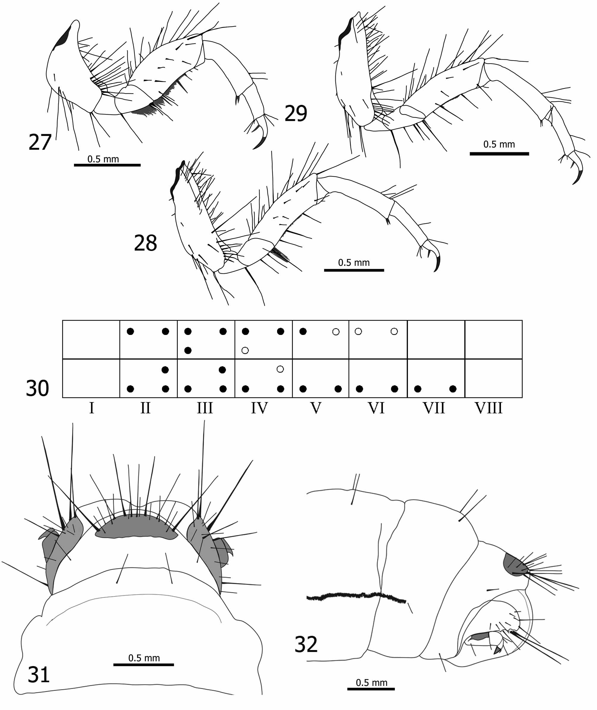
Legs yellow-brown to dark brown or black, with dark ventral and dorsal margins ( Figs 16–18 View FIGURES 16 – 21 ). Foreleg coxae with dark setae on ventral and dorsal edges. Foreleg trochanters without dorsal setae, each with few light-yellow setae on ventral margin and distally with dense ros of short, fine, yellowish setae (trochanteral brush) continuing onto basoventral margin of femur ( Fig. 27 View FIGURES 27 – 32 ). Foreleg femora each with dark setae on dorsal edge, few stout, light-yellow setae on ventral margin in addition to basoventral dense row of short, fine, yellowish setae ( Fig. 27 View FIGURES 27 – 32 ). Mid- and hind leg coxae and femora with dark setae on both ventral and dorsal edges, midleg trochanters with few, fine, yellowish setae apicoventrally ( Figs 28–29 View FIGURES 27 – 32 ). Additional setae present on anterior and posterior faces of all femora. Setae on dorsal edges of tibiae present only distally in all legs. Tarsae each with claw and basal seta, and tibial spurs light brown, almost yellowish. Foreleg coxae and femora wide compared to those of mid- and hind legs ( Figs 27–29 View FIGURES 27 – 32 ). Mid- and hind-legs similar in shape and size ( Figs 28–29 View FIGURES 27 – 32 ), with slender coxae, trochanters, and femora.
Abdomen
Abdominal segment I with well-developed dorsal and lateral humps (protuberances). Numerous setae present anterior and lateral to dorsal hump. It is not possible to distinguish between setal areas sa 1 and sa 2 on dorsal side. With numerous ventral setae, some of them with small sclerites at bases. Lateral humps with few setae.
Single filamentous gills ( Fig. 17 View FIGURES 16 – 21 ) present on segments II–VII ( Fig. 30 View FIGURES 27 – 32 ). Dorsal pre- and post-segmental gills present on segments II–VI; ventral pre- and post-segmental gills present on segments II–VII. Lateral presegmental gills present on segments III–IV and lateral post-segmental gills on segments II–IV ( Fig. 30 View FIGURES 27 – 32 ). In some specimens dorsal pre-segmental gills on segment VI, post-segmental gills on segments V and VI, and lateral pre- and post-segmental gills on segment IV are not present ( Fig. 30 View FIGURES 27 – 32 ). Number of dorsal setae on abdominal segment VIII varies (2–6). Lateral fringe extends from last third of segment II to first half of segment VIII; in some specimens, only few setae forming lateral fringe on segment II or setae not visible.
Segment IX bearing irregular, semicircular, light brown dorsal sclerite, generally with 8 long, dark setae on posterior margin and several shorter, light setae on posterior half of sclerite ( Fig. 31 View FIGURES 27 – 32 ). Anal prolegs typical of limnephilids ( Fig. 32 View FIGURES 27 – 32 ), each with lateral sclerite longitudinally prolonged, sickle-shaped, yellowish, with small setae scattered over posterior 2/3rds and 2 large, dark setae at posterior end ( Fig. 32 View FIGURES 27 – 32 ). Anal claws brown.


No known copyright restrictions apply. See Agosti, D., Egloff, W., 2009. Taxonomic information exchange and copyright: the Plazi approach. BMC Research Notes 2009, 2:53 for further explanation.