
Astyanax vermilion, Zanata & Camelier, 2009
|
publication ID |
https://doi.org/ 10.1590/S1679-62252009000200007 |
|
persistent identifier |
https://treatment.plazi.org/id/03AF9165-8B70-6F56-17D6-FEB48616FE62 |
|
treatment provided by |
Carolina |
|
scientific name |
Astyanax vermilion |
| status |
sp. nov. |
Astyanax vermilion View in CoL , new species
Figs. 1-3 View Fig View Fig View Fig
Holotype. MZUSP 101243 View Materials , 39.4 mm SL, Brazil, Bahia State, Floresta Azul, rio Salgado under bridge on road BA 130, rio Cachoeira drainage, 14°51’18.0”S 39°39’41”W, 189 m alt., 12 Feb 2008, A. M. Zanata, P. Camelier, R. Burger & A. B. A. Góes. GoogleMaps
Paratypes. All from Brazil, Bahia State. ANSP 189328 About ANSP , 10 About ANSP , 35.4 About ANSP - 39.0 mm SL, MZUSP 101244 View Materials , 10 View Materials , 37.4-38.7 mm SL, UFBA 4344 , 76 , 30.5-40.8 mm SL (12, 30.5-40.8 mm SL), collected with holotype. UFBA 4342 , 31 , 26.9-39.2 mm SL (12, 3 c&s, 26.9- 39.2 mm SL), Floresta Azul, streamlet tributary of rio Salgado drainage on road between Floresta Azul and Almadina, rio Cachoeira drainage, 14°48’31”S 39°39’13”W, 188 m alt., 12 Feb 2008, Zanata et al. UFBA 4343 , 56 , 35.1-40.3 mm SL (12, 35.1- 40.3 mm SL), Itajuípe , rio Almada on Fazenda Luanda, 14°40’53”S 39°24’24”W, 258 m alt., 12 Feb 2008, Zanata et al. UFBA 4813 , 24 , 25.7-42.8 mm SL, Ilhéus , district of Rio do Braço , rio do Braço , rio Almada basin, in front of Fazenda Norma , between roads BR 415 and BA 262,14°41’10’’S 39°16’27’’W, 80 m alt., 24 Feb 2009, Zanata et al. UFBA 4905 , 32 , 32.4-39.5 mm SL, Ilhéus , rio Santana , on road between Ilhéus and Buerarema, rio Cachoeira basin, 14°54’42’’S 39°08’36’’W, 53 m alt., 24 Feb 2009, Zanata et al GoogleMaps .
Diagnosis. Astyanax vermilion can be distinguished from all other Astyanax species known from northeastern Brazilian drainages ( A. brevirhinus , A. epiagos , A. fasciatus , A. intermedius , A. jacobinae , A. lacustris , A. pelecus , A. rivularis , A. taeniatus , and A. turmalinensis ) by having distal portion of pelvic fin dark, a combination of sexually dimorphic characters, the posteroventral portion of body and all fins (except pectoral) reddish in life, and an inconspicuous humeral and caudal spots. Differs further by having highest body depth just anterior to dorsal-fin origin (vs. body highest on a vertical around middle or posterior portion of pectoral fin in A. epiagos , A. intermedius , A. jacobinae , and A. rivularis ), 32-34 lateral-line scales (vs. 35 or more in A. brevirhinus , A. intermedius , A. jacobinae , A. pelecus , A. rivularis ), five series of scales between dorsalfin origin and lateral line (vs. six or more in A. brevirhinus , A. fasciatus , A. lacustris ), three series of scales between lateral line and pelvic-fin origin (vs. 4.5 or more, respectively, in A. brevirhinus , A. lacustris , A. turmalinensis , A. intermedius ), presence of one or two maxillary teeth (vs. none in A. lacustris or three or more in A. brevirhinus and A. fasciatus ). Astyanax vermilion can be further diagnosed from A. epiagos and A. pelecus by having 20-23 branched anal-fin rays (vs. 13-17 and 16-18, respectively), from A. jacobinae and A. lacustris by having one or two maxillary teeth (vs. three and none, respectively), from A. taeniatus by having no more than five cusps and dentary teeth abruptly decreasing in size posteriorly (vs. usually seven cusps on dentary and premaxillary teeth and dentary teeth gradually smaller posteriorly). This new species differs further from A. turmalinensis by having 32-34 lateral-line scales (vs. 34-36) and dorsal fin usually ii,8,i (vs. dorsal fin always with ii,9 rays). Astyanax vermilion differs from A. burgerai on its overall body shape and color, including an inconspicuous humeral spot (vs. humeral blotch conspicuous and vertically elongated), posteroventral portion of body reddish when alive (vs. absence of red pigmentation on body), branched anal-fin rays 20-23 (vs. 18-19), dorsal-fin rays usually ii,8,i (vs. ii,9), and 8-12 scales aligned on base of anterior anal-fin rays (vs. 3-6).
Description. Morphometric data of holotype and paratypes are presented in Table 1. Body somewhat compressed.
Greatest body depth along vertical through dorsal-fin origin. Dorsal profile of head convex from upper lip to vertical through anterior nostrils; straight to slightly convex from latter point to tip of supraoccipital spine and nearly straight to slightly convex from this point to dorsal-fin origin. Body profile straight and posteroventrally slanted along dorsal-fin base; straight from dorsal-fin base terminus to adipose fin, and slightly concave along caudal peduncle. Ventral profile of head and body strongly convex anteriorly and convex from region below eye to anal-fin origin. Body profile along analfin base straight and posterodorsally slanted. Ventral profile of caudal peduncle nearly straight to slightly concave.
Head somewhat pointed to rounded anteriorly in lateral profile; mouth terminal. Posterior terminus of maxilla extending slightly beyond vertical through anterior margin of orbit. Premaxillary teeth somewhat narrow, distributed in two rows. Outer row with 3 (7), 4 (24), or 5* (4) teeth bearing 3 cusps. Inner row with 4 (1), 5* (32), or 6 (3) teeth bearing 3, 4 or 5 cusps. Symphyseal tooth of inner series narrow, asymmetrical, with one cusp on anteromedial side, one larger central cusp and two smaller on lateral side; second tooth the larger, with 5 cusps; last teeth with 3 cusps. Maxilla with 2 (1) or 3 (2) teeth bearing one or 3 cusps. Dentary with 10 (1) or 11 (2) symmetrical teeth; 5 anterior teeth larger, with 5 cusps, followed by 6 teeth unicuspidate; posterior smaller teeth abruptly smaller than anterior ones in one c&s specimen and somewhat decreasing gradually in size posteriorly in the other two.
Scales cycloid, circuli absent on exposed area of scales, with few slightly divergent radii extending to posterior margin of scales. Lateral line slightly decurved anteriorly, completely pored from supracleithrum to base of caudal fin, with 32 (7), 33* (8), or 34 (6) perforated scales. Horizontal scale rows between dorsal-fin origin and lateral line 5 (36), not including scale of predorsal series situated just anterior to first dorsalfin ray. Horizontal scale rows between lateral line and pelvicfin insertion 3 (36). Scales along middorsal line between tip of supraoccipital process and origin of dorsal fin 9 (6), 10* (24), or 11 (2). Horizontal scale rows around caudal peduncle 14 (13). Base of anteriormost anal-fin rays covered by a series of 8-12 scales.
Dorsal-fin rays ii,8 (1), ii,8,i* (29), or ii,9 (6). Distal margin of dorsal fin straight. Dorsal-fin origin situated at vertical approximately at or slightly posterior to middle of standard length. Base of last dorsal-fin ray posterior to vertical through anal-fin origin. First dorsal-fin pterygiophore inserting behind neural spine of 10 th (3) vertebra. Adipose fin present. Anal-fin rays iii,20 (9), 21 (16), 22* (10), or 23 (1); cleared and stained specimens revealed 5 unbranched rays (3). Distal margin of anal fin in males slightly concave to nearly straight and females with anterior portion of fin distinctly concave. First anal-fin pterygiophore inserting behind haemal spine of 16 th (3) vertebra. Pectoral-fin rays i,9 (1), 10* (6), 11 (25), or 12 (4). Tip of pectoral fin usually reaching vertical through pelvic-fin insertion. Pelvic-fin rays i,6 (1) or 7* (35); in males, tip of pelvic fin usually trespasses insertion of first anal-fin rays, while in females the pelvic fin does not reach or barely reaches the anal-fin origin. Caudal fin forked, lobes pointed, similar in size. Principal caudal-fin rays 10+9 (3). Eight (2) or 9 (1) dorsal procurrent caudal-fin rays, and 9 (3) ventral procurrent caudalfin rays. First gill arch with 7 (2) or 8 (1) + 1 (3) + 13 (3) rakers. Vertebrae 33 (3). Supraneurals 4 (1) or 5 (2).
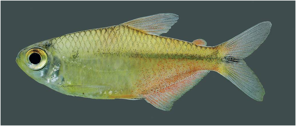
Color in alcohol. Overall ground color yellow ( Figs. 1-2 View Fig View Fig ). Guanine restricted to part of infraorbitals, preopercle, and opercle. Anteriormost portion of head dark; upper and lower lips completely dark, followed by areas with lower concentration of chromatophores, usually forming a black border on anterior portion of mouth. Head dorsum moderately dark, with relatively sparse dark chromatophores; central area between eyes less pigmented. Dark chromatophores sparsely distributed over anterior half of maxillary bone. Infraorbitals, preopercle and opercle clear. Ventral portion of head clear, unless by presence of scattered dark chromatophores on its anteriormost portion.
Scales of dorsalmost four longitudinal series and middorsal series with dark chromatophores concentrated along its posterior margin resulting in reticulate pattern; center of scales usually clear, except for some more pigmented specimens that have chromatophores on central portion of scales, masquerading the reticulate pattern. Scales below the lateral line less or no pigmented on anteriormost portion of body and with sparse dark chromatophores on posterior half of body; dark lines usually present along myosepts above anal-fin base.Abdominal region without dark chromatophores.
Humeral region with a roughly rounded and inconspicuous humeral spot, formed by underlying dark chromatophores; spot over third and fourth scales of horizontal series immediately above lateral line. Dark and narrow stripe extending usually from humeral spot to caudal peduncle; stripe more conspicuous behind vertical through origin of dorsal fin. Stripe usually enlarged at the end of caudal peduncle, forming an inconspicuous spot, extending to end of median caudal-fin rays.
All fin rays dark. Dorsal fin with dark chromatophores over membranes on area close to borders of rays and pectoral fin with chromatophores over borders of rays, forming dark lines along borders. Anal fin similarly colored, with dark chromatophores over lateral borders of rays and also with concentration of chromatophores close to distal border of fin, over rays and interradial membranes, forming an inconspicuous dark margin on fin; dark margin usually more visible on males, more evident on longest unbranched and first branched rays. Pelvic fins with dark chromatophores over borders of rays, mainly on unbranched and first four branched rays; distal portion of rays usually darker; males with distal portion of first pelvic-fin rays distinctly darker than females ( Figs. 1-2 View Fig View Fig vs. Fig. 3 View Fig ). Caudal fin usually darker than remaining fins, with chromatophores over rays and interradial membranes, apparently more concentrated on its distal half. Adipose fin clear, with a few dark chromatophores on its margin.
Color in life. Life color pattern taken from photographs of specimens soon after fixation ( Fig. 2 View Fig ). Overall body coloration silver to yellowish. Head silvery, at least on infraorbitals, preopercle, and opercle. Scales on dorsal half of body with borders dark, forming a reticulate pattern. Posteroventral portion of body, above anal fin, reddish; red pigment on body more intense in males. Midlateral line of body darkened, formed apparently mostly by underlying dark pigment, and extending from area below dorsal fin to caudal peduncle. All fins reddish, except pectoral, with scattered dark chromatophores; red pigmentation more intense in males. Caudal fin somewhat darker, with dark chromatophores concentrated on its median rays.
Sexual dimorphism. Males of Astyanax vermilion are easily recognized by the presence of bony hooks on anal fin rays of all males examined. Hooks are distributed over the two posteriormost unbranched and all branched rays, except for the last two. Usually 8 to 10 large and dorsally curved hooks are present on most rays, decreasing in number and size towards posterior branched rays. Hooks absent on remaining fins. Males and females can be also distinguished externally by the shape of the anal fin and coloration of the pelvic fin ( Figs. 1-3 View Fig View Fig View Fig ). Anal-fin border of males somewhat straight to slightly concave whereas in females it is concave on its anterior half. Males also have distal portion of first pelvic-fin rays distinctly darker than females. Some morphometric data also showed to be dimorphic, including males with slightly higher body depth than females (35.7-40.3%, X = 38.1% vs. 30.5-40.8%, X = 37.3%), longer pectoral fin (22.4-26.3%, X = 24.6% vs. 21.0-23.9%, X = 22.5%), longer pelvic fin (19.1-22.0%, X = 20.3% vs. 17.3-19.7%, X = 18.5%), and shorter anal-fin lobe (15.2-19.1%, X = 17.5% vs. 17.8-22.0%, X =19.9%). Examined immature males (27.1 and 31.7 mm SL) also show hooks on fins, although less developed, and also somewhat elongated pelvic fin.
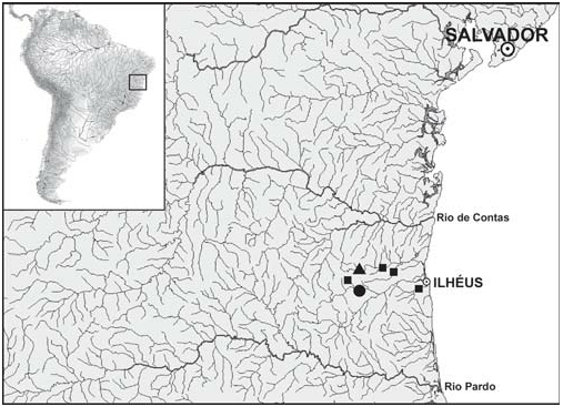
Geographic distribution and ecological notes. Astyanax vermilion is known from rio Salgado, a tributary of rio Cachoeira, and also from rio Almada ( Fig. 4 View Fig ). Both are relatively small Eastern Brazilian coastal drainages, with estuaries around Ilhéus, Bahia State. The species was collected in clear water streams, running over rocky, pebbles and sand bottom at low altitudes (188-258 m), in habitats characterized by slow to moderate water current, less than one meter deep ( Fig. 5 View Fig ). The surrounding vegetation is mainly composed by remains of the Atlantic Forest, with trees, cocoa trees and grass. Astyanax vermilion was collected syntopically with several fish species, including Nematocharax venustus , Lignobrycon myersi , Astyanax aff. bimaculatus , Leporinus sp. , Oligosarcus acutirostris , Steindachnerina elegans , Parotocinclus sp. , and Geophagus sp ..
The analysis of the stomach contents of three specimens revealed the presence of two distinct forms of filamentous algae, fragments of vascular plants and seeds, larvae of Diptera (Chironomidae) , adults of Hemiptera and Coleoptera (Chrysomelidae) , fragments of Hymenoptera (Formicidae) and of other unidentified arthropods, and organic debris.
Etymology. The name vermilion comes from the Latin word vermiculus (small worm which yields a red dye), meaning an orangish red pigment, originally derived from the powdered mineral cinnabar. Used herein in reference to the red coloration of posterior body portion of this fish when alive.
(vs. four teeth) and fins homogeneously darkened by chromatophores (vs. dark chromatophores concentrated on edges of rays). Astyanax burgerai differs from A. vermilion on its overall body shape and color, including a conspicuous and vertically elongated humeral blotch (vs. humeral spot poorly defined), absence of red pigmentation on body when alive (vs. posteroventral portion of body reddish), 18-19 branched analfin rays (vs. 20-23), dorsal-fin rays ii,9 (vs. usually ii,8,i), and 3- 6 scales on base of anterior anal-fin rays (vs. 8-12).


| R |
Departamento de Geologia, Universidad de Chile |
No known copyright restrictions apply. See Agosti, D., Egloff, W., 2009. Taxonomic information exchange and copyright: the Plazi approach. BMC Research Notes 2009, 2:53 for further explanation.
|
Kingdom |
|
|
Phylum |
|
|
Class |
|
|
Order |
|
|
Family |
|
|
Genus |