
Prolivatis gorochovi Emeljanov
|
publication ID |
https://doi.org/10.5281/zenodo.196171 |
|
DOI |
https://doi.org/10.5281/zenodo.6203214 |
|
persistent identifier |
https://treatment.plazi.org/id/03AF0519-0278-C945-FF3C-FE94F2C4F92F |
|
treatment provided by |
Plazi |
|
scientific name |
Prolivatis gorochovi Emeljanov |
| status |
|
Prolivatis gorochovi Emeljanov View in CoL
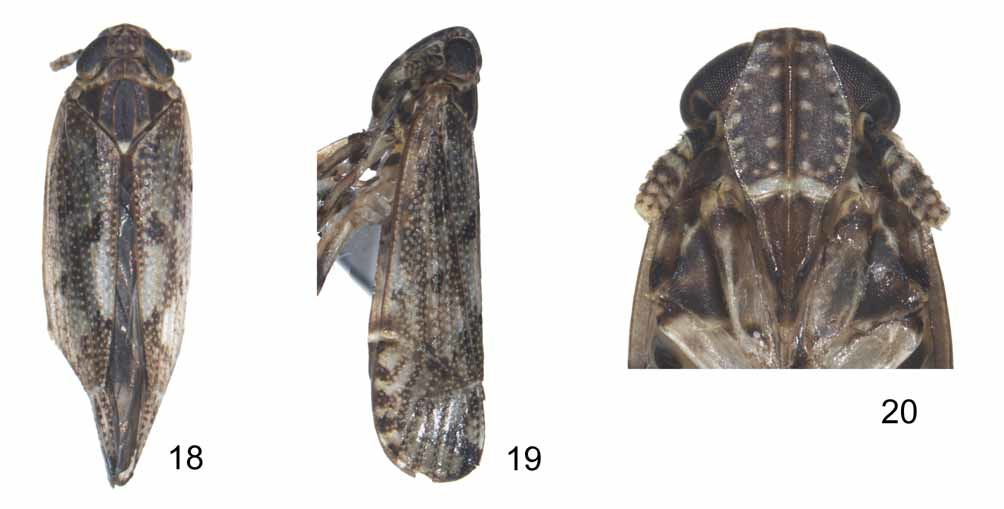
( Figs 18–35 View FIGURES 18 – 20 View FIGURES 21 – 35 )
Prolivatis gorochovi Emeljanov, 1995: 781 View in CoL ; 1996: 138–139.
Description. Body length: male (macropterous, N=1) 3.75 mm; female (macropterous, N=5) 4.57–4.59 mm.
Colour. General color brown. Vertex blackish brown. Frons brown, with light arched transverse stripe at apex. Postclypeus brownish yellow. Eyes black. Ocelli yellowish. Vertex, frons, genae, pronotum and mesonotum with whitish spots. Antennal segment I black basally and apically, segment II with black ring subbasally, apical half black adorned with yellowish brown sensory fields. Forewings brown, with blackish markings as figured, its surface covered with pale granules with long setae accompanied on both sides of longitudinal veins, hind wing pale fuliginous, subhyaline, veins brown. Legs sordid yellowish, ante- and middle tibiae with blackish patches basally and submedially, hind femur blackish brown at apex, hind tibiae with black patches at bases of lateral teeth, tips of apical teeth on hind tibiae and tarsi black. Venter of abdomen yellowish brown, dorsum of abdomen tawny brown with irregular patches laterally at each segment. Female with same color as male except frons and mesonotum blackish brown, postclypeus brown to blackish brown, pronotum with blackish spots along posterior margins. Ante- and middle femur with longitudinal blackish stripes. Venter of abdomen sordid brownish adorned with irregular black patches. Ovipositor black.
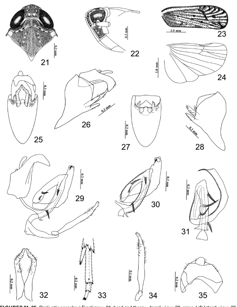
Head. Including eyes slightly narrower than pronotum about 0.92: 1 ( Figs 18 View FIGURES 18 – 20 , 21 View FIGURES 21 – 35 ). Vertex in dorsal view about 1.30 times wider than long, nearly as wide at base as apex, lateral carinae subparallel, posterior margin concave medially ( Figs 18 View FIGURES 18 – 20 , 21 View FIGURES 21 – 35 ), isosceles triangle at base of vertex distinctly depressed; Y-shaped carina faint except common stem ridged posteriorly ( Figs 18 View FIGURES 18 – 20 , 21 View FIGURES 21 – 35 ). Median carina of frons simple, basally uniting with convergent apex of the elevated plane, frons in midline longer than wide (about 1.31: 1), widest at level of antennal bases, slightly wider at apex than at base (about l.12: 1), lateral frontal carinae ridged and distinctly convex. Postclypeus with same width as frons at apex, about half as long as frons (about 0.53: 1) and ca. 1.54 times longer than anteclypeus, median carina of post- and anteclypeus apparently ridged, together approximately 0.86x length of frons ( Fig. 20 View FIGURES 18 – 20 ), in profile distinctly convex ( Figs 19 View FIGURES 18 – 20 , 22 View FIGURES 21 – 35 ). Rostrum rather long, reaching hind trochanters. Antennal segments cylindrical, elongate, reaching near apex of postclypeus, segment I 1.38–1.41 times wider than apical width, narrow at base and apparently broadening towards apex, apex distinctly broad, segment II about 2.25 times longer than I ( Figs 19–20 View FIGURES 18 – 20 ).
Thorax. Pronotum in dorsal view approximately 0.71x length of vertex, anterior margin transverse, anterior lateral areas strongly sloping laterad, posterior margin weakly arched inwardly, lateral carinae sinuate, diverging posterolaterally, nearly parallel with hind margin of eyes, not reaching posterior margin ( Figs 18 View FIGURES 18 – 20 , 21 View FIGURES 21 – 35 ), pronotum width 1.16–1.27 mm (n=6), length 0.19–0.26 mm (n=6). Mesonotum medially ca. in 1.85 times longer than vertex and pronotum together, with five carinae, median carina straight, obsolete apically, the inner pair slightly arched laterad, attaining posterior margin, outer pair nearly straight, extending to posterior margin ( Figs 18 View FIGURES 18 – 20 , 21 View FIGURES 21 – 35 ), Tegmina 4.67–4.94 mm long, 2.9 times longer than wide, widest near apex, rounded apically ( Figs 19 View FIGURES 18 – 20 , 23 View FIGURES 21 – 35 ). Legs with ante- and middle femur compressed, hind tibiae 1.53–1.70 mm long, with three lateral teeth on outer edge, five apical teeth with outer one largest, middle one smallest, metatarsomere I with five teeth apically, four spines in row and the fifth (middle one) displaced more basad, metatarsomere II with three apical spines (the middle one minute) ( Fig. 33 View FIGURES 21 – 35 ), metatarsomere I (0.50–0.63) slightly longer than tarsomere II (0.20–0.23) + III (0.27–0.30) combined. Posttibial spur (0.37–0.40) shorter than metabasitarsus, spine-like and without teeth on inner margin ( Fig. 33 View FIGURES 21 – 35 ).
Male genitalia. Male pygofer in caoduventral view elongate, strongly expanded apically and gradually narrowed towards base, ventrocaudal margin strongly excavated with large median process, apex rounded ( Figs 25, 27 View FIGURES 21 – 35 ), lateroventral margin with lanceolate lobe with tapered prolonged tip at each side ( Figs 25–28 View FIGURES 21 – 35 ), lateral margins of male pygofer asymmetrical, the left side protruding angularly while the right side is rounded does not protrude ( Figs 26, 28 View FIGURES 21 – 35 ). Parameres fairly slender and narrow, contiguous and divergent from bases, slightly narrowing apicad in caoduventral aspect, apical 1/4 slightly broadened, strongly bent mesad, almost meeting at rounded apex ( Figs 25, 26, 29, 32, 34 View FIGURES 21 – 35 ). Aedeagus 3-segmented, in caudoventral view first segment with spinous process subapically at right side, second with two spinous processes in basal half, distal segment arched clockwise (from base curved to left), with spinous process near base, subapically expanded, with numerous marginal teeth at left side, at right side with teeth in middle portion, apex of aedeagus sharply narrowed and spine-like, gonopore subapical on dorsal surface ( Figs 29–31 View FIGURES 21 – 35 ). Anal segment short, broad, in caudal aspect its anterior margin distinctly sinuate and laterodistal angle strongly produced at left side, in profile process bent ventrad ( Figs 25, 26, 29, 35 View FIGURES 21 – 35 ).
Material examined. 1 male (macropterous), China: Hainan Province, Mt. Diaoluo, 10 Apr. 2008, 900 m; 1 female (macropterous), 2 Mar. 2008, 2 females (macropterous), 0 7 May 2007, 800 m, 1 female (macropterous), 9 May 2008, 800 m, Hainan Province, Jianfengling; 1 female (macropterous), Hainan Province, Tongguling, 24 Apr. 2008, 280 m. All specimens were collected by Qiulei Men.
Distribution. Southern China (Hainan Province), Vietnam (Zyalay-Kontum Province).
Remarks. The holotype of P. gorochovi Emeljanov was collected from Vietnam (Zyalay-Kontum Province) and now it is deposited in the Zoological Institute, Russian Academy of Sciences, St. Petersburg, Russia. The Prolivatis specimens from Hainan Province match Emeljanov’s (1995) description and illustrations of body and male pygofer except the lateral margins of the pygofer are asymmetrical, which differs from specimens studied by Emeljanov (1995) (symmetrical in holotype). We regard this difference to be intraspecific individual variations in P. gorochovi and the specimens from southern China are therefore placed in this species.
No known copyright restrictions apply. See Agosti, D., Egloff, W., 2009. Taxonomic information exchange and copyright: the Plazi approach. BMC Research Notes 2009, 2:53 for further explanation.
|
Kingdom |
|
|
Phylum |
|
|
Class |
|
|
Order |
|
|
Family |
|
|
Genus |
Prolivatis gorochovi Emeljanov
| Qin, Dao-Zheng & Zhang, Ya-Lin 2010 |
Prolivatis gorochovi
| Emeljanov 1995: 781 |