
Choeras Mason, 1981
|
publication ID |
https://doi.org/ 10.11646/zootaxa.3754.5.1 |
|
publication LSID |
lsid:zoobank.org:pub:F63008D1-5612-4E75-A701-AD0BB7C398AE |
|
DOI |
https://doi.org/10.5281/zenodo.3507534 |
|
persistent identifier |
https://treatment.plazi.org/id/03AC87D6-FF95-5958-58FF-A0B607C3D26B |
|
treatment provided by |
Plazi |
|
scientific name |
Choeras Mason, 1981 |
| status |
|
Key to species of subgenus Choeras Mason, 1981 from China
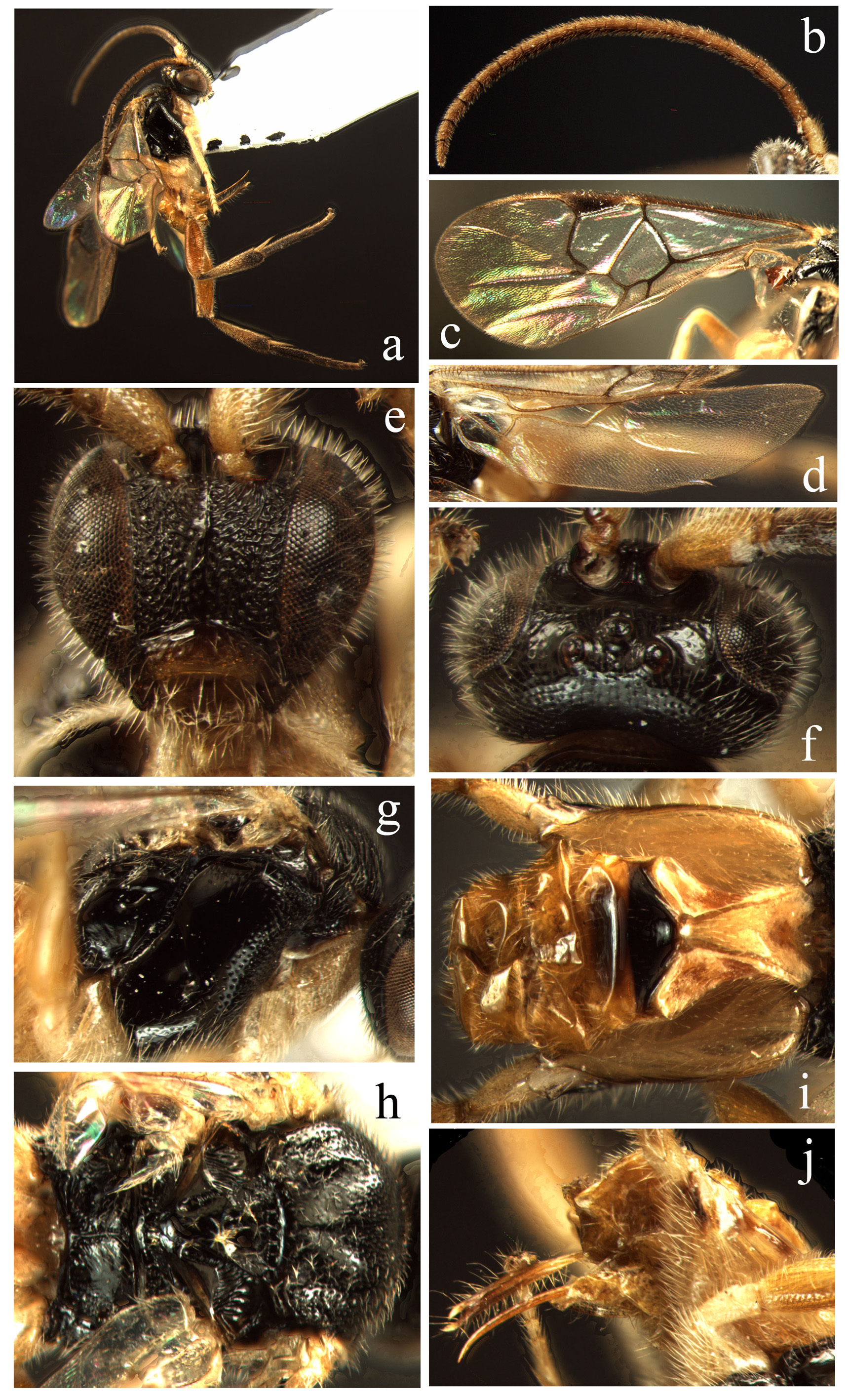
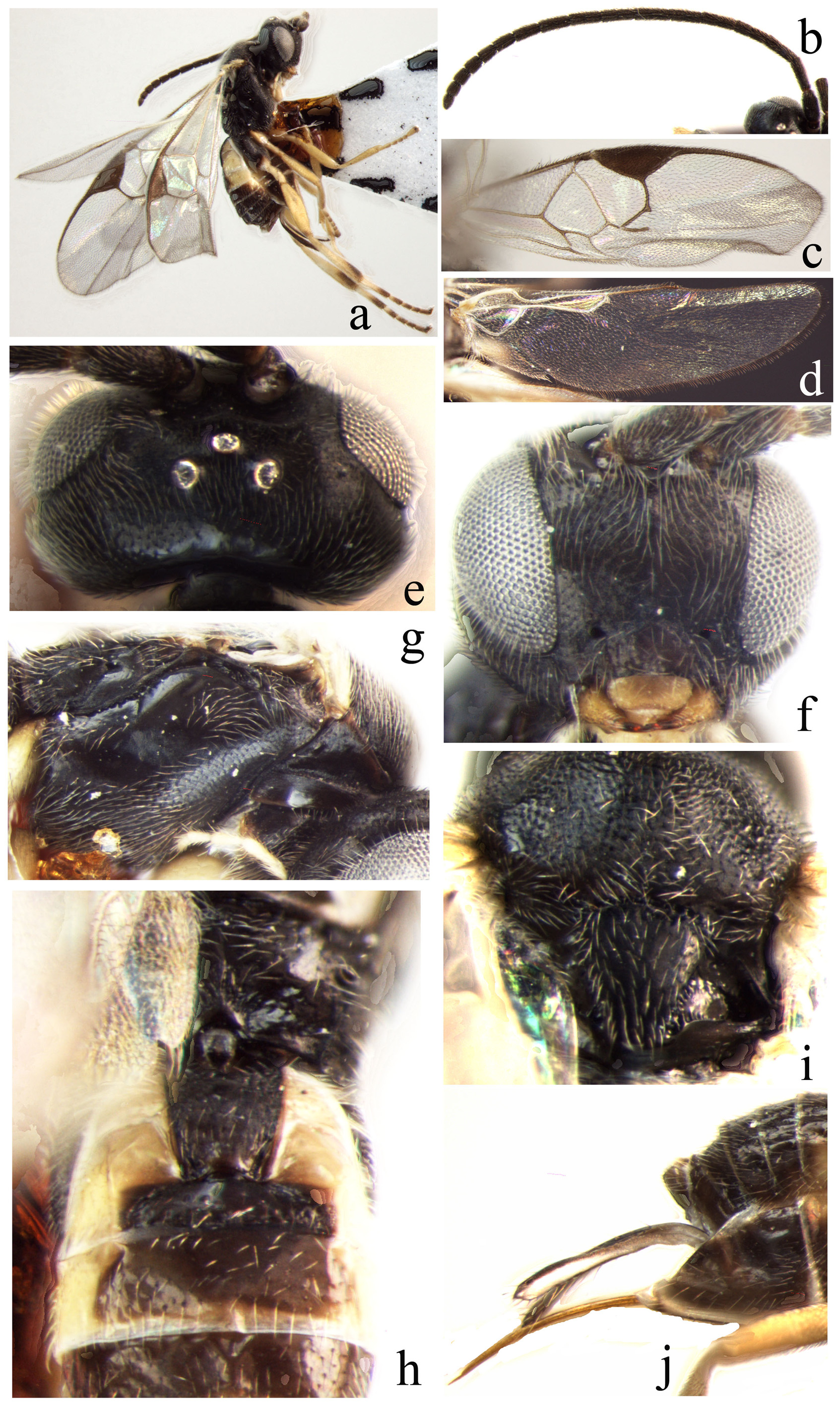
1. Vein r-m well developed, areolet completely closed, large or small ( Figs. 3c View FIGURE 3. a – j , 6c View FIGURE 6. a – k , 9c View FIGURE 9. a – j , 10c View FIGURE 10. a – j , 13c View FIGURE 13. a – j , 15c View FIGURE 15. a – i , 16c View FIGURE 16. a – j , 17c View FIGURE 17. a – j , 18c View FIGURE 18. a – j )......... 2
- Stub of vein r-m of fore wing very short or absent, resulting in an open second areolet, areolet absent ( Figs. 1c View FIGURE 1. a – j , 2d View FIGURE 2. a – j , 4c View FIGURE 4. a – i , 5c View FIGURE 5. a – j , 7c View FIGURE 7. a – j , 8c View FIGURE 8. a – i , 11c View FIGURE 11. a – j , 12c View FIGURE 12. a – j , 14c View FIGURE 14. a – j , 19c View FIGURE 19. a – j )................................................................................. 10
2. Areolet very small and triangular, vein r-m arising from vein 2-SR at a considerable distance proximal to the junction of vein r and 2-SR ( Figs. 3c View FIGURE 3. a – j , 6c View FIGURE 6. a – k , 10c View FIGURE 10. a – j , 13c View FIGURE 13. a – j , 15c View FIGURE 15. a – i , 18c View FIGURE 18. a – j ).................................................................. 3
- Areolet very large and quadrangular or subtriangular, triangular or subtriangular, vein r-m arising from the junction of vein r and 2-SR ( Figs. 9c View FIGURE 9. a – j , 16c View FIGURE 16. a – j , 17c View FIGURE 17. a – j )............................................................................ 8
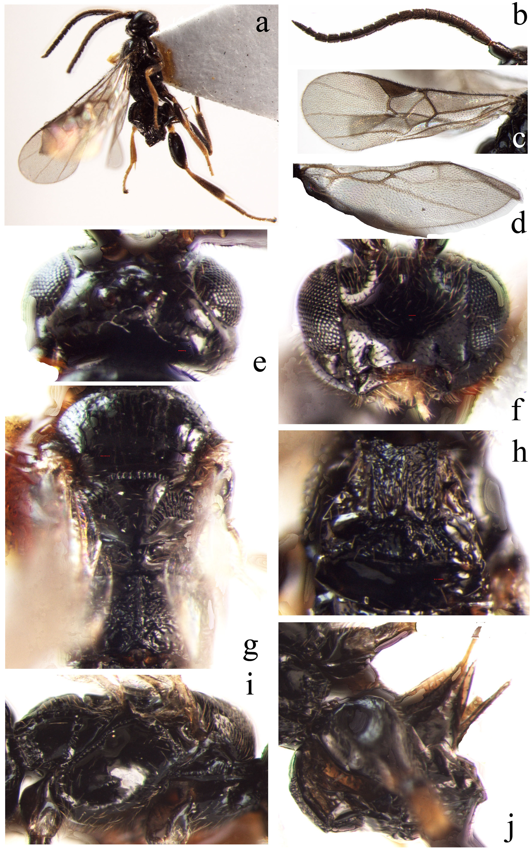
3. Ocelli in a very high triangle, the posterior transverse tangent to the median ocellus leaving far from the posterior pair ( Fig. 15d View FIGURE 15. a – i ). Propodeum mainly smooth, only medio-posteriorly with some rugae, without a median carina ( Fig. 15f View FIGURE 15. a – i ). Tergite I distinctly quadrangular, almost parallel-sided ( Fig. 15i View FIGURE 15. a – i )........................................... A. tenuialatus sp. n.
- Ocelli in a moderately low triangle, the posterior transverse tangent to the median ocellus slightly touching the posterior pair ( Figs. 3f View FIGURE 3. a – j , 6f View FIGURE 6. a – k , 10e View FIGURE 10. a – j , 13e View FIGURE 13. a – j , 18e View FIGURE 18. a – j ). Propodeum largely smooth and with a strong median longitudinal carina which bifurcates into several short oblique carinae, more distinct at its base ( Figs. 3h View FIGURE 3. a – j , 6j View FIGURE 6. a – k , 10g View FIGURE 10. a – j , 13g View FIGURE 13. a – j , 18g View FIGURE 18. a – j ). Tergite I narrow apically ( Figs. 3i View FIGURE 3. a – j , 6g View FIGURE 6. a – k , 10h View FIGURE 10. a – j , 13g View FIGURE 13. a – j , 18h View FIGURE 18. a – j )............................................................................................ 4
4. Side of scutellum with rather narrow crenulate depression, lunula large-sized and semicircular ( Figs. 3h View FIGURE 3. a – j , 6i View FIGURE 6. a – k , 13g View FIGURE 13. a – j ).......... 5
- Side of scutellum with relatively wide crenulate depression, lunula small-sized and obtusely triangular ( Figs. 10g View FIGURE 10. a – j , 18g View FIGURE 18. a – j ).... 7
5. Length of 16th segment shorter than 15th segment ( Figs. 3b View FIGURE 3. a – j , 13b View FIGURE 13. a – j ). Tergite II subtriangular ( Figs. 3i View FIGURE 3. a – j , 13i View FIGURE 13. a – j ). Ovipositor sheath short, its length (total length) shorter than hind tibia ( Figs. 3j View FIGURE 3. a – j , 13j View FIGURE 13. a – j )............................................... 6
- Length of 16th segment equal to 15th segment ( Fig. 6b View FIGURE 6. a – k ). Tergite II quadrangular ( Fig. 6g View FIGURE 6. a – k ). Ovipositor sheath relatively very long, its length (total length) much longer than hind tibia ( Fig. 6h View FIGURE 6. a – k )................................ A. fujianensis sp. n.
6. Pterostigma relatively wide, 2.4 times as long as wide ( Fig. 13c View FIGURE 13. a – j ). Vein r (actually r plus part of 2-SR because of low position of r-m) distinctly curved ( Fig. 13c View FIGURE 13. a – j ). Vein 2-SR straight ( Fig. 13c View FIGURE 13. a – j ). Vein 1-CU1 longer than vein 2-CU1 ( Fig. 13c View FIGURE 13. a – j ). Vein 1-SR pointing to the junction of vein 1-CU1 and 2-CU1 ( Fig. 13c View FIGURE 13. a – j ). Vein cu-a distinctly curved ( Fig. 13d View FIGURE 13. a – j ). Edge of vannal lobe beyond its widest part more or less straight and here with a fringe of hairs ( Fig. 13d View FIGURE 13. a – j ). Apical width of tergite I longer than median length of tergite II ( Fig. 13i View FIGURE 13. a – j )....................................................... A. semilunatus sp. n.
- Pterostigma relatively narrow, 3.3 times as long as wide ( Fig. 3c View FIGURE 3. a – j ). Vein r straight ( Fig. 3c View FIGURE 3. a – j ). Vein 2-SR curved ( Fig. 3c View FIGURE 3. a – j ). Vein 1- CU1 equal to 2-CU1 ( Fig. 3c View FIGURE 3. a – j ). Vein cu-a straight ( Fig. 3d View FIGURE 3. a – j ). Edge of vannal lobe beyond its widest part very slightly concave and without a hair-fringe ( Fig. 3d View FIGURE 3. a – j ). Apical width of tergite I half of median length of tergite II ( Fig. 3i View FIGURE 3. a – j )....................................................................................... A. compressifemur ( Chen & Song, 2004) View in CoL
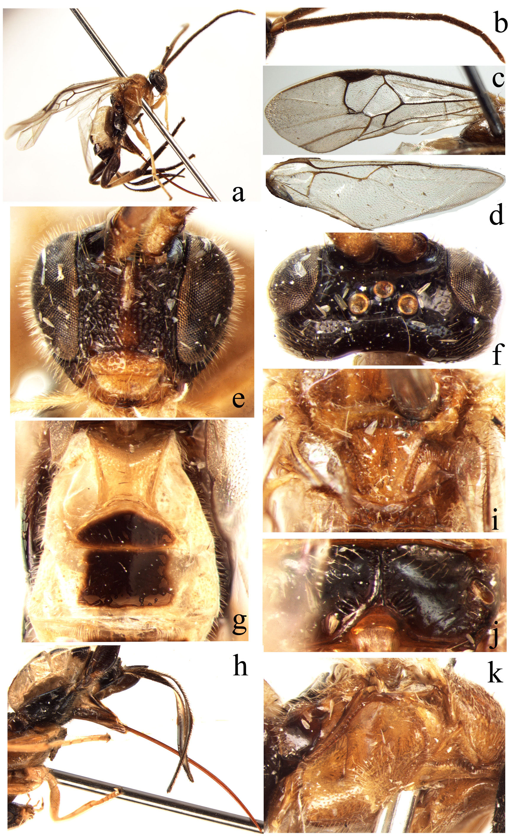
7. Vein 1-R1 longer, about 1.5 times as long as pterostigma ( Fig. 10c View FIGURE 10. a – j ). Vein r slightly curved ( Fig. 10c View FIGURE 10. a – j ). 1-CU1 longer than 2- CU1 ( Fig. 10c View FIGURE 10. a – j ). Tergite I subtriangular, strongly narrowed apically ( Fig. 10h View FIGURE 10. a – j ). Apical width of tergite I half of median length of second tergite ( Fig. 10h View FIGURE 10. a – j ). Length of ovipositor sheath (total length) much longer than hind tibia ( Fig. 10j View FIGURE 10. a – j )... A. longus sp. n.
- Vein 1-R1 relatively short, almost equal to length of pterostigma ( Fig. 18c View FIGURE 18. a – j ). Vein r straight ( Fig. 18c View FIGURE 18. a – j ). 1-CU1 shorter than 2- CU1 ( Fig. 18c View FIGURE 18. a – j ). Tergite I distinctly quadrangular, slightly narrow apically ( Fig. 18h View FIGURE 18. a – j ). Apical width of tergite I almost equal to median length of tergite II ( Fig. 18h View FIGURE 18. a – j ). Length of ovipositor sheath (total length) slightly shorter than hind tibia ( Fig. 18j View FIGURE 18. a – j )........................................................................................... A. varicolor sp. n.
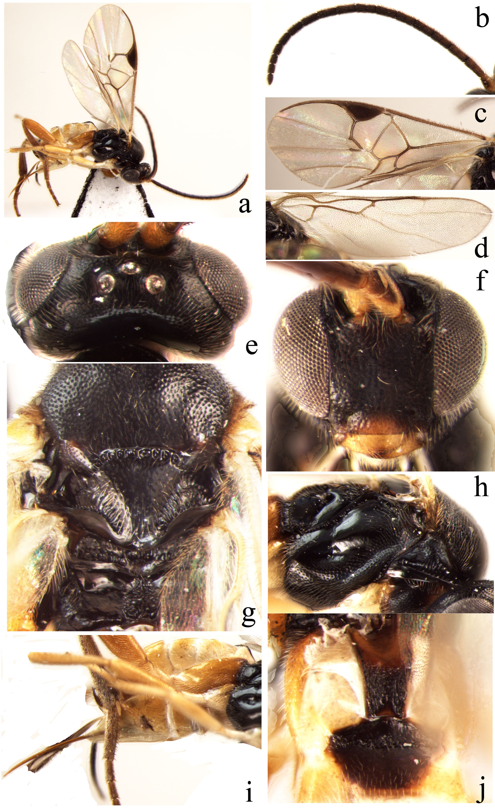
8. Propodeum largely smooth and shiny, only rugose laterally ( Fig. 9g View FIGURE 9. a – j ). Tergite I sharply narrowed apically, tergites I and II largely smooth, but only small area of them rugose ( Fig. 9i View FIGURE 9. a – j ). Tergite II subtriangular ( Fig. 9i View FIGURE 9. a – j )........ A. longitergitus sp. n.
- Propodeum strongly rugose ( Figs. 16g View FIGURE 16. a – j , 17g View FIGURE 17. a – j ). Tergite I distinctly quadrangular and parallel-sided, tergites I and II largely and strongly rugose ( Figs. 16h View FIGURE 16. a – j , 17h View FIGURE 17. a – j ). Tergite II quadrangular ( Figs. 16h View FIGURE 16. a – j , 17h View FIGURE 17. a – j ).......................................... 9
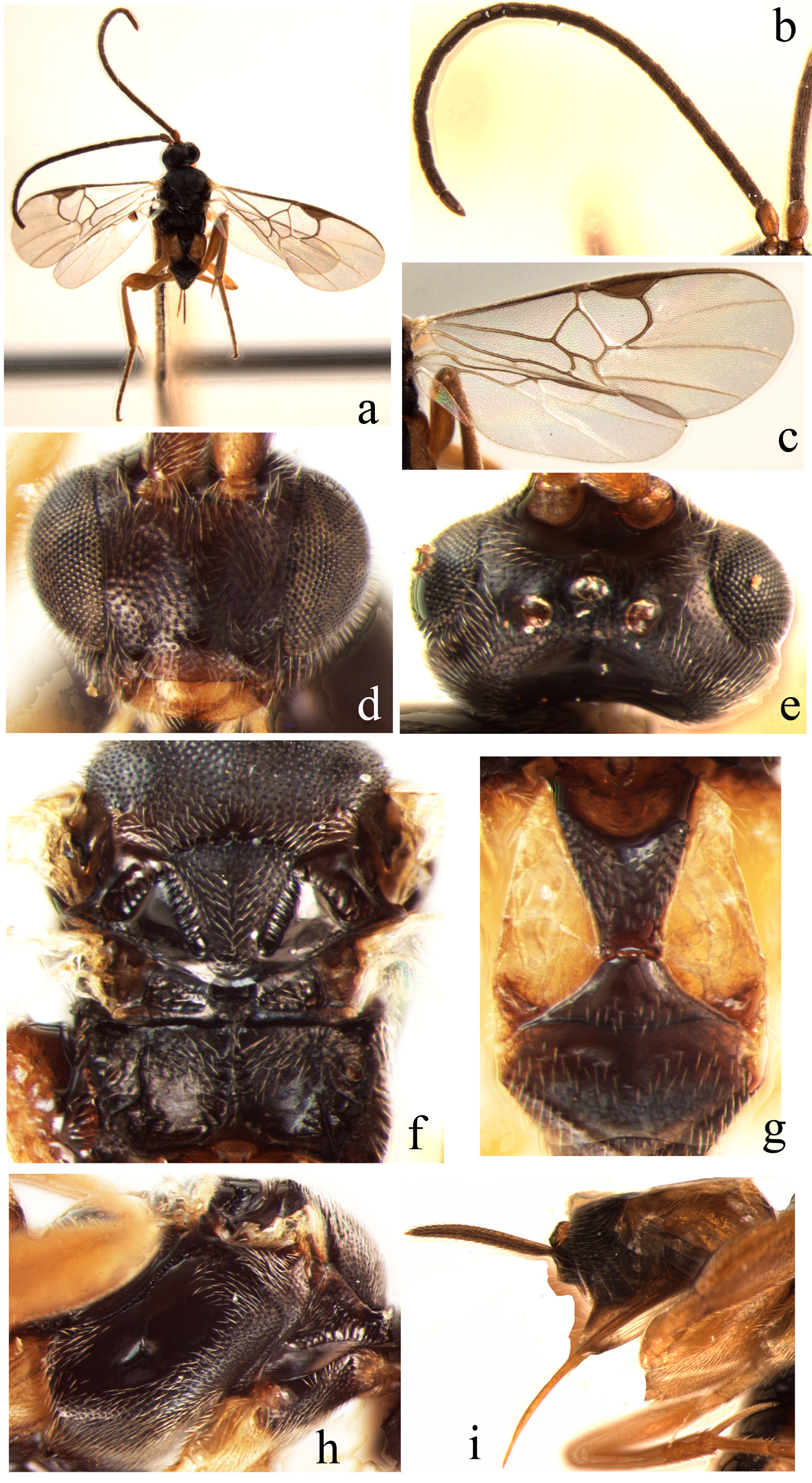
9. Head robust, 1.5 times as wide as long, wider than mesoscutum ( Fig. 16e View FIGURE 16. a – j ). Ocelli in a moderately high triangle, the posterior transverse tangent to the median ocellus hardly touching the posterior pair ( Fig. 16e View FIGURE 16. a – j ). Face transverse, convex and without a median longitudinal carina ( Fig. 16f View FIGURE 16. a – j ). Vein 2-SR straight ( Fig. 16c View FIGURE 16. a – j ). Vein 1-CU1 equal to 2-CU1 ( Fig. 16c View FIGURE 16. a – j ). A. tumidus sp. n.
- Head 2.1 times as wide as long, narrower than mesoscutum ( Fig. 17e View FIGURE 17. a – j ). Ocelli in a moderately low triangle, the posterior transverse tangent to the median ocellus slightly touching the posterior pair ( Fig. 17e View FIGURE 17. a – j ). Face with a distinctly and incompletely longitudinal carina medially near the antennal sockets, its length about 0.3 times as long as face ( Fig. 17f View FIGURE 17. a – j ). Vein 2-SR curved ( Fig. 17c View FIGURE 17. a – j ). Vein 1-CU1 shorter than 2-CU1 ( Fig. 17c View FIGURE 17. a – j ).......................................... A. validicarinatus sp. n.
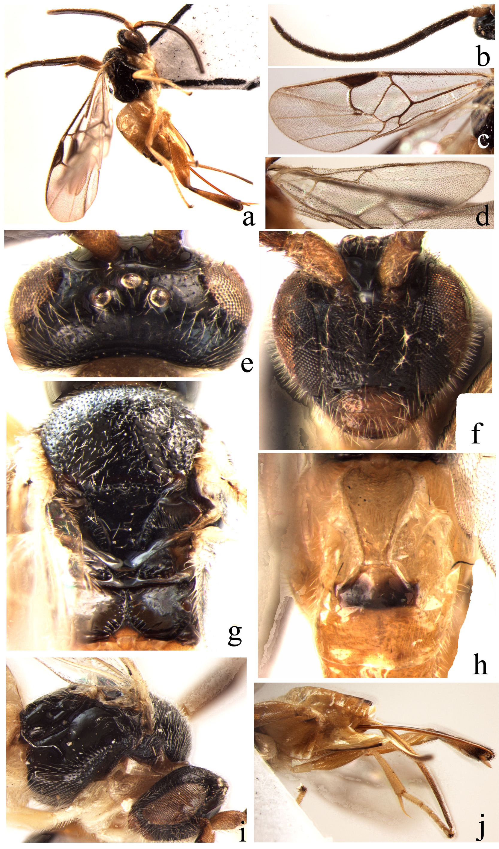
10. Propodeum without a median longitudinal carina ( Figs. 1g View FIGURE 1. a – j , 7j View FIGURE 7. a – j , 11h View FIGURE 11. a – j , 19i View FIGURE 19. a – j )......................................... 11
- Propodeum with a strong or an incomplete median longitudinal carina ( Figs. 2h View FIGURE 2. a – j , 4f View FIGURE 4. a – i , 5h View FIGURE 5. a – j , 8f View FIGURE 8. a – i , 12h View FIGURE 12. a – j , 14g View FIGURE 14. a – j ).................. 14
11. Pterostigma relatively narrow ( Fig. 1c View FIGURE 1. a – j ). Vein r longer than vein 2-SR ( Fig. 1c View FIGURE 1. a – j ). Side of scutellum with rather narrow crenulate depression, lunula large-sized and semicircular ( Fig. 1g View FIGURE 1. a – j )......................................... A. angustus sp. n.
- Pterostigma relatively wide ( Figs. 7c View FIGURE 7. a – j , 11c View FIGURE 11. a – j , 19c View FIGURE 19. a – j ). Vein r shorter than vein 2-SR ( Figs. 7c View FIGURE 7. a – j , 11c View FIGURE 11. a – j , 19c View FIGURE 19. a – j ). Side of scutellum with relatively wider crenulate depression, lunula small-sized and almost linear ( Figs. 7g View FIGURE 7. a – j , 11i View FIGURE 11. a – j , 19g View FIGURE 19. a – j )........................... 12
12. Tergite I very long and narrow, sharply narrowed apically. Vein r straight ( Fig. 19i View FIGURE 19. a – j )................. A. yunnanensis sp. n.
- Tergite I short and wide, slightly narrowed apically, even parallel-sided. Vein r slightly curved ( Figs. 7j View FIGURE 7. a – j , 11h View FIGURE 11. a – j )............ 13
13. Subapical antennal segments slightly elongated ( Fig. 11b View FIGURE 11. a – j ). Edge of vannal lobe beyond its widest part very slightly convex ( Fig. 11d View FIGURE 11. a – j ). Width of scutellar sulcus about equal to length of scutellum ( Fig. 11i View FIGURE 11. a – j ). Apical width of tergite I about 2.0 times as long as median length of tergite II ( Fig. 11h View FIGURE 11. a – j ). Length of ovipositor sheath (total length) slightly shorter than hind tibia ( Fig. 11j View FIGURE 11. a – j ). Hypopygium with narrow longitudinal creases laterally, and acute apically ( Fig. 11j View FIGURE 11. a – j )............ A. parviocellus sp. n.
- Subapical antennal segments moniliform and cubic ( Fig. 7b View FIGURE 7. a – j ). Edge of vannal lobe beyond its widest part straight ( Fig. 7d View FIGURE 7. a – j ). Width of scutellar sulcus slightly shorter than length of scutellum ( Fig. 7g View FIGURE 7. a – j ). Apical width of tergite I about 4.0 times as long as median length of tergite II ( Fig. 7j View FIGURE 7. a – j ). Length of ovipositor sheath (total length) slightly longer than length of hind tibia ( Fig. 7i View FIGURE 7. a – j ).
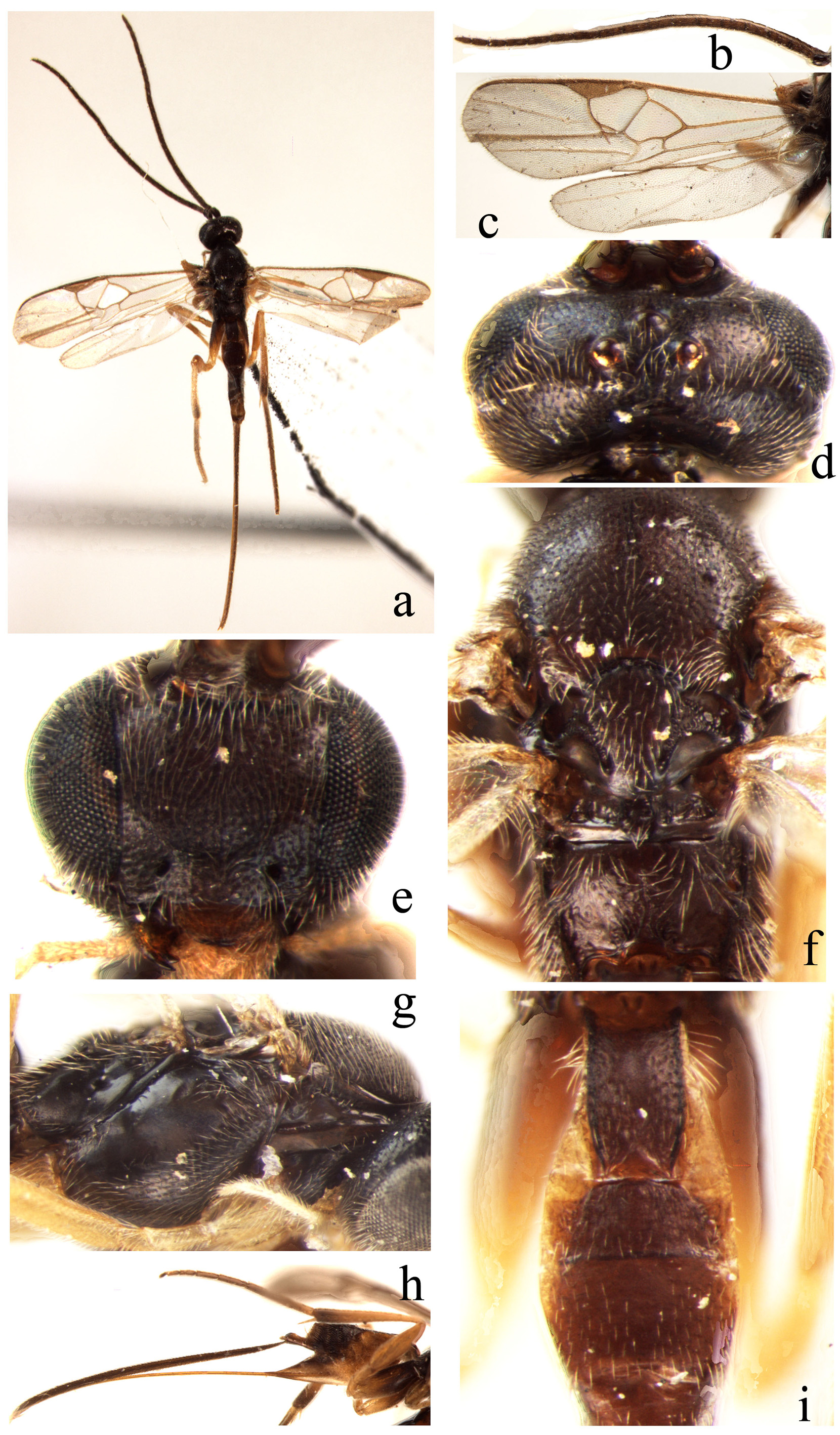
Hypopygium without longitudinal creases laterally and acute apically ( Fig. 7i View FIGURE 7. a – j )................. A. grammatitergitus sp. n. 14. Ocelli relatively small, in a moderately high triangle, the posterior transverse tangent to the median ocellus hardly touching the posterior pair ( Figs. 5e View FIGURE 5. a – j , 12f View FIGURE 12. a – j )............................................................................ 15
- Ocelli relatively large, in a very low triangle, the posterior transverse tangent to the median ocellus cutting into the posterior pair ( Figs. 2e View FIGURE 2. a – j , 4e View FIGURE 4. a – i , 8d View FIGURE 8. a – i , 14e View FIGURE 14. a – j ).............................................................................. 16
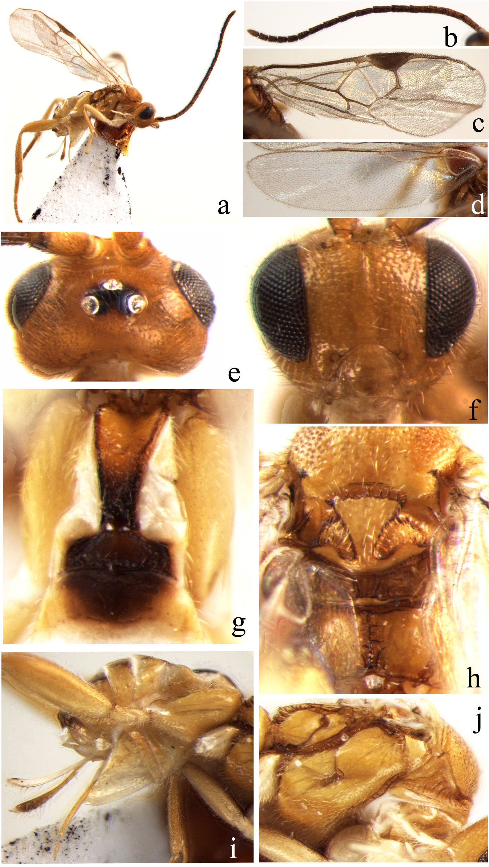
15. Body largely yellow, subapical antennal segments slightly elongated ( Figs. 5a, 5b View FIGURE 5. a – j ). Width of scutellar sulcus slightly longer than length of scutellum ( Fig. 5h View FIGURE 5. a – j ). Apical width of tergite I equal to median length of tergite II ( Fig. 5g View FIGURE 5. a – j )....................................................................................................... A. flavicorpus sp. n.
- Body black, subapical antennal segments moniliform and subquadrate ( Figs. 12a, 12b View FIGURE 12. a – j ). Width of scutellar sulcus slightly shorter than length of scutellum ( Fig. 12h View FIGURE 12. a – j ). Apical width of tergite I longer than median length of tergite II ( Fig. 12i View FIGURE 12. a – j )............................................................................................. A. rugulosus sp. n.
16. Side of scutellum with rather narrow crenulate depression, lunula large-sized and semicircular ( Figs. 2h View FIGURE 2. a – j , 4f View FIGURE 4. a – i , 8f View FIGURE 8. a – i ).......... 17
- Side of scutellum with relatively wide crenulate depression, lunula small-sized and obtusely triangular ( Fig. 14g View FIGURE 14. a – j ).............................................................................................. A. semirugosus sp. n.
17. Pterostigma relatively narrow ( Fig. 4c View FIGURE 4. a – i ). Vein r shorter than Vein 2-SR ( Fig. 4c View FIGURE 4. a – i ). Vein 2-SR curved ( Fig. 4c View FIGURE 4. a – i ). Ovipositor sheath relatively shorter, its length (total length) slightly longer than half of hind tibia ( Fig. 4i View FIGURE 4. a – i ).............. A. curvinervus sp. n.
- Pterostigma relatively wide ( Figs. 2d View FIGURE 2. a – j , 8c View FIGURE 8. a – i ). Vein r longer than Vein 2-SR ( Figs. 2d View FIGURE 2. a – j , 8c View FIGURE 8. a – i ). Vein 2-SR straight ( Figs. 2d View FIGURE 2. a – j , 8c View FIGURE 8. a – i ). Ovipositor sheath very long, its length (total length) longer than hind tibia ( Figs. 2j View FIGURE 2. a – j , 8h View FIGURE 8. a – i )................................ 18
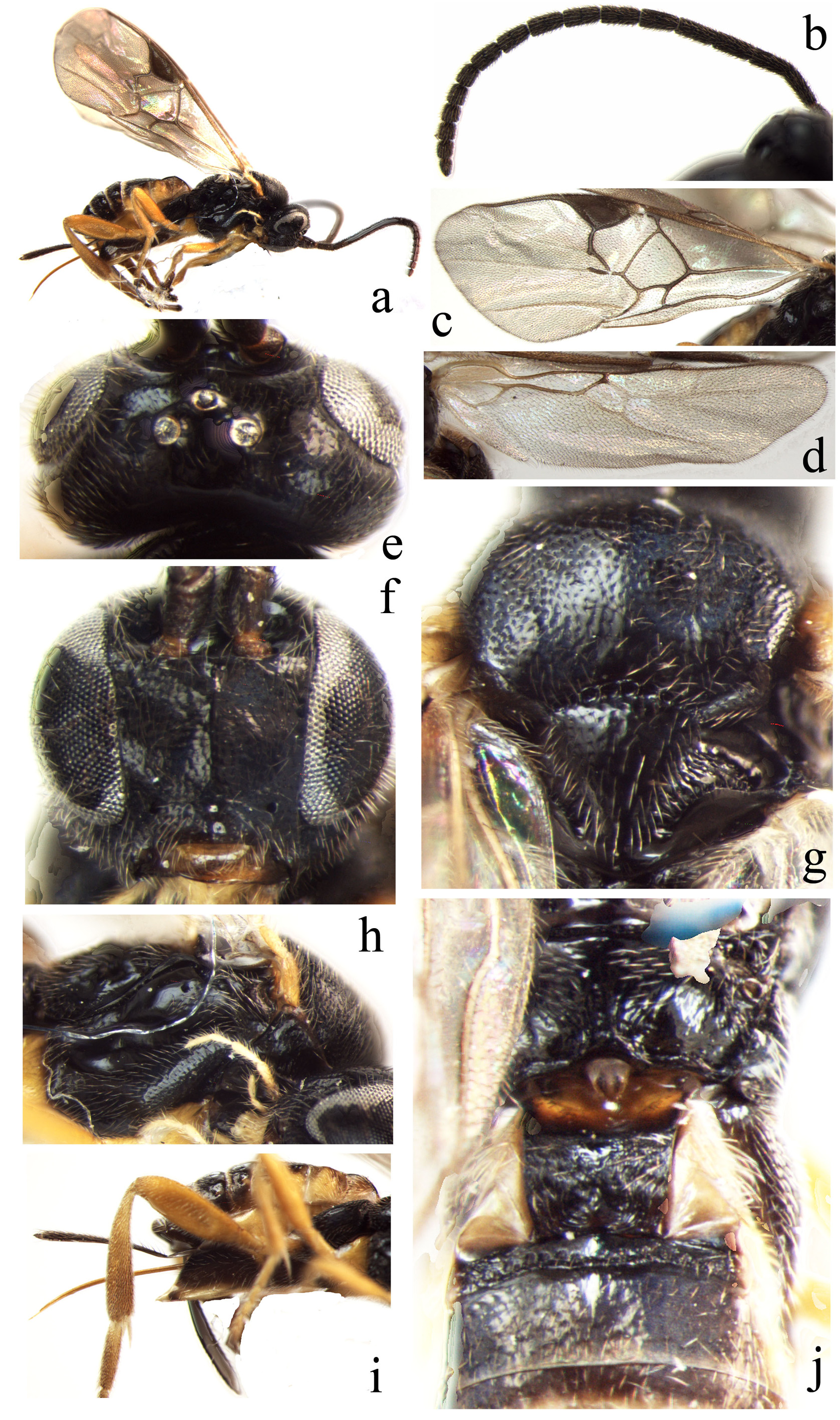
18. Mandible relatively wide and large ( Fig. 2f View FIGURE 2. a – j ). Vein 1-SR of fore wing pointing to the junction of vein 1-CU1 and 2-CU1 ( Fig. 2d View FIGURE 2. a – j ). Vein 1-CU1 equal to 2-CU1 ( Fig. 2d View FIGURE 2. a – j ). Vein cu-a of hind wing curved ( Fig. 2c View FIGURE 2. a – j ). Edge of vannal lobe beyond its widest part concave ( Fig. 2c View FIGURE 2. a – j ). Width of scutellar sulcus slightly shorter than length of scutellum ( Fig. 2h View FIGURE 2. a – j )......... A. brevinervus sp. n.
- Mandible normal ( Fig. 8e View FIGURE 8. a – i ). Vein 1-SR of fore wing pointing to vein 2-CU1 ( Fig. 8c View FIGURE 8. a – i ). Vein 1-CU1 shorter than 2-CU1 ( Fig. 8c View FIGURE 8. a – i ). Vein cu-a of hind wing straight ( Fig. 8c View FIGURE 8. a – i ). Edge of vannal lobe beyond its widest part straight( Fig. 8c View FIGURE 8. a – i ). Width of scutellar sulcus equal to length of scutellum ( Fig. 8f View FIGURE 8. a – i )................................................... A. infirmicarinatus sp. n.


No known copyright restrictions apply. See Agosti, D., Egloff, W., 2009. Taxonomic information exchange and copyright: the Plazi approach. BMC Research Notes 2009, 2:53 for further explanation.
|
Kingdom |
|
|
Phylum |
|
|
Class |
|
|
Order |
|
|
Family |
|
|
SubGenus |
Choeras |