
Plautia himechabane, Ishikawa & Moriya, 2019
|
publication ID |
https://doi.org/ 10.11646/zootaxa.4564.2.8 |
|
publication LSID |
lsid:zoobank.org:pub:171E7B4D-773A-4A16-8C0F-3D23F6947A |
|
DOI |
https://doi.org/10.5281/zenodo.5922546 |
|
persistent identifier |
https://treatment.plazi.org/id/03A687D8-FFD1-FFDF-E69C-9679FBD8FF05 |
|
treatment provided by |
Plazi |
|
scientific name |
Plautia himechabane |
| status |
|
Plautia himechabane Ishikawa & Moriya, sp. nov.
( Figs. 4 View FIGURES 1–5 , 17 View FIGURES 14–18 , 26 View FIGURES 19–27 , 31 View FIGURES 28–32 , 36 View FIGURES 33–37 , 41 View FIGURES 38–42 , 46 View FIGURES 43–47 , 51 View FIGURES 48–52 , 56 View FIGURES 53–57 , 61 View FIGURES 58–62 , 66 View FIGURES 63–67 , 71 View FIGURES 68–72 , 76 View FIGURES 73–77 , 83 View FIGURES 78–84 )
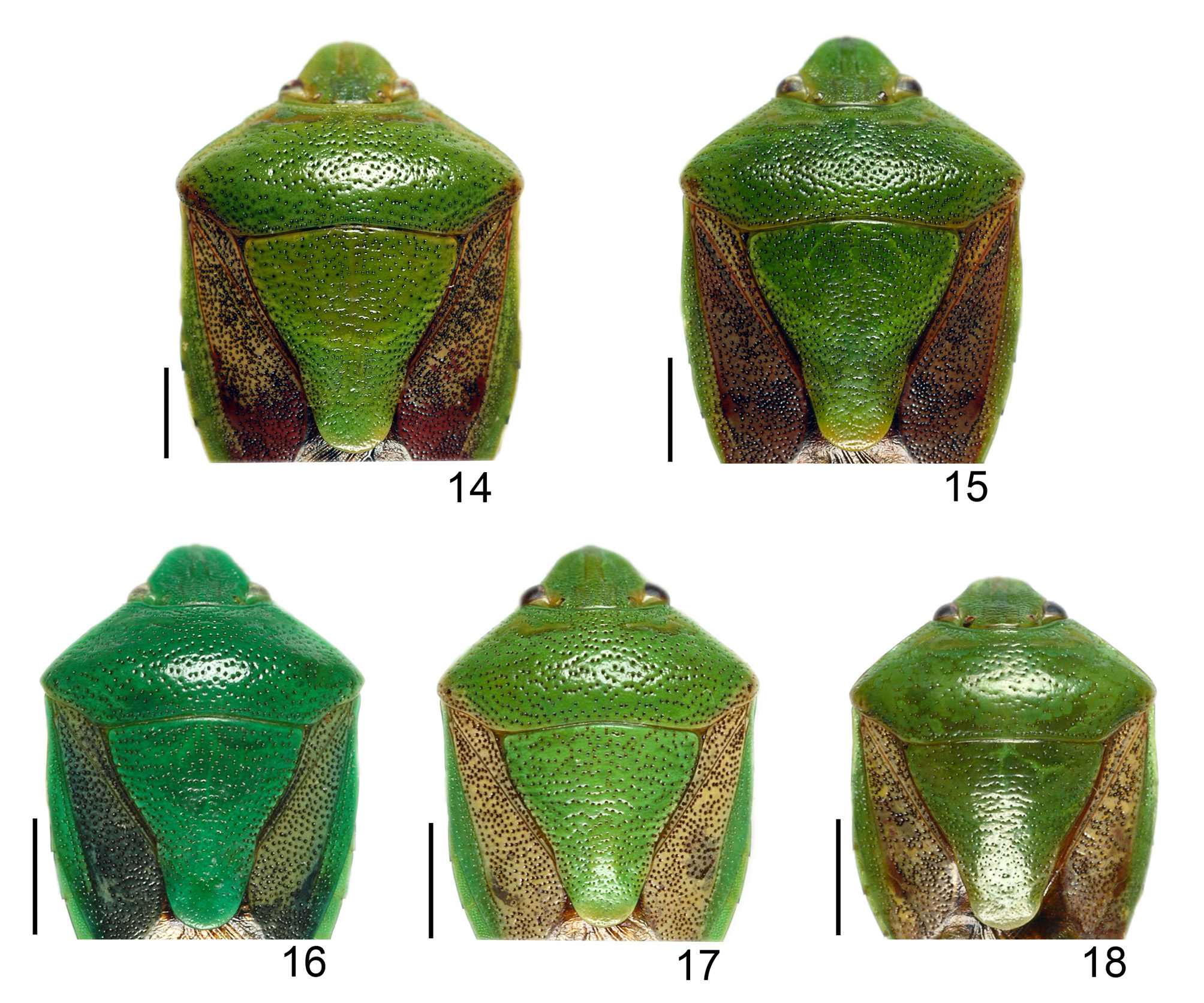
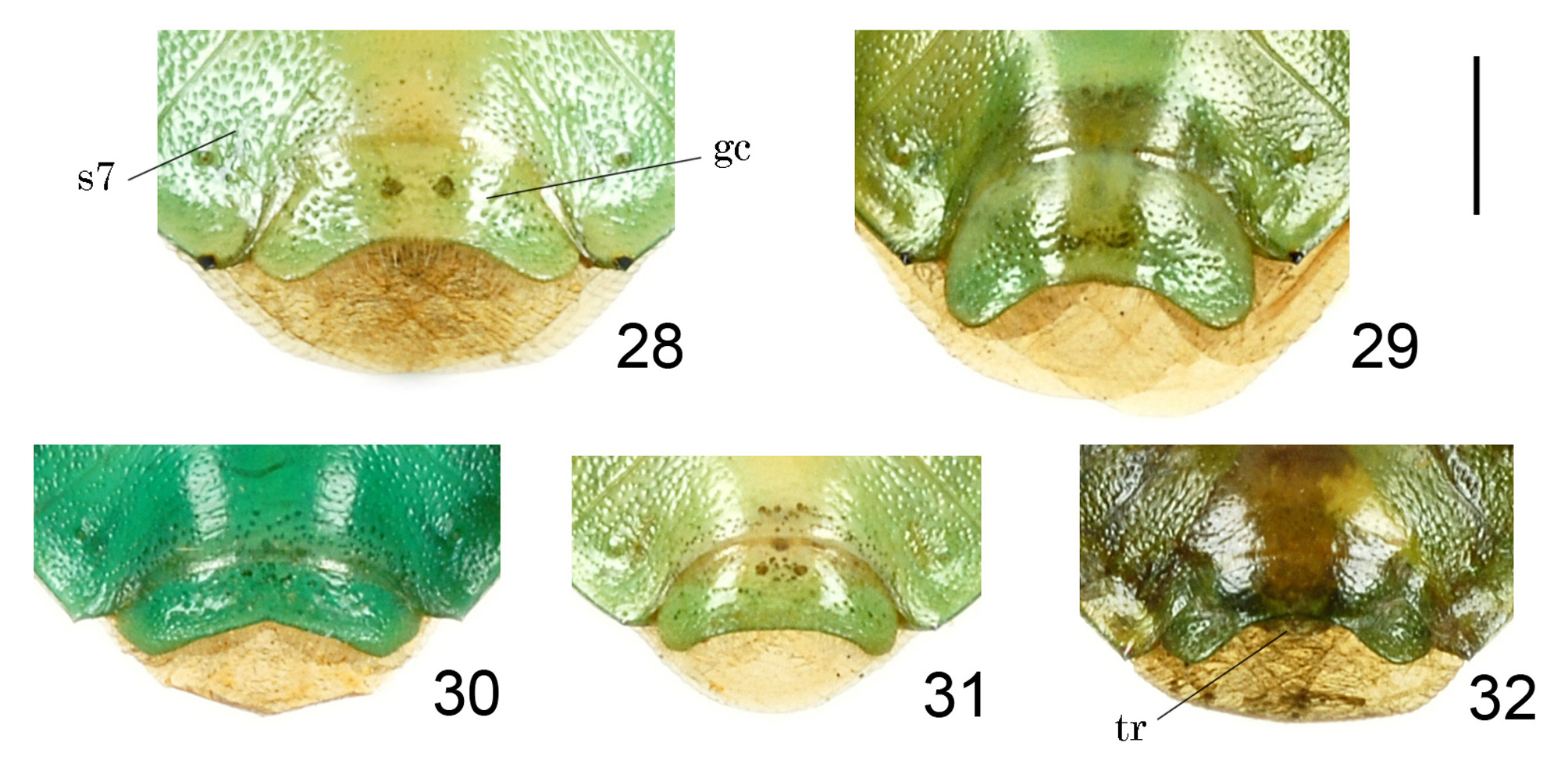
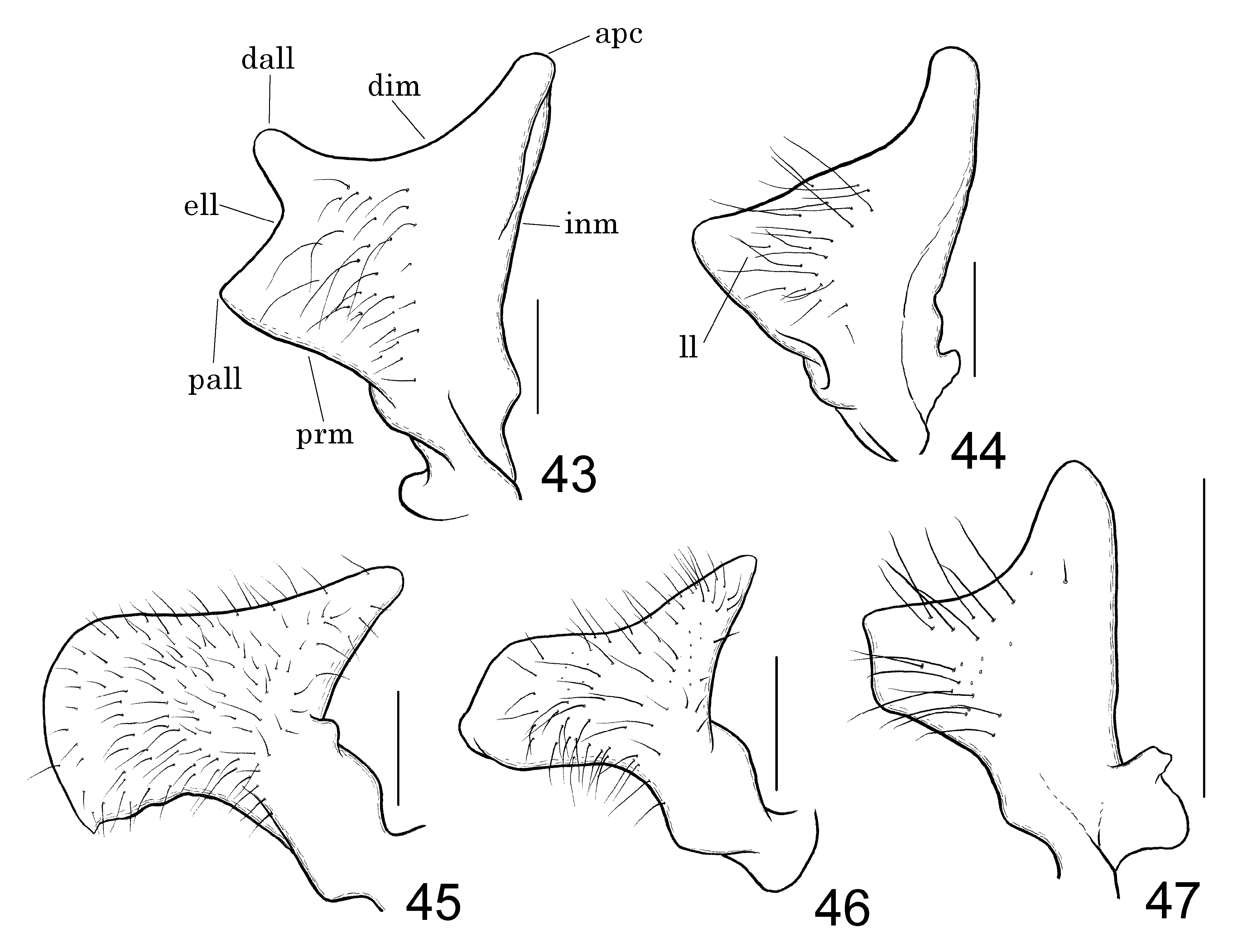
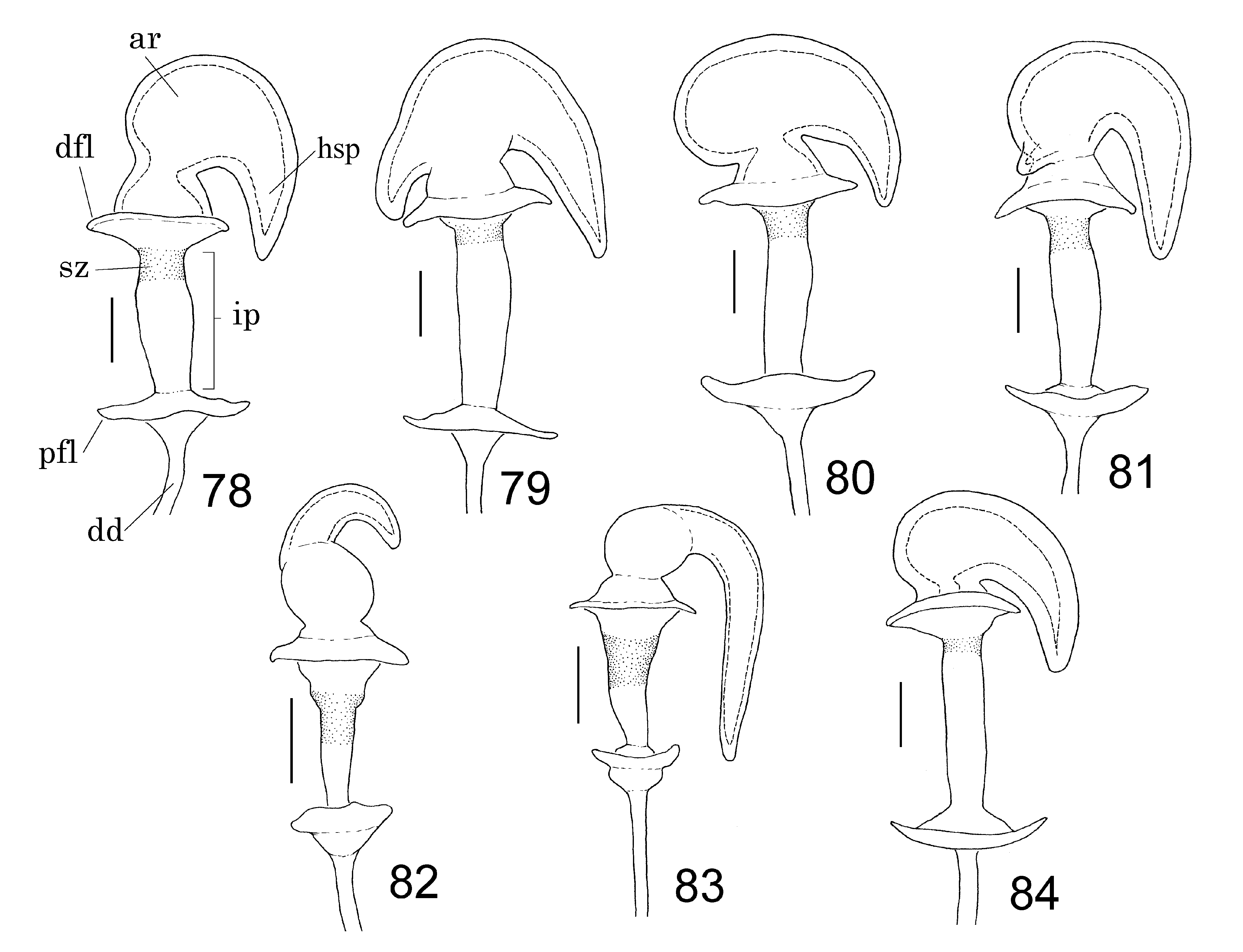
Plautia splendens (non Distant 1900): Esaki (1932: 1581) (record from Honshu (the mainland of Japan), redescription) and subsequent Japanese authors; Liu & Zheng (1995: 231) (new record from China, description and figures of male genitalia). Diagnosis. Among the species of Plautia View in CoL , this pentatomid is recognized by the following combination of characters: body 6.7–9.8 mm long; antennal segment III blackish in apical 1/3 ( Fig. 4 View FIGURES 1–5 ); antennal segment IV blackish in apical half ( Fig. 4 View FIGURES 1–5 ); punctures on disc of pronotum and scutellum blackish and strong, almost same in size and color as punctures on coria of fore wings ( Fig. 17 View FIGURES 14–18 ); lateral margins of pronotum pale without dark line ( Fig. 26 View FIGURES 19–27 ); ventral rim of genital capsule widely, shallowly and roundly concave ( Figs. 31 View FIGURES 28–32 , 36 View FIGURES 33–37 ); crown of paramere drop-shaped ( Fig. 46 View FIGURES 43–47 ); lateral lobe of crown roundly and strongly extended laterad ( Fig. 46 View FIGURES 43–47 ); conjunctival processes of endosoma long, tapering ( Figs. 51 View FIGURES 48–52 , 56 View FIGURES 53–57 , 61 View FIGURES 58–62 , 66 View FIGURES 63–67 ); processes of vesica roughly Y-shaped in ventral view, semirectangular in outline in posterior view ( Figs. 56 View FIGURES 53–57 , 66 View FIGURES 63–67 ); valvifer VIII often with oval oblique brownish spot apically ( Fig. 71 View FIGURES 68–72 ); horn-shaped process of apical receptacle of spermatheca long, produced laterad of apical receptacle, reaching proximal flange ( Fig. 83 View FIGURES 78–84 ); sclerotized zone of intermediate part of spermatheca approximately half as long as intermediate part ( Fig. 83 View FIGURES 78–84 ).
Description. Male. Coloration. Body generally yellowish green ( Fig. 4 View FIGURES 1–5 ). Punctures on pronotum, scutellum and coria generally blackish ( Fig. 17 View FIGURES 14–18 ). Head with short longitudinal blackish line above anteniferous tubercle ( Fig. 26 View FIGURES 19–27 ). Antennal segments I and II yellowish green; segment III yellowish green in basal 1/3, brownish yellow from basal 1/3 to apical 1/3, and blackish in apical 1/3; segment IV brownish yellow in basal half and blackish in apical half; segment V brownish yellow in basal 2/5 and in apical 1/5, and blackish from basal 2/5 to apical 1/5. Labium brownish yellow, with blackish apical part. Pronotum pale along lateral margins ( Fig. 26 View FIGURES 19–27 ). Apical part of scutellum whitish ( Figs. 4 View FIGURES 1–5 , 17 View FIGURES 14–18 ). Corium of fore wing except antero-marginal area greyish pale yellow, with a few obscure irregular markings ( Figs. 4 View FIGURES 1–5 , 17 View FIGURES 14–18 ); membrane of fore wing transparent, tinged with pale brown ( Fig. 4 View FIGURES 1–5 ). Legs yellowish green; tarsi yellowish green, with yellowish tinge in apical part of tarsomere III; claws brownish yellow in basal half and black in apical half; pulvilli dark brown. Posterolateral corners of abdominal segments III to VII narrowly and distinctly black.
Structure. Head finely punctate, weakly and densely rugose, a little more than 3/4 as long as width across eyes; interocular space approximately 3.2 times as wide as eye in dorsal view. Bucculae roundly convex ventrad in anterior 1/3, pointed at anterior 1/4, gradually tapering posteriad in posterior 2/3. Antenna a little more than 3/5 as long as body length (to apex of abdomen), sparsely covered with short, erect and suberect setae in segments I, II and basal half of III, and densely in segments IV, V and apical half of III; approximate proportion of segments I to V 1.0: 1.7: 2.2: 2.8: 2.8. Labium reaching abdominal segment III or IV; approximate proportion of segments I to IV 1.0: 1.8: 1.5: 1.3.
Pronotum ( Fig. 17 View FIGURES 14–18 ) along midline approximately 1.1 times as long as head, approximately 2/5 as long as humeral width; disc of pronotum densely punctate; these punctures distinct and strong ( Fig. 17 View FIGURES 14–18 ); width across humeri 2.1 times as much as width across eyes; humeral angle round, slightly projected laterad; lateral margin straight, smooth, ecarinate, with sparse fine setae. Scutellum ( Fig. 17 View FIGURES 14–18 ) as long as its basal width, punctate as in pronotum. Fore wings exceeding apex of abdominal segment VII by approximately 1/10 of its length ( Fig. 4 View FIGURES 1–5 ); punctures of coria denser than and almost same in size as punctures of pronotum and scutellum ( Fig. 17 View FIGURES 14–18 ).
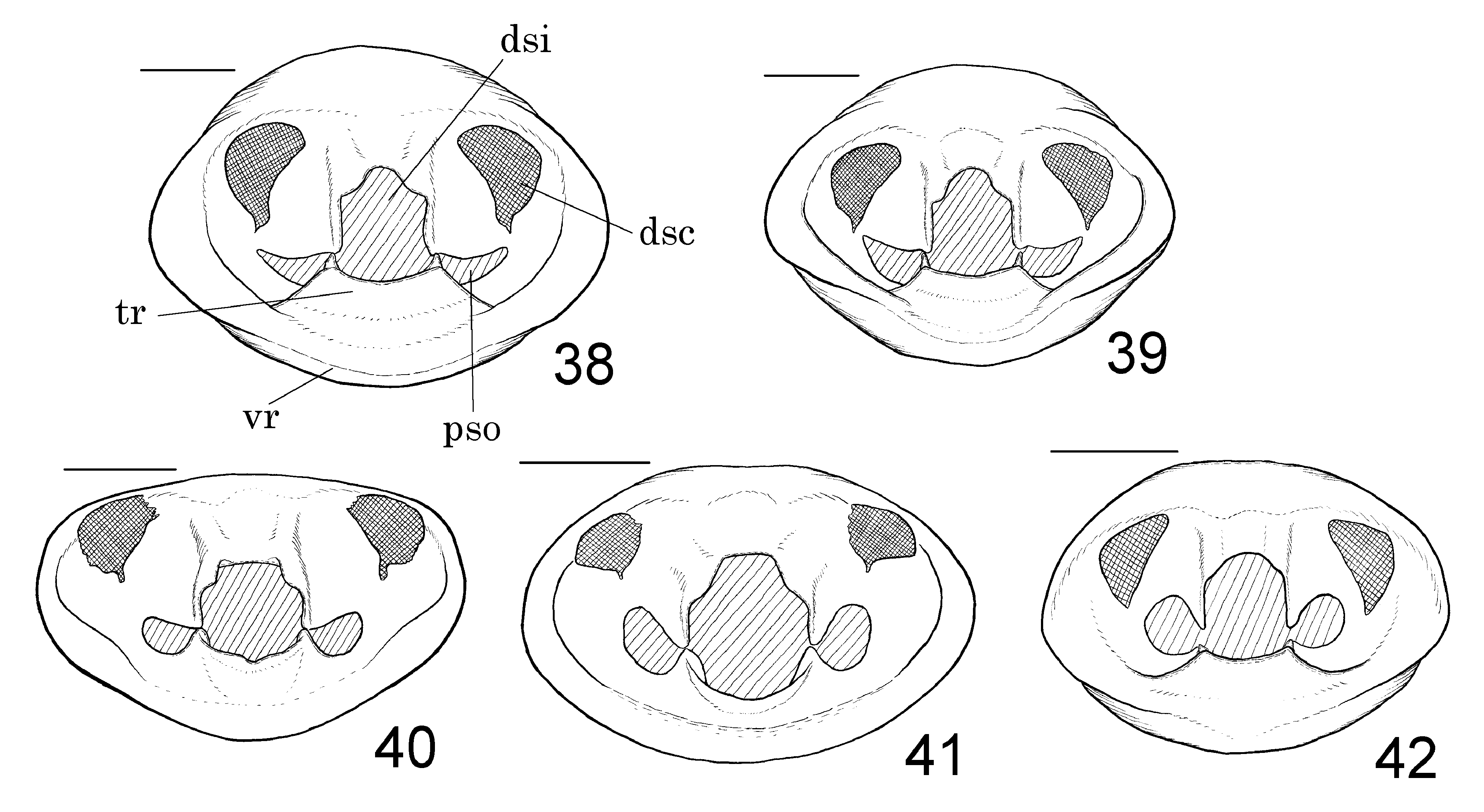
Abdomen (from base of segment III to apex of segment VII) approximately 7/10 as long as its maximum width. Genital capsule ( Figs. 36 View FIGURES 33–37 , 41 View FIGURES 38–42 ) approximately 2.5 times as wide at maximum as its basal width, approximately 3/5 as long as its maximum width, roundly produced at posterior angles; ventral rim ( Figs. 31 View FIGURES 28–32 , 36 View FIGURES 33–37 ) widely, shallowly and roundly concave, almost straight in middle part; concavity of ventral rim approximately 1/15 as deep as its maximum width ( Fig. 36 View FIGURES 33–37 ); transverse ridge well developed, invisible in ventral view, with roundly and deeply concave dorsal margin in posterior view ( Fig. 41 View FIGURES 38–42 ); dorsal sclerites elliptic, angularly extended outward, with small projection at ventral angle ( Fig. 41 View FIGURES 38–42 ); dorsal sinus suboctagonal in posterior view ( Fig. 41 View FIGURES 38–42 ); paramere sockets elliptic in posterior view ( Fig. 41 View FIGURES 38–42 ). Crown of paramere ( Fig. 46 View FIGURES 43–47 ) drop-shaped, covered with short to long setae for most part, obtuse at apex; lateral lobe roundly and strongly extended laterad with widely round distal and proximal angles; distal margin gently concave; proximal margin roundly concave. Conjunctival processes of endosoma ( Figs. 51 View FIGURES 48–52 , 56 View FIGURES 53–57 , 61 View FIGURES 58–62 , 66 View FIGURES 63–67 ) simple, long, almost straight, directed distad, tapering, blunt at apex. Vesica short, straight in lateral view, directed dorsoposteriad ( Fig. 61 View FIGURES 58–62 ). Processes of vesica relatively large, roughly Y-shaped in ventral view ( Fig. 56 View FIGURES 53–57 ), elliptic in lateral view ( Fig. 61 View FIGURES 58–62 ), semirectangular in outline in posterior view ( Fig. 66 View FIGURES 63–67 ).
Female. Similar to the male. Body relatively larger than male. Valvifer VIII roughly triangular, generally yellowish green, often with oval oblique brownish spot apically ( Fig. 71 View FIGURES 68–72 ), covered with short to long setae on disc and along posterior and mesal margins ( Fig. 76 View FIGURES 73–77 ); apical angle a little more obtuse than right angle ( Fig. 76 View FIGURES 73–77 ). Spermatheca long; apical receptacle ( Fig. 83 View FIGURES 78–84 ) spherical, with 1 horn-shaped process; horn-shaped process long, produced laterad of apical receptacle, abruptly curved basad, reaching proximal flange; intermediate part ( Fig. 83 View FIGURES 78–84 ) gradually narrowed basad, approximately 3 times as long as its width at middle, with sclerotized zone apically; apical sclerotized zone of intermediate part approximately half as long as intermediate part ( Fig. 83 View FIGURES 78–84 ); distal flange 1.5 times as wide as proximal flange ( Fig. 83 View FIGURES 78–84 ).
Measurements [in mm, ♂ (n=56) / ♀ (n=53). Body length 6.7–8.6 (holotype 8.1) / 7.1–9.8; width across eyes 2.1–2.6 / 2.2–2.9; width across humeral width 4.3–5.8 / 4.7–6.4; maximum width of abdomen 4.2–5.6 / 4.7–6.2.
Type series. Holotype: ♂ ( Fig. 4 View FIGURES 1–5 ), “ JAPAN the Ryukyus, Ishigaki-jima Is., Hirae, 24.38904N 124.19505E, 23. IX. 2014, light trap, Kiichi Shimizu” (LETUA IC 2018-00024) ( TUA). GoogleMaps
A total of 108 paratypes (55♂ 53♀, LETUA IC 2018-00025–00132, TUA) are here designated from the following localities: Okinawa-jima Is., Ishigaki-jima Is., Iriomote-jima Is., and Yonaguni-jima Is. of the Ryukyu Islands, Japan.
Other material examined. In addition to the type specimens, 163 specimens (ELEU, ELKU, NIAES, NSMT, OMNH, TUA) were examined from the following localities: JAPAN: Honshu (Kanagawa, Aichi, Wakayama, Ishikawa, Okayama, Hiroshima, and Yamaguchi Prefectures), Shikoku (Tokushima, Kagawa, Ehime, and Kochi Prefectures), Kyushu (Miyazaki and Kagoshima Prefectures), and the Ryukyu Islands (Yaku-shima Is., Nakanoshima Is. of the Tokara Islands, Amami-oshima Is., Tokuno-shima Is., Yoron-to Is., Okinawa-jima Is., Minamidaito-jima Is., Miyako-jima Is., Ishigaki-jima Is., Taketomi-jima Is., Iriomote-jima Is., and Yonaguni-jima Is.).
Distribution. Japan (Honshu, Shikoku, Kyushu, and the Ryukyu Islands), China (Southeastern Territory). Based on locality data, it is likely that in Honshu, Shikoku and Kyushu, Japan, this species only inhabits regions within a few kilometers of the coast.
Etymology. The specific epithet is named after the Japanese common name for this new species ‘Himechabane-ao-kamemushi’; a noun in apposition.
Remarks. This new species has been misidentified for more than 85 years. The first error of this nature was made by Esaki (1932) who used Plautia splendens to describe the population of the mainland Japan (Honshu). Subsequent Japanese heteropterists such as Syôiti Miyamoto and Hitoshi Hasegawa followed Esaki’s identification, using the name P. splendens for populations from other areas of Japan (Shikoku, Kyushu, and the Ryukyu Islands) and for Chinese populations ( Liu & Zheng 1995). There were likely two factors responsible for the misidentification. Firstly, the type locality of P. splendens was described by Distant (1900) as simply “ Japan ” based on specimens likely collected from the Ogasawara Islands of Japan. Secondly, the general appearances of P. splendens and P. himechabane are similar to each other except for the overall body color. Esaki (1932) would likely have been unable to determine that the “ Japan ” referred to by Distant (1900) was in fact the Ogasawara Islands, which are 1,000 km away from the mainland, supposing that the differences in body color were simply due to intraspecific variation. The comprehensive fieldwork and detailed morphological examinations provided here clarify that P. splendens is a species found only in the Ogasawara Islands (furthermore, it is a senior synonym of P. cyanoviridis , as mentioned above) and that any species referred to as P. splendens found in the mainland of Japan, the Ryukyu Islands, or China do not correspond to any species described until now.
This new species is closely related with P. splendens Distant, 1900 . However, P. himechabane sp. nov. is distinguished from P. splendens by the yellowish green body ( Fig. 4 View FIGURES 1–5 ) [vs. dark blue green body ( Fig. 3 View FIGURES 1–5 )], antennae with blackish areas ( Fig. 4 View FIGURES 1–5 ) [vs. lacking blackish area ( Fig. 3 View FIGURES 1–5 )], the apical part of scutellum whitish ( Figs. 4 View FIGURES 1–5 , 17 View FIGURES 14–18 ) [vs. concolorous with the remaining parts of scutellum ( Figs. 3 View FIGURES 1–5 , 8, 10 View FIGURES 6–11 , 16 View FIGURES 14–18 )], the ventral rim of genital capsule widely and shallowly concave ( Figs. 31 View FIGURES 28–32 , 36 View FIGURES 33–37 ) [vs. V-shaped ( Figs. 30 View FIGURES 28–32 , 35 View FIGURES 33–37 )], the dorsal sclerites of genital capsule angularly extended outward ( Fig. 41 View FIGURES 38–42 ) [vs. roundly extended outward ( Fig. 40 View FIGURES 38–42 )], paramere sockets elliptic in the posterior view ( Fig. 41 View FIGURES 38–42 ) [vs. semicircular ( Fig. 40 View FIGURES 38–42 )], the lateral lobe of the crown of paramere with widely round proximal angle ( Fig. 46 View FIGURES 43–47 ) [vs. with acutely angulated proximal angle ( Fig. 45 View FIGURES 43–47 )], processes of vesica semirectangular in outline in the posterior view ( Fig. 66 View FIGURES 63–67 ) [vs. oblong ( Fig. 65 View FIGURES 63–67 )], valvifer VIII roughly triangular ( Fig. 76 View FIGURES 73–77 ) [vs. pentagonal ( Fig. 75 View FIGURES 73–77 )], and a horn-shaped process of the apical receptacle of spermatheca long and produced laterad of the apical receptacle ( Fig. 83 View FIGURES 78–84 ) [vs. short and produced dorsad of apical receptacle ( Fig. 82 View FIGURES 78–84 )].


No known copyright restrictions apply. See Agosti, D., Egloff, W., 2009. Taxonomic information exchange and copyright: the Plazi approach. BMC Research Notes 2009, 2:53 for further explanation.
|
Kingdom |
|
|
Phylum |
|
|
Class |
|
|
Order |
|
|
Family |
|
|
Genus |