
Acartiella kempi Sewell, 1914
|
publication ID |
https://doi.org/10.12782/sd.20.2.167 |
|
DOI |
https://doi.org/10.5281/zenodo.5737547 |
|
persistent identifier |
https://treatment.plazi.org/id/03A5E838-FF9C-FFC9-8593-FA4E562EFD16 |
|
treatment provided by |
Felipe |
|
scientific name |
Acartiella kempi Sewell, 1914 |
| status |
|
( Figs 2–4 View Fig View Fig View Fig , 10A View Fig )
Acartiella kempi Sewell, 1914: 246 , pls 20–21, figs 1–5 (original description); 1919: 17; 1947: 324, 429; Wellershaus 1969: 269–270, figs 53–54.
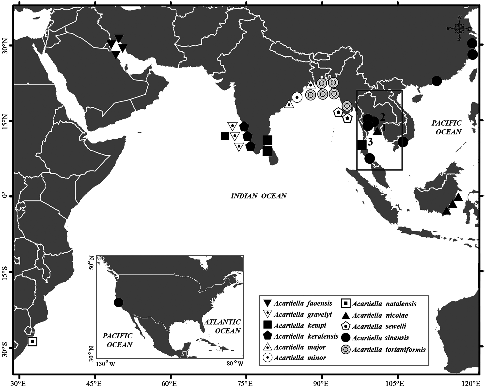
Material examined. Ten females and 13 males, Kraburi Estuary, Andaman Sea (station 3 in Fig. 1 View Fig ), November 7, 2011 (BIMS-Zoo-0262).
Female. Total length 0.80–0.96 mm (mean±SD=0.88± 0.05 mm, N=10); prosome length 0.57–0.64 mm (0.60± 0.03 mm); prosome width, 0.19–0.23 mm (0.20± 0.01 mm).
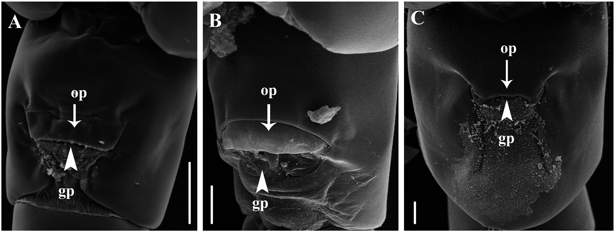
Body ( Fig. 2A, B View Fig ) slender; cephalosome and first pedigerous somite separate; cephalosome anteriorly round in dorsal view; rostrum absent; pedigers four and five fused; prosomal ends symmetrical and round posterolaterally. Urosome composed of three somites; symmetrical in dorsal view ( Fig. 2B View Fig ); genital double-somite about 2.5 times longer than anal somite; genital operculum trapezoidal, located slightly behind ventro-central midpoint ( Fig. 10A View Fig ); caudal rami asymmetrical, right ramus longer than left, with five plumose setae and one small seta. Both rami fused basally with anal somite.
Antennule ( Fig. 2C View Fig ) reaching beyond posterior end of genital double-somite, symmetrical, 23-segmented; segments 2–4 incompletely fused; segments 2, 3, 4, 5, 7, 12, 23 each with aesthetasc (ae). Armature elements as follow: 1=1, 2(2–4)=3+ae, 3=1+ae, 4=2+ae, 5=1+ae, 6=(1 spiniform element), 7=2+ae, 8–9=0, 10=1, 11=1+(1 spiniform element), 12=1+(2 spiniform elements)+ae, 13=1+(2 spiniform elements), 14=1+(2 spiniform elements), 15=1+(2 spiniform elements), 16=1+(2 spiniform elements), 17=1+(3 spiniform elements), 18=1+(2 spiniform elements), 19=1, 20=2, 21=2, 22=2, 23=4+ae.
Antenna ( Fig. 2D View Fig ) bearing thick-walled setae along anterior margin of coxa, basis, and endopod; coxa with single seta; basis fused to both rami with two setae at midlength and a terminal seta; free endopodal segment with two setae at midlength and five terminal setae; exopod completely fused to basis, with four long and two medium long setae.
Mandible ( Fig. 2E View Fig ) with two cuspidate processes and two small teeth on gnathobase; basis with one seta at midlength, two terminal setae, and short row of fine setules near median margin; first endopodal segment fused to basis and, second segment with four setae terminally; proximal part of exopod also fused to basis, with two inner setae, free exopodal segment, with three distal setae.
Maxillule ( Fig. 2F View Fig ) with 7 praecoxal arthrite bearing (1 slightly long thick seta, 4 strong setae, and a thin anterior seta); coxal endite with thick seta; coxal epipodite with six long setae; basal exite with single seta; exopod partly fused with basis and bearing one proximal seta, two terminal setae, and row of fine hairs along inner margin. Endopod totally reduced.
Maxilla ( Fig. 2G View Fig ) robust with unarmed praecoxa; syncoxal endite with three long, one medium, and four short setae; basis with one stout serrate seta; endopod with one short and five long serrate setae.
Maxilliped ( Fig. 3A View Fig ) highly reduced; syncoxa with three endites, first endite with two strong setae, second with one medium and one long setae, third endite with medium long seta; basis with small seta at midlength and row of fine hairs along inner margin; endopod unisegmented, with three inner spines and inwardly curved terminal claw.
Legs 1–4 ( Fig. 4A− D View Fig ) biramous, with 2-segmented endopod and 3-segmented exopod; coxa unarmed. Seta and spine formula as shown in Table 1.
Legs 5 ( Fig. 4E View Fig ) nearly symmetrical; coxa unarmed, separated from intercoxal sclerite by suture; basis with outer plumose seta; endopod represented by conical process with fine serration at tip; exopod about 3.6 times longer than endopod, bearing outer seta at distal one-third of its length and fine teeth along terminal on both sides margin.
Male. Total length 0.79–0.90 mm (0.82± 0.03 mm, N=13); prosome length 0.51–0.64 mm (0.60± 0.13 mm); prosome width 0.16–0.18 mm (0.17± 0.007 mm).
Body ( Fig. 3B, C View Fig ) similar to that of female except for 5-segmented urosome.
Right antennule ( Fig. 3D View Fig ) geniculate, incompletely 18-segmented; segments 2–3 and 9–12 partly fused; armature elements as follows: 1=1, 2 (2–4)=3+ae, 3=1, 4=2, 5=1, 6=1, 7=1+(1 spiniform element), 8–9=0, 10=1+(1 spiniform element), 11=1, 12=1+ae, 13=1, 14=1, 15=1+(1 process), 16=2+(1 process), 17=1+(1 spiniform element)+(1 process), 18=8+(2 spiniform elements)+ae. Left antennule incompletely 18-segmented, reaching beyond posterior end of genital somite. Armature elements as in female.
Legs 5 ( Fig. 4F View Fig ) asymmetrical and uniramous; coxae fused to each other. Right leg basis with sub-terminal plumose seta; exopod 2-segmented, first segment with small seta, second segment with small spine on inner margin and one terminal spine. Left leg with thickened basis with large acuminate proximomedial projection, also bearing single plumose seta on outer margin and row of small spinules on inner terminal margin. Exopod comprising of two segments; first segment unarmed, longer than wide; second segment with irregular margins, a small spine and fine hairs, outer proximal margin with crested projection, inner segment with a small spine at central, and three unequal spines on tip.
Remarks. Acartia kempi was originally described from the Gulf of Mannar in the Indian Ocean by Sewell (1914). The present specimens differ from the original description in the following respects: (1) the antennules of the present females are 23-segmented with an incomplete fusion of segments 2–4, while those in the original description had 21 separate segments, with an incomplete fusion of segments 2–6; (2) the left caudal ramus is slightly shorter than the right in males in the present study ( Fig. 2A, B View Fig ) whereas, although the original paper did not describe the caudal rami in the text, the drawings suggest that the caudal rami were of equal length; (3) the body lengths of the females (range 0.80–0.96 mm, mean 0.88 mm) and males ( 0.79–0.90 mm, 0.82 mm) examined in the present study are slightly less than those from the Gulf of Mannar (1.0mm, 0.9 mm).
Barthélémy (1999) observed the internal and external structures of the female reproductive system of six species of the genus Acartiella and found that the relative positions of the opercular pad and gonopores on the genital doublesomite are species-specific. The present study represents the first report of these being located slightly behind mid-length of the ventral surface of the genital double-somite in A. kempi (see Fig. 10A View Fig ).
Distribution. This species has so far been reported from the waters of Paumben and Kilakarai in the Gulf of Mannar, southern India ( Sewell 1914, 1919) and the Cochin Backwater on the southwestern coast of India ( Wellershaus 1969) (see Fig. 1 View Fig ). The specimens in the present study were collected from the Kraburi Estuary in the Andaman Sea during the dry season (November, 2011), when the water temperature and salinity ranges were 31.4–32.1°C and 13–22 ‰, respectively.


No known copyright restrictions apply. See Agosti, D., Egloff, W., 2009. Taxonomic information exchange and copyright: the Plazi approach. BMC Research Notes 2009, 2:53 for further explanation.
|
Kingdom |
|
|
Phylum |
|
|
Class |
|
|
Order |
|
|
Family |
|
|
Genus |
Acartiella kempi Sewell, 1914
| Srinui, Khwanruan & Ohtsuka, Susumu 2015 |
Acartiella kempi
| Wellershaus, S. 1969: 269 |
| Sewell, R. B. S. 1914: 246 |