
Corydoras guapore Knaack, 1961
|
publication ID |
https://doi.org/ 10.1590/1982-0224-20150018 |
|
DOI |
https://doi.org/10.5281/zenodo.4776243 |
|
persistent identifier |
https://treatment.plazi.org/id/03A487BC-B704-9131-FC7F-D488FEDDFDA4 |
|
treatment provided by |
Carolina |
|
scientific name |
Corydoras guapore Knaack, 1961 |
| status |
|
Corydoras guapore Knaack, 1961 View in CoL
( Figs. 1-5 View Fig View Fig View Fig View Fig View Fig ; Table 1 View Table 1 )
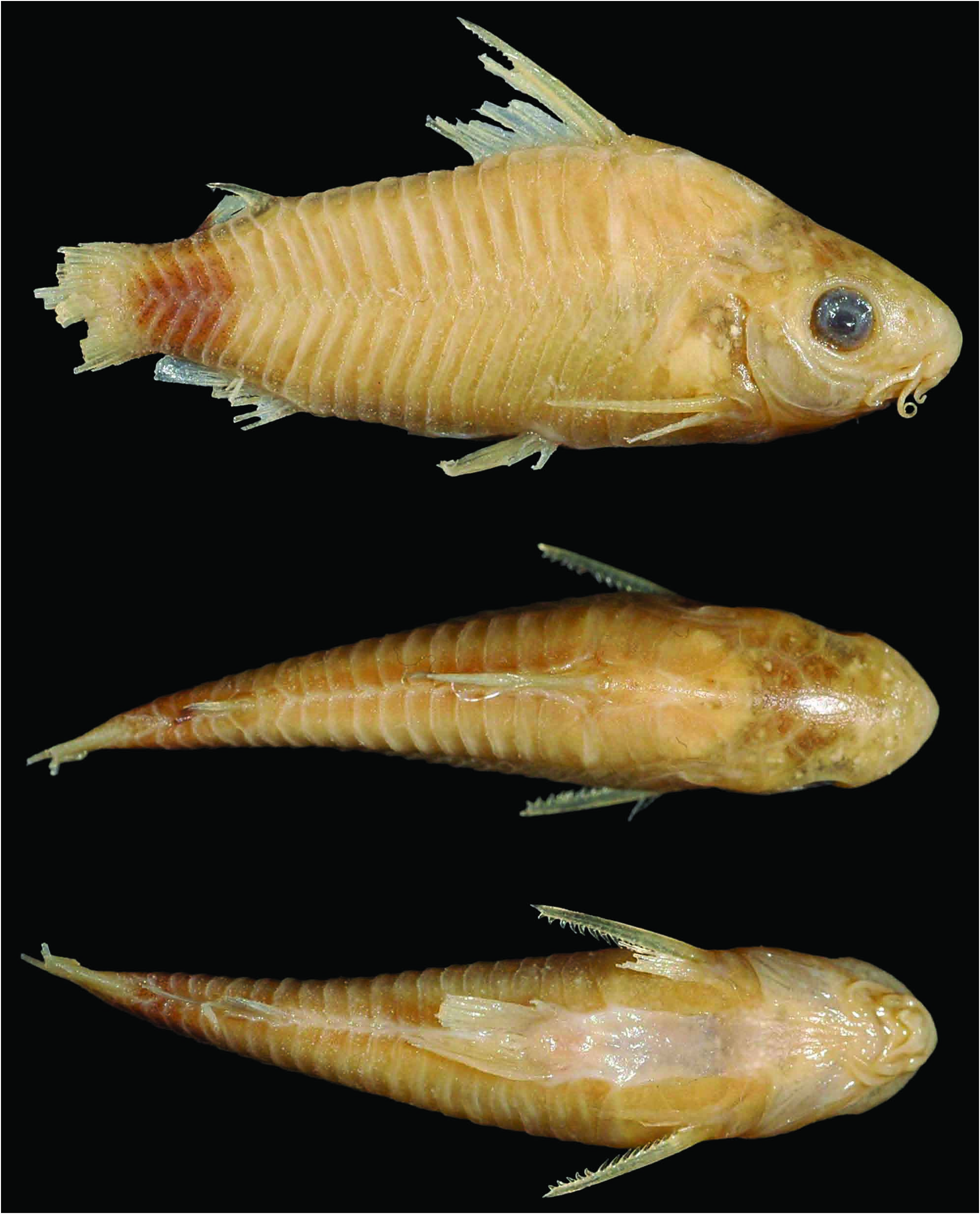
Diagnosis. Corydoras guapore can be distinguished from its congeners, with exception of C. bilineatus , C. elegans , C. gracilis , C. mamore , C. nanus , C. napoensis , C. nijsseni , C. paucerna and C. undulatus , by having the following unique combination of features: mesethmoid short, with anterior tip poorly developed (vs. long, with well-developed anterior tip); serrations directed towards pectoral-spine origin (vs. perpendicularly inserted; or directed towards pectoral-spine tip); and conical serrations on posterior margin of pectoral spine (vs. laminar). Corydoras guapore can be distinguished from C. bilineatus , C. elegans , C. gracilis , C. mamore , C. nanus , C. napoensis , C. nijsseni , C. paucerna and C. undulatus by the presence of dorso- and ventrolateral body plates with vertically elongated or irregular brown blotches anteriorly to adipose fin, and lateral portion of caudal peduncle almost entirely blackened (vs. with two or three longitudinal black stripes in C. bilineatus , C. elegans , C. napoensis , C. undulatus ; a thickened black stripe on dorsolateral body plates, ventrolateral body plates with irregular black spots in C. gracilis ; irregular small black spots in C. mamore and C. paucerna ; upper portion of dorsolateral body plates with intense black pigmentation, becoming diffuse toward ventrolateral body plates in C. nijsseni ). Additionally, C. guapore can be distinguished from C. hastatus and C. pygmaeus by the presence of adipose fin with anterior portion hyaline and posterior portion blackened (vs. entirely hyaline); and the absence of a longitudinal black stripe on midline of flank (vs. presence of a slender diffuse longitudinal black stripe in C. hastatus ; and a thicker conspicuous longitudinal black stripe in C. pygmaeus ).
Description. Morphometric data presented in Table 1 View Table 1 . Head compressed with convex dorsal profile; somewhat pentagonal in dorsal view. Snout short and rounded. Head profile convex from tip of snout to anterior nares; and slightly concave from this point to the tip of posterior process of parieto-supraoccipital. Dorsal profile of body slightly convex along dorsal-fin base. Body profile nearly straight from posterior portion of dorsal-fin to adiposefin spine; markedly concave from this point to caudal-fin base. Ventral profile of body slightly convex from isthmus to pelvic girdle; nearly straight from pelvic girdle to base of first anal-fin ray; abruptly concave from this point to caudal-fin base. Body roughly elliptical in cross section at pectoral girdle, gradually becoming more compressed toward caudal fin.
Eye rounded, located meso-laterally on head; orbit delimited dorsally by lateral ethmoid, frontal and sphenotic,ventrallybyinfraorbitals.Anteriorandposterior nares close to each other, only separated by flap of skin. Anterior naris tubular. Posterior naris relatively distant to antero-dorsal margin of orbit, separated from it by distance equal to twice the diameter of naris. Mouth small, subterminal, width nearly equal to bony orbit diameter. Maxillary barbel long in size, reaching anteroventral limit of gill opening. Outer mental barbel slightly smaller than maxillary barbel. Inner mental barbel fleshy, its base slightly separated from its counterpart. Small rounded papillae covering entire surface of all barbels, upper and lower lips, and isthmus.
Mesethmoid short; anterior tip thickened and poorly developed, smaller than 50% of the bone length; with poorly-developed lateral cornua; posterior portion widened, partially exposed and bearing minute odontodes. Nasal slender, curved laterally, with inner margin laminar; posterior portion of outer margin laminar; mesial border contacting frontal and mesethmoid. Frontal elongated, narrow, with width slightly larger than half of entire length; anterior projection short, size smaller than nasal length. Frontal fontanel large, slender; posterior tip extension slightly entering anterior margin of parietosupraoccipital. Parieto-supraoccipital wide, posterior process long and contacting nuchal plate; region of contact between posterior process and nuchal plate covered by thick layer of skin.
Two laminar infraorbitals with minute odontodes; infraorbital 1 large, ventral laminar expansion very reduced; anterior portion with poorly developed expansion ( Fig. 2 View Fig ); infraorbital 2 small, thickened; with posterior laminar expansion well developed; posteroventral margin contacting posterodorsal ridge of hyomandibula, dorsal tip contacting sphenotic and compound pterotic ( Fig. 3 View Fig ). Posterodorsal ridge of hyomandibula close to its articulation with opercle conspicuously slender; exposed, very reduced and bearing small odontodes; dorsal ridge of hyomandibula between compound pterotic and opercle covered by posterodorsal laminar expansion of infraorbital 2. Interopercle almost entirely exposed, somewhat triangular, anterior projection well developed. Preopercle slender, elongated, with minute sparse odontodes on external surface. Opercle dorsoventrally elongated, width equal or smaller than length; free margin slightly convex, without serrations and covered by small odontodes. Anteroventral portion of cleithrum and posterolateral portion of scapulocoracoid exposed. Anteroventral and posteroventral suture between cleithrum and scapulocoracoid exposed; moderately developed odontodes sparse on exposed areas. Vertebral count 22(2); ribs 7(2), first pair conspicuously larger; complex vertebra slender in shape. Neural and haemal spines with laminar expansions on anterior margin of proximal region; expanded in distal tips.
Four branchiostegals rays decreasing in size posteriorly. Hypobranchial 2 somewhat triangular, tip ossified and directed towards anterior portion, posterior margin cartilaginous; ossified portion well developed, about twice size of cartilaginous portion. Five ceratobranchials with expansions increasing posteriorly; ceratobranchial 1 with small process on anterior margin of mesial portion; ceratobranchial 3 notched on posterolateral margin; ceratobranchial 5 toothed on posterodorsal surface, 26 to 29(2) teeth aligned in one row. Four epibranchials with similar size; epibranchial 2 slightly larger than others, with small pointed process on laminar expansion of posterior margin; epibranchial 3 with triangular uncinate process on laminar expansion of posterior margin. Two wide pharyngobranchials (3 and 4), pharyngobranchial 3 with large triangular laminar expansion on posterior margin. Upper tooth plate oval; 26 to 31(2) teeth aligned in two rows on posteroventral surface.
Lateral-line canal entering neurocranium through compound pterotic, splitting into two branches before entering sphenotic: pterotic, with single pore, and preoperculo mandibular, with two pores. Sensory canal continuing through compound pterotic, entering sphenotic as temporal canal, which splits into two branches:one branch giving rise to infraorbital canal, other branch entering frontal through supraorbital canal, both with a single pore. Supraorbital canal not branched, running through nasal bone. Epiphyseal pore opening at supraorbital main canal, slightly directed towards frontal fontanel. Nasal canal with two pores. Infraorbital canal running through entire second infraorbital, extending to infraorbital 1 and opening into two pores. Preoperculo mandibular branch giving rise to preoperculo-mandibular canal, which runs through entire preopercle with three openings, leading to pores 3, 4, and 5, respectively.
Dorsal fin triangular, located just posterior to second dorsolateral body plate. Dorsal-fin rays II,7(2), II,8(18), posterior margin of dorsal-fin spine with 12 to 13 serrations directed towards dorsal-fin spine tip; serrations absent only on proximal region of posterior margin. Nuchal plate relatively large; exposed, with minute odontodes; spinelet short; spine relatively long, adpressed distal tip surpassing last dorsal-fin branched ray origin; anterior margin with small odontodes. Pectoral fin triangular, its origin just posterior to gill opening. Pectoral-fin rays I,7(14), I,8(6); posterior margin of pectoral spine with 14 to 17 well-developed conical serrations along its entire length; serrations directed towards pectoral-spine origin ( Fig. 4 View Fig ). Pelvic fin oblong, located just below second ventrolateral body plate, and at vertical through second branched dorsal-fin ray. Pelvic-fin rays i,5. Adipose fin roughly triangular, separated from base of last dorsal-fin ray by typically seven dorsolateral body plates. Anal fin triangular, located just posterior to 12 th ventrolateral body plates, and at vertical through anterior margin of adiposefin spine. Anal-fin rays ii,5(1), ii,6(19). Caudal-fin rays i,12,i, generally four dorsal and ventral procurrent rays; bilobed, lobes with similar size.
Two laterosensory canals on trunk; first ossicle tubular and second ossicle laminar. Body plates with minute odontodes scattered over exposed area, a conspicuous line of odontodes confined on posterior margins; dorsolateral body plates 23(2), 24(16), 25(2); ventrolateral body plates 21(17), 22(3); dorsolateral body plates along dorsal fin base 6; dorsolateral body plates between adipose and caudal fin 6(2), 7(8), 8(10); preadipose platelets 1(14), 2(6); small platelets covering base of caudal-fin rays; small platelets disposed dorsally and ventrally between junctions of lateral plates on posterior portion of caudal peduncle. Dorsal portion of snout, lateral ethmoid region, and upper lip region covered with small platelets. Ventral surface of trunk without platelets.
Color in alcohol. Ground color of the body yellowish, with top of the head and snout dark brown. Top of the head and snout, infraorbitals, opercle, preopercle, interopercle, compound pterotic, cleithrum, upper lip, maxillary and outer mental barbels covered by dark brown chromatophores. Dorso- and ventrolateral body plates with vertically elongated or irregular brown blotches anteriorly to adipose fin; lateral portion of caudal peduncle almost entirely blackened. Dorsal fin with diffuse black spots on dorsal-fin rays, generally restricted to the upper half of the dorsal fin. Pectoral, pelvic and anal fins with black chromatophores on rays. Adipose fin with anterior portion hyaline; posterior portion darkened. Caudal fin with four to nine transversal black bars ( Fig. 2 View Fig ).
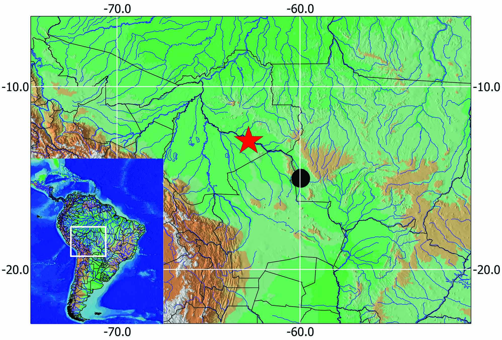
Geographic distribution. Corydoras guapore is only known from the upper rio Guaporé basin in Brazil ( Fig. 6 View Fig ).
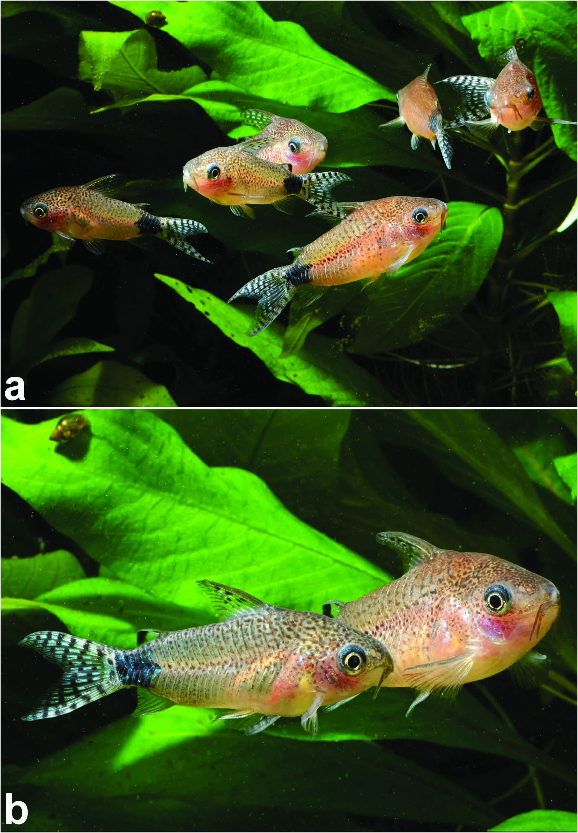
Color in life. Similar to preserved specimens but with ground color of the body rosy. Top of the head and snout, infraorbitals, opercle, preopercle, interopercle, compound pterotic and cleithrum with irregular striated brownish dots. Fins whitish; black spots on dorsal-fin rays more evident. Body covered by a yellowish green iridescent coloration ( Fig. 5 View Fig ).
Sexual dimorphism. Additionally to the presence of lanceolate genital papilla in males, which is common to all Corydoradinae (see Nijssen & Isbrücker, 1980; Britto, 2003), the males are generally smaller than females ( Fig. 5b View Fig ).
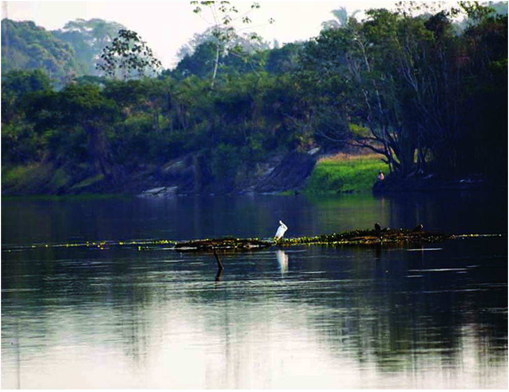
Ecological notes. Corydoras guapore is a freeswimming species, which occupies the middle of the water column in a small group when they feel safe ( Fig. 5a View Fig ), similar to the observed in C. hastatus . They form small breeding groups of up to 20 specimens associated to aquatic macrophytes, like Eichhornia . Unlike most of the Corydoras species which generally inhabit streams or the main channel of rivers, C. guapore is generally captured in lentic habitats as ponds and lakes (Hans- Georg Evers, pers. comm.). The specimens examined herein were captured close to the banks of the rio Guaporé, in Mato Grosso State (Cláudio Oliveira and Markos Alexandrou, pers. comm.) ( Fig. 7 View Fig ).
Material examined. All from Brazil, Mato Grosso State, Municipality of Vila Bela da Santíssima Trindade, rio Guaporé , rio Madeira basin. LBP 10089, 80 , 24.4-30.8 mm SL. ZUFMS-PIS 4000 , 5 , 26.9-33.6 mm SL, 2 c&s, 28.8- 29.2 mm SL .
Conservation status. Despite the fact that the species is known only from its type-locality (rio Guaporé) it is probably widespread in the surroundings and no imminent threat is suspected, therefore, according to the International Union for Conservation of Nature (IUCN) categories and criteria (IUCN Standards and Petitions Subcommittee, 2014), Corydoras guapore can be classified as Least Concern (LC).


No known copyright restrictions apply. See Agosti, D., Egloff, W., 2009. Taxonomic information exchange and copyright: the Plazi approach. BMC Research Notes 2009, 2:53 for further explanation.