
Molgula hodgsoni Herdman, 1910
|
publication ID |
https://doi.org/10.11646/zootaxa.3920.1.9 |
|
publication LSID |
lsid:zoobank.org:pub:C6BB0C5A-3317-4119-9C41-02F0EC487A9E |
|
DOI |
https://doi.org/10.5281/zenodo.6097442 |
|
persistent identifier |
https://treatment.plazi.org/id/039E0352-FFBE-FF89-33DD-F88CCE1A9A49 |
|
treatment provided by |
Plazi |
|
scientific name |
Molgula hodgsoni Herdman, 1910 |
| status |
|
Molgula hodgsoni Herdman, 1910
Molgula hodgsoni Herdman, 1910: 11 –12 pl. 3,figs 7–13. Hartmeyer 1909 –11. Monniot & Monniot: 1983 and synonymy; 1994: 34. Tatián et al. 2005: 211. Primo & Vázquez: 2007: 1805. Molgula angulata Ärnbäck-Christie-Linde: 1938.
Material: Terre Adélie:
66°563322 S–141°255738 E, 360– 151 m; 66°001305 S–142°0143 E, 240 m; 66°003882 S–143°7160085 E, 430 m; 66°516823 S–140°001423 E, 175–262 m; 66°38878 S–140°428852 E, 791– 684 m; 66° 0 0 3882 S–142° 313777 E, 233 m; 66°40072 S–139°49647 E, 129 m; 66°661100 S–140°081 E, 140 m; 66°6060281 S–140° 0 85 E, 140 m; 66° 6039662 S–139°982E, 140 m.
Antarctic Peninsula, South Orkney Islands,Wilkes Land, Ross Sea: numerous specimens from 50 to 500 m (for stations see Monniot & Monniot: 1983)
Bransfield Strait: 62° 26.66 S –56° 17.35 W, 438 m, 27/01/2013 “Polarstern” exp. 81.
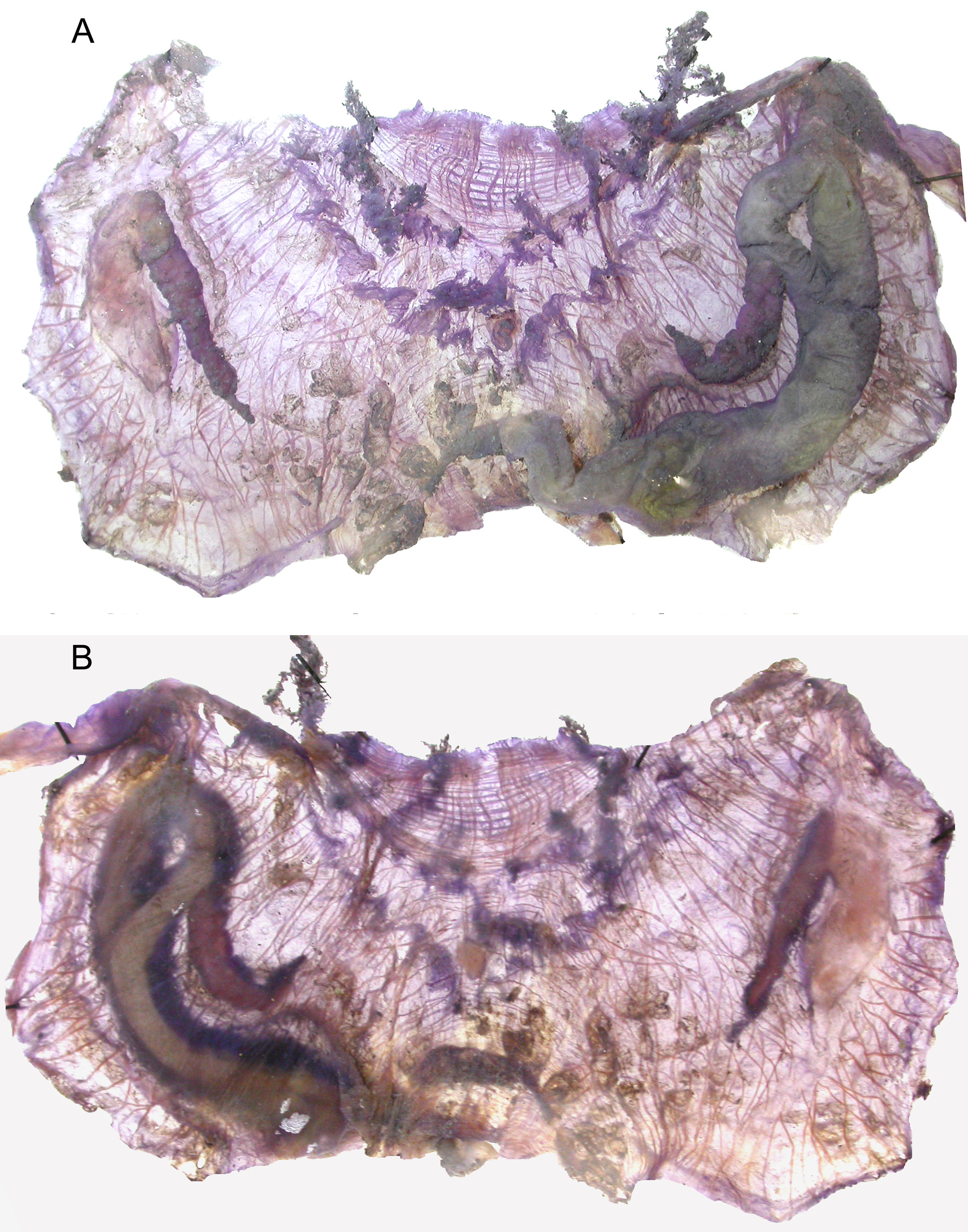
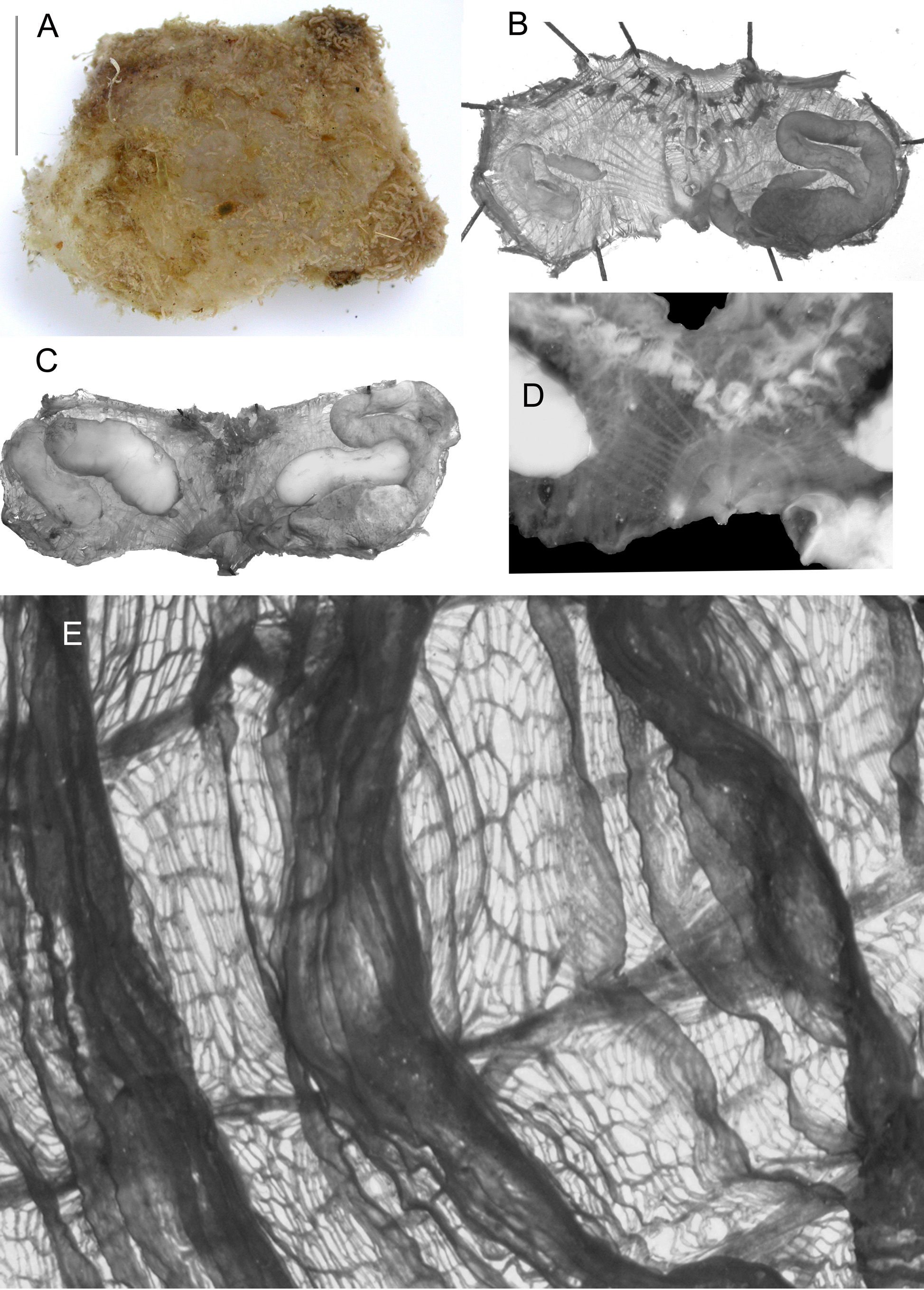
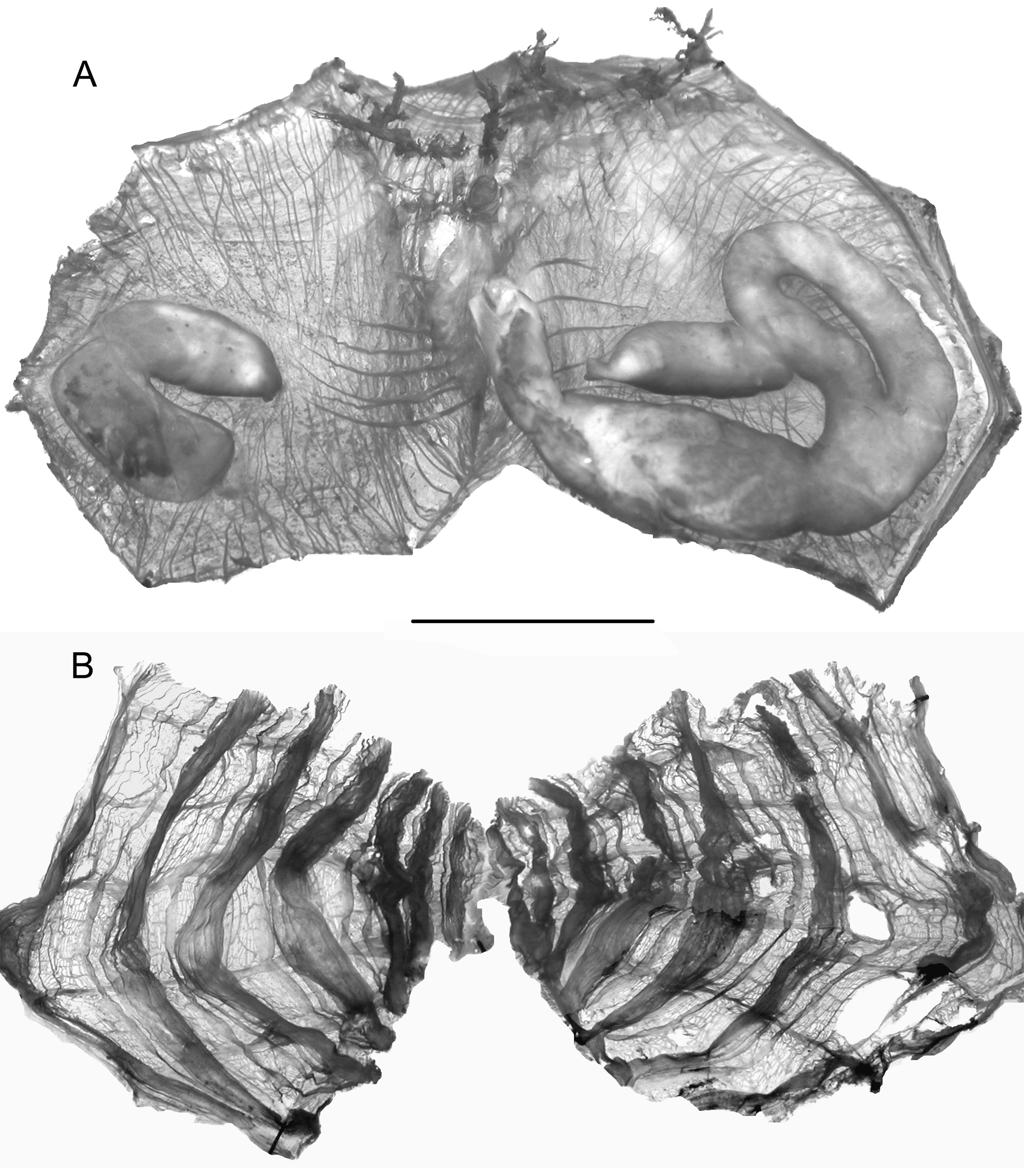
Oval or triangular in outline, the body is 2.5 to 4cm long, coated with dense filaments holding to the sediment ( Fig. 17 View FIGURE 17 A). There is no peduncle. The not protruding siphons are located at the anterior corners of the body. The tunic is resistant but thin. Removed from the tunic, the rim of the siphon is thinly dentate. The body wall is thin ( Figs 16 View FIGURE 16 B,C; 18A) with crossed muscular ribbons interrupted over the gut and the right gonad in the middle of each body side. In addition, 8 to 10 parallel strong fibres issued from the atrial siphon extend on both sides until the gonads level without ramification; they make an obvious particular fan-shaped design ( Fig. 18 View FIGURE 18 A). There are 8 large tentacles of unequal size alternating with smaller ones; the most ventral is often the longest. A velum is present in both siphons. The dorsal tubercle is located in a large V of the peri-pharyngeal band; it is C-shaped and opens to the left or more or less posteriorly, its horns curved but not rolled ( Fig. 17 View FIGURE 17 A).
The branchial tissue is thin, with 7 folds on each side ( Fig. 18 View FIGURE 18 B). The dorsal lamina is high, smooth edged, prolonged farther than the oesophagus on the left and linked to the end of the branchial folds. There is generally one or two longitudinal vessels between the folds, 16–17 vessels on the median folds in the largest specimens and 9 -14 vessels only in a specimen 2.5cm large; the thinner longitudinal vessels (more or less distant from each other) are counted as belonging to a fold but the higher intermediate vessels are more separated from the base of the fold ( Fig. 17 View FIGURE 17 E). The longitudinal vessels are gathered by contraction at the level of the 5 wide transverse vessels which delimit large branchial meshes. The stigmata are straight and crossed by many parastigmatic vessels in all directions ( Fig. 17 View FIGURE 17 E); they are curved at the top of the most ventral folds. The digestive tract makes a deep curved loop ( Figs 17 View FIGURE 17 B,C; 18 A). The stomach is spindle-shaped, separated from the intestine by a constriction and covered with flat hepatic vesicles. The intestine draws a closed loop with a pronounced secondary bend inside which is located the left gonad. The anus has a smooth but thick rim rarely with 2 low lobes. The two gonads ( Figs 17 View FIGURE 17 B,C; 18A) lie transversally on each side, they are oval and thick with a central ovary covered by dense small testis vesicles. The oviduct is short and terminal. Three to 8 male papillae in short buttons are lined along the gonad, hardly visible when it is not mature. The gonads are linked by their proximal pole to the intestine and to the kidney ( Fig. 18 View FIGURE 18 A). The kidney is bean-shaped and sometimes contains granular material.
COI sequences of the barcode region were obtained for three specimens. They are available for reference in the BOLD under specimen numbers S3 MOLA 380, S3 MOLA 381, S3 MOLA 383 (Dumont d'Urville Sea), and represent the first sequence references for this species. All sequences represent distinct haplotypes, and the maximum divergence between sequences was 0.17% (p-distance), with S3 MOLA 380 and S3 MOLA 381 having identical sequences. The species most similar in sequence in BOLD is Molgula pedunculata , but it is clearly distinct with an average of 19% divergence in p-distance.
Remarks. The characters described above are common to all specimens collected from both Terre Adélie and the Antarctic Peninsula. They correspond well to the description given by Tatián et al (2005) for specimens from the Scotia Arc. The precise descriptions of Caesira maxima by Hartmeyer (1911) and of M. angulata Ärnbäck- Christie-Linde, 1938 indicate that they are synonyms of M. hodgsoni .
The figure 34A, page 47 in Monniot et al. (2011) was erroneously inserted in the publication, it represents a dissection of M. hodgsoni which was present in the collection from Terre Adélie but was not described in the publication.
No known copyright restrictions apply. See Agosti, D., Egloff, W., 2009. Taxonomic information exchange and copyright: the Plazi approach. BMC Research Notes 2009, 2:53 for further explanation.
|
Kingdom |
|
|
Phylum |
|
|
Class |
|
|
Order |
|
|
Family |
|
|
Genus |
Molgula hodgsoni Herdman, 1910
| Monniot, Françoise & Dettai, Agnès 2015 |
Molgula hodgsoni
| Primo 2007: 1805 |
| Primo 2007: 2007 |
| Tatian 2005: 211 |
| Monniot 1983: 1983 |
| Herdman 1910: 11 |