
Parentocirrus brasiliensis, Paiva & Silva-Neto, 2004
|
publication ID |
https://doi.org/10.11646/zootaxa.504.1.1 |
|
DOI |
https://doi.org/10.5281/zenodo.5248610 |
|
persistent identifier |
https://treatment.plazi.org/id/039D9C24-445A-FFAC-6879-F9076830B891 |
|
treatment provided by |
Felipe |
|
scientific name |
Parentocirrus brasiliensis |
| status |
sp. nov. |
Description of Parentocirrus brasiliensis sp. n.
Derivatio nominis
Because of the country in which this species was found.
Locus typicus
Samples of activated sludge from Estação de Tratamento de Esgoto da Penha (ETE – Penha), a wastewater treatment plant from Companhia Estadual de Águas e Esgotos (CEDAE/RJ), located in Penha, Rio de Janeiro, Brazil.
Diagnosis
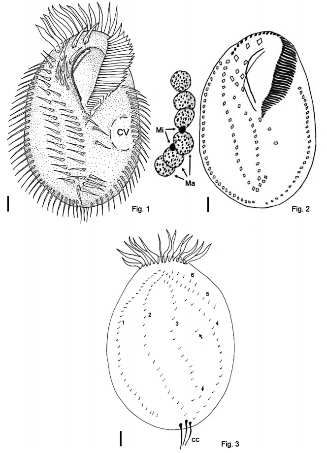
Size in vivo about 110 x 75µm. Body elliptical, with a single round shaped contractile vacuole in the equatorial region, close to the left margin. The ventral ciliature is composed of three distinct enlarged anterior frontal cirri, followed by a set of 2–3 buccal cirri and 3– 5 posterior frontal cirri of the same size; two marginal cirral rows, two rows of ventral cirri ending in an oblique row of 3–4 transverse cirri, and 2–4 postperistomial cirri. Macronuclear apparatus composed of 4–6 macronuclear nodules and 1–6 micronuclei. Dorsal side shows 6 (rarely 5) kineties, with very few scattered kinetids between kineties 3 and 4. Kineties 1, 2 and 4 ends each one in a caudal cirrus. Feeds on smaller protists and bacteria.
Morphological Characterization
This new species has conspicuous adoral zone of membranelles, composed of 32–49 membranelles and occupies about 43% of average cell length, the membranelles in the transversal region are about 18µm long. The first 2 or 3 proximal membranelles are engulfed by an oral lip. The paroral and endoral membranes virtually intercept each other in their middle section, displaying the usual “oxytricha pattern” ( Berger & Foissner, 1997).
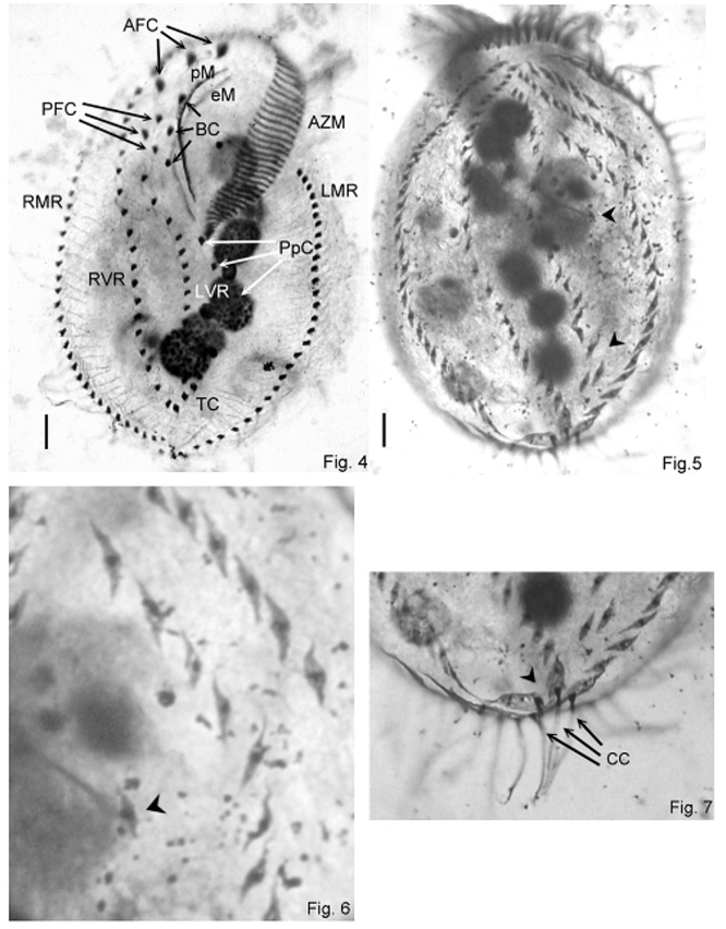
All studied specimens show three enlarged anterior frontal cirri, which are easily recognizable from the rest of the ventral ciliature, due to their position and constant size. Below this cirral set is a complex of buccal and posterior frontal cirri that are somewhat difficult to distinguish. In fact, we tried to follow the terminology adopted by Voss (1997), but we found that it may be inaccurate for P. brasiliensis , due to the variable number of cirri in this region, as well as their organization, which is different from P. hortualis . This complex is composed by 2–3 buccal cirri, in which the anteriormost is placed right and adjacent to the anterior section of the paroral membrane, and 3–5 posterior frontal cirri, which we considered as the cirri below the anterior frontal cirri, that are not placed adjacent to the undulating membranes and whose the distance from the first cirrus of the ventral rows is larger than the distance observed between the cirri on such rows ( Figs. 1, 2 View FIGURES 1–3. 1 , 4 View FIGURES 4–7 ).
The left ventral cirral row commences right of the posterior section of the endoral membrane, usually below the rightmost posterior frontal cirrus, extending to the transverse cirri set. The right ventral cirral row commences right of the rightmost anterior frontal cirri and ends in the rightmost cirrus of the transverse set, which is composed by an oblique row of 3–4 cirri. When in 4, the 2 leftmost transverse cirri are placed left of the ventral rows.
The left marginal cirral row begins below the middle region of the adoral zone of membranelles. The right marginal cirral row begins at the level of the uppermost buccal cirrus. Both marginal rows are convergent in the anterior end of cell, but do not fusionate, terminating at the same level. The cirri in the marginal rows measure about 15µm in the equatorial region of the cell.
In the dorsal surface, we observed the occurrence of six ( 5 in one specimen) dorsal rows of kinetids. Row three begins anterior to row two, and its orientation in the anterior end is opposite to that observed in P. hortualis . Also, there are few scattered kinetids present between rows 3 and 4, usually in small number (3 or 4 in most specimens). The posterior scattered kinetids seems to compose a loose short kinety that merges with the posterior region of kinety 3 ( Figs. 5 View FIGURES 4–7 , 10 View FIGURES 8–10 ). Kineties 5 and 6 are shortened, ending before the equatorial region of cell. Kineties 1, 2 and 4 bears each one a caudal cirrus about 15µm long ( Figs. 3–7 View FIGURES 1–3. 1 View FIGURES 4–7 ).
The nuclear apparatus is composed of a row of 4–6 macronuclear nodules, which sometimes are distinctly spread into two sets. These sets are not linked by a connection bridge, as in P. hortualis . The nodules are spheroid in shape, and measure 12µm in average diameter. One to six micronuclei are attached to the macronuclear nodules. Their average diameter in the studied population is 2.5µm ( Fig. 2 View FIGURES 1–3. 1 ). The whole nuclear apparatus is located along the central longitudinal axis of the body, tending to form an arch, in which the convex side bends to the left region of body.
Typification
A slide with the holotype and several paratypes of Parentocirrus brasiliensis was deposited in the collection of Lab. de Protistologia, Dept. de Zoologia, Inst. de Biologia , Universidade Federal do Rio de Janeiro – UFRJ. Access number: 0007–1.
Discussion
The presence of very few kinetids scattered on the dorsal surface may represent a close relationship with genus Apoamphisiella Foissner, 1997 , a genus with both similar ventral ciliature organization and the presence of numerous scattered kinetids in the dorsal surface. Other possible related genera include Paraurostyla Borror, 1972 and Territricha Berger and Foissner, 1988 , however, in Paraurostyla , the ventral pattern shows increased number of ventral cirral rows, although still resembles the organization present in Parentocirrus and Apoamphisiella , and in contrast, Territricha has two ventral cirral rows which are arranged in a single set of paired cirri.
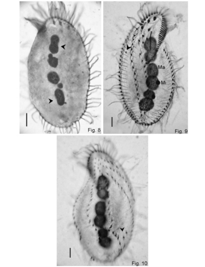
Another observation is that in some specimens measuring 90µm or less, the nuclear apparatus is composed of four or five macronuclear nodules instead of six, and that in these specimens, the nodules in both ends of the macronuclear row are elongated and constricted at the equatorial region ( Fig. 8 View FIGURES 8–10 ). Rare intrapopulational variants with anomalous nuclear apparatus are known to occur in some species of spirotrichous ciliates. Kattar (1970) observed it in populations of Urostrongylum caudatum (Kahl, 1932) .
In early dividers, we observed that the morphogenesis of the dorsal kineties occurred in the same way as described by Voss (1997) for P. hortualis , but we did not observe the occurrence of parental cirri rows, except for an uncommon isolated cirrus that was observed in only two interphasic specimens. When present, this cirrus was located between the right marginal row and right ventral row, above level of the basis of adoral zone of membranelles ( Fig. 9 View FIGURES 8–10 ). The presence of parental rows reported by Voss in specimens of P. hortualis was considered by Berger (1999) as a very rare condition.
We regard this population as a novel species different from P. hortualis because of the arrangement of dorsal kineties, the presence of scattered kinetids between kineties 3 and 4, the relative position of the posterior frontal cirri, the average number of macronuclear nodules, the number of postperistomial cirri, and the average body size. This species probably is conspecific with another population of P. hortualis found by Blatterer (unpublished data) and mentioned in Berger (1999), which was found in activated sludge samples from a papermill in Salzburg, Austria. This population of P. hortualis shows similarities with Parentocirrus brasiliensis sp. n. in organization of the posterior frontal cirri and the number of postperistomial cirri. However, the number of macronuclear nodules exceeds by three the maximum we observed ( Table 1 View TABLE 1 ), and the contractile vacuole is located above the equatorial region of the cell, almost adjacent to the adoral zone of membranelles. Due to the lack in sufficient data about this population (there is only a single drawing of a living specimen showing the ventral surface available), we consider it incipient to synonymize them.
Furthermore, because of the dorsal morphogenesis pattern, the ventral cirral configuration, and the overall similarities with Apoamphisiella and related genera, we agree with Berger (1999) that this genus should be placed within the Oxytrichidae , instead of the Kahliellidae , as originally proposed by Voss (1997).
| UFRJ |
Universidade Federal Rural do Rio de Janeiro |
No known copyright restrictions apply. See Agosti, D., Egloff, W., 2009. Taxonomic information exchange and copyright: the Plazi approach. BMC Research Notes 2009, 2:53 for further explanation.
|
Kingdom |
|
|
Phylum |
|
|
Class |
|
|
Order |
|
|
Family |
|
|
Genus |