
Hydryphantes clypeatus (Thor, 1899)
|
publication ID |
https://doi.org/10.11646/zootaxa.3869.2.2 |
|
publication LSID |
lsid:zoobank.org:pub:407C7804-ABAB-4419-A4E3-478856A3A57B |
|
DOI |
https://doi.org/10.5281/zenodo.6132174 |
|
persistent identifier |
https://treatment.plazi.org/id/039B87E2-FF87-FFF7-2494-FC214823FA10 |
|
treatment provided by |
Plazi |
|
scientific name |
Hydryphantes clypeatus (Thor, 1899) |
| status |
|
Hydryphantes clypeatus (Thor, 1899)
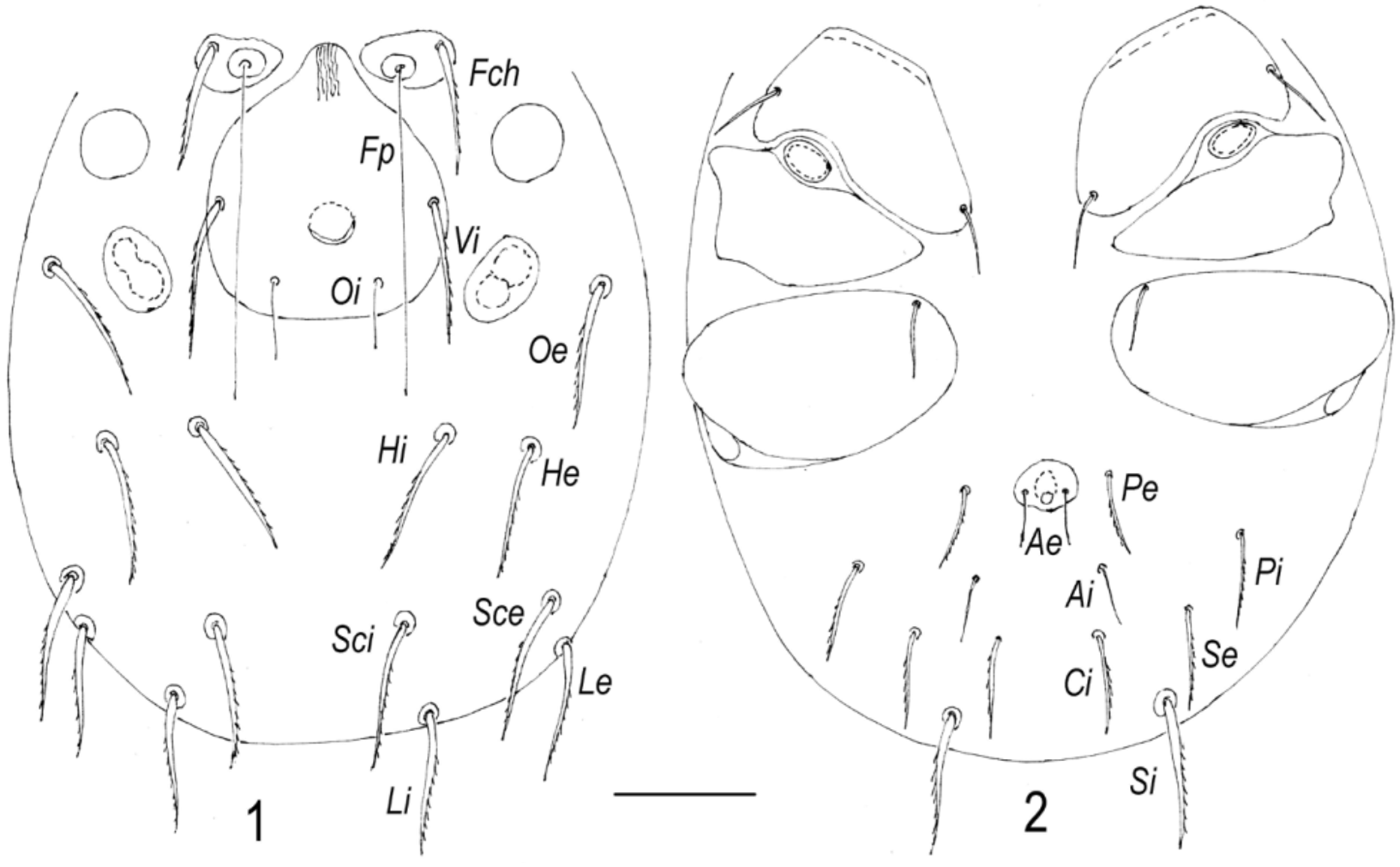
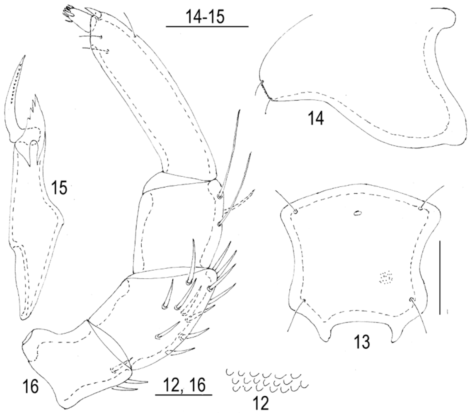
( Figs 1–16 View FIGURES 1 – 2 View FIGURES 12 – 16 )
Material examined. Larvae (n = 26) were reared from four females collected in a sedge bog near village Postyltsevo, Nekouz District, Yaroslavl Province: two females 25 May 2000, one female 1 June 2002 and one female 10 June 2004. The duration of the embryonic period was 12–15 days.
Diagnosis. Larva. Distance between bases of trichobothria Oi larger than their length; all dorsal hysterosomal setae subequal; excretory pore plate wider than long; urstigma wider than long; posterior margin of coxal plate II convex; P-5 with long solenidion; I–II-Leg-4 solenidia 2.0–2.5 times longer than eupathidia; I–II-Leg-5 with unequal solenidia; I-Leg-6 de shorter than ds.
Description. Colour red. Anterior pair of platelets small, more or less triangular, posterior plate relatively large, anteriorly narrow, posteriorly widening; median eye weakly developed and situated between setae Vi ( Fig. 1 View FIGURES 1 – 2 ). Both pairs of trichobothria thin, Fp very long, Oi short. Distance between bases of trichobothria Oi larger than their length. Simple proterosomal setae ( Fch and Vi) thick, but Fch slightly shorter than Vi. Other dorsal setae ( Oe, Hi, He, Sci, Sce, Li and Le) thick and approximately equal in length. lateral eyes with anterior lenses large and circular, posterior lenses elongate.
Coxal plates II triangular, coxal plates I and III more or less trapezoidal and rounded medially ( Fig. 2 View FIGURES 1 – 2 ); all coxal setae relatively short and subequal. Urstigma rather large, wider than long. Setae Si slightly longer than other ventral idiosomal setae. Setae Se, Ci, Pi and Pe subequal and slightly longer and thicker than both pairs of anal setae. Excretory pore plate small and usually wider than long (Figs 3, 5). Bases of setae Ae near middle or in posterior portion of excretory pore plate.
Chelicera (Fig. 6) with large basal segment and small stylet. Basal segment of chelicera with rather wide strips.
Pedipalps stout (Fig. 7): P-1 short, without seta; P-2 large with convex dorsal margin and a single dorsal seta in proximal part; P-3 with two subequal setae (proximal and distal); P-4 with three thin subequal setae and large dorsodistal claw; P-5 small conical, with a single moderately long solenidion and seven setae, five long and thick, two short and thin.
Legs 6-segmented. Shape and arrangement of specialized setae on terminal leg segments as shown in Figs 8– 10. I–II-Leg-4 solenidia 2.0–2.5 times longer than eupathidia; I-Leg-5 and II-Leg-5 with two proximal unequal solenidia; I-Leg-6 de shorter than ds; II-Leg-6 solenidion in proximal, eupathidium in submedial position. Claws of legs III slightly larger than claws of legs I and II. Lateral claws shorter and thinner than the strong empodial claw (Fig.11).
Measurements, n=10. Dorsal plate L 45–48, W 41–44; setae Fch L 21–23, setae Fp L 62–69, setae Vi L 28–32, setae Oi L 8 –10, setae Oe, Hi, He, Sci, Sce, Li, Le and Si L 26–28; setae Se, Ci, Pi and Pe L 16–18, setae Ai and Ae L 13–14; distance between setae Vi– Vi 31–35, distance between setae Oi-Oi 17–20; excretory pore plate L 6–9, W 10–12; basal segments of chelicera L 68–73, cheliceral stylet L 13–14; strips on basal segment of chelicera W 0.5 –0.7; pedipalpal segments (P-1–5) L: 6–7, 27–29, 22–25, 15–19, 10–11; leg segments L: I-Leg-1–6: 23–27, 18–24, 15–19, 25–28, 32–37, 62–65; II-Leg-1–6: 22–25, 17–19, 11–13, 16–19, 30–32, 46–50; III-Leg-1–6: 22–25, 17–23, 12–15, 20–23, 33–37, 45–48.
Female. Integument soft with rather long rounded papillae ( Fig. 12 View FIGURES 12 – 16 ). Frontal plate ( Fig. 13 View FIGURES 12 – 16 ) compact and nearly quadrate, anterior margin slightly convex or obtuse-angled, lateral margins concave, posterior margin straight, lateral and posterior extensions short. Median eye small and situated slightly posterior to anterior setae.
Capitulum ( Fig.14 View FIGURES 12 – 16 ) with equally S-shaped ventral margin and rather long rostrum (base of capitulum/rostrum L ratio 4.4–5.1). Chelicera ( Fig.15 View FIGURES 12 – 16 ) slender, basal segment with obtuse-angled hump near middle, cheliceral stylet moderate in length.
Pedipalp slender ( Fig. 16 View FIGURES 12 – 16 ): P-1 with 3–4 dorsodistal setae; P-2 with 10–13 setae; P-3 usually with two long dorsodistal setae (occasionally an additional short one); P-4 slender with three thin distal setae and a short, thick dorsodistal spine.
Measurements, n=5. Idiosoma L 1100–1400; dorsal plate L 360–400, W 360–390; genital flap L 235–250, W 110–125; genital acetabula (ac.1–ac.3) L 35–42, 25–30, 55–63; capitulum L 275– 290, rostrum L 55–63, chelicera L 360–375, cheliceral stylet L 120–125; pedipalpal segments (P-1–5) L: 62–75, 110–120, 60–90, 185–200, 25–30; leg segments L: I-Leg-1–6: 75–90, 110–135, 135–140, 210–220, 235–250, 250–265; II-Leg-1–6: 100–105, 125–140, 160–175, 275–290, 325–340, 325–350; III-Leg-1–6: 95–105, 125–140, 170–180, 385–300, 325–340, 330–340; IV-Leg-1–6: 180–190, 185–200, 250–265, 400–415, 285–400, 335–350.
Remarks. Lundblad (1962) considered H. clypeatus , as well as the species and subspecies H. bayeri Pisarovic, 1896 , H. bayeri nonundulatus Viets, 1919 , H. affinis Sokolow, 1931 and H. ruber tuxeni Motas, 1961 , as junior synonyms of H. planus . However, clear differences can be found both in the larvae and females of H. clypeatus .
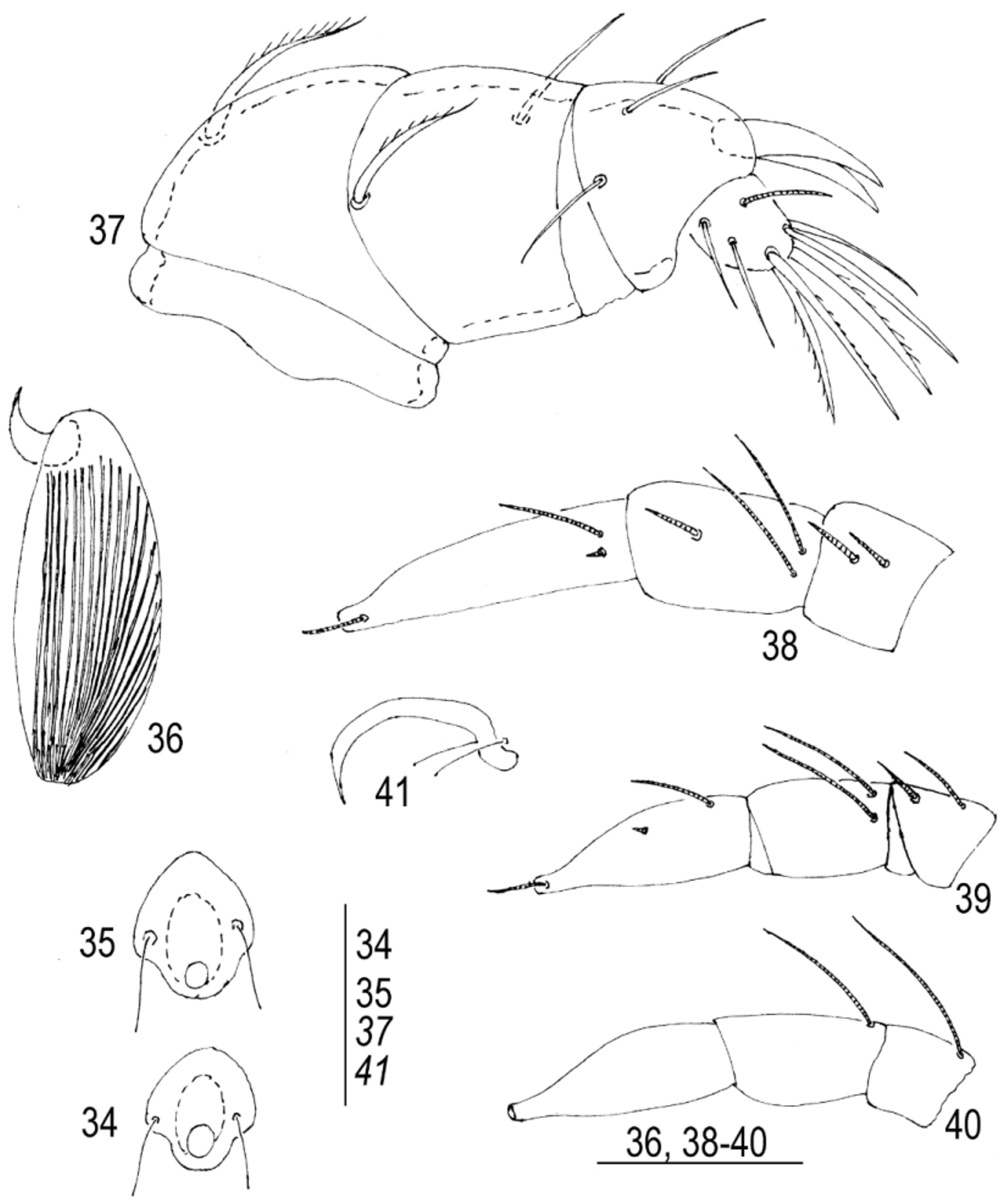
The larva is similar to the larva of H. planus (see description of this species). However, the latter differs from H. clypeatus in: (1) the posterior margin of the coxal plate II straight ( Fig. 33 View FIGURES 32 – 33. H ), not convex; (2) the excretory pore plate is longer than wide ( Fig. 34 View FIGURES 34 – 41. H –55); (3) P-5 with short solenidion ( Fig. 37 View FIGURES 34 – 41. H ); (4) I-Leg-4 solenidion slightly shorter than eupathidium ( Fig. 38 View FIGURES 34 – 41. H ), I-Leg-6 de as long as ds.

No known copyright restrictions apply. See Agosti, D., Egloff, W., 2009. Taxonomic information exchange and copyright: the Plazi approach. BMC Research Notes 2009, 2:53 for further explanation.
|
Kingdom |
|
|
Phylum |
|
|
Class |
|
|
Order |
|
|
Family |
|
|
Genus |