
Pseudophacopteron fuscivenosum, Malenovský & Burckhardt & Tamesse, 2007
|
publication ID |
https://doi.org/10.1080/00222930701515488 |
|
persistent identifier |
https://treatment.plazi.org/id/039A87A6-FFD7-FF96-FE23-C1EAFC71FD19 |
|
treatment provided by |
Felipe |
|
scientific name |
Pseudophacopteron fuscivenosum |
| status |
sp. nov. |
Pseudophacopteron fuscivenosum View in CoL sp. n.
(Figures 1G, H, 4C, 5F, 6D, 7D, 8D, 9E, 10D, 12A, 13D, 16, 21C, 22A, B)
Description
Adult. Colour: body off-white to ochreous. Vertex and pronotum laterally and on either side of median line with dark brown markings. Mesopraescutum with two oblong diverging dark brown macules in anterior two-thirds. Mesoscutum with four dark bands. Genae, frons, and clypeus uniformly ochreous to light brown. Antenna off-white, segments 4–8 infuscated or dark brown apically, segments 9–10 entirely black, terminal setae white. Legs off-white to ochreous with dark markings on femora and tibiae; metacoxa, metafemur, and basal half of metatibia frequently almost entirely dark brown; metatarsus infuscate to brown. Fore wing membrane clear, transparent, except for dark brown infuscation extending along veins R, basal half of M, entire Cu1b, and basal half of Cu1a, and indistinct infuscations in the cells c+sc and basal half of cu2 ( Figure 6D View Figure 6 ). Veins light, off-white, except for portions covered by the infuscation, C+Sc, apical half of R+M+Cu1, base and two spots on anal vein, which are dark brown. Hind wing clear, transparent, C+Sc fuscous. Abdominal tergites entirely dark brown to black, with orange markings on dorsum of tergites 4–5. Sternites dark brown to black, caudal margins slightly lighter. Male terminalia ochreous, ventral side of subgenital plate and base of proctiger dark brown. Female terminalia dark brown to black.
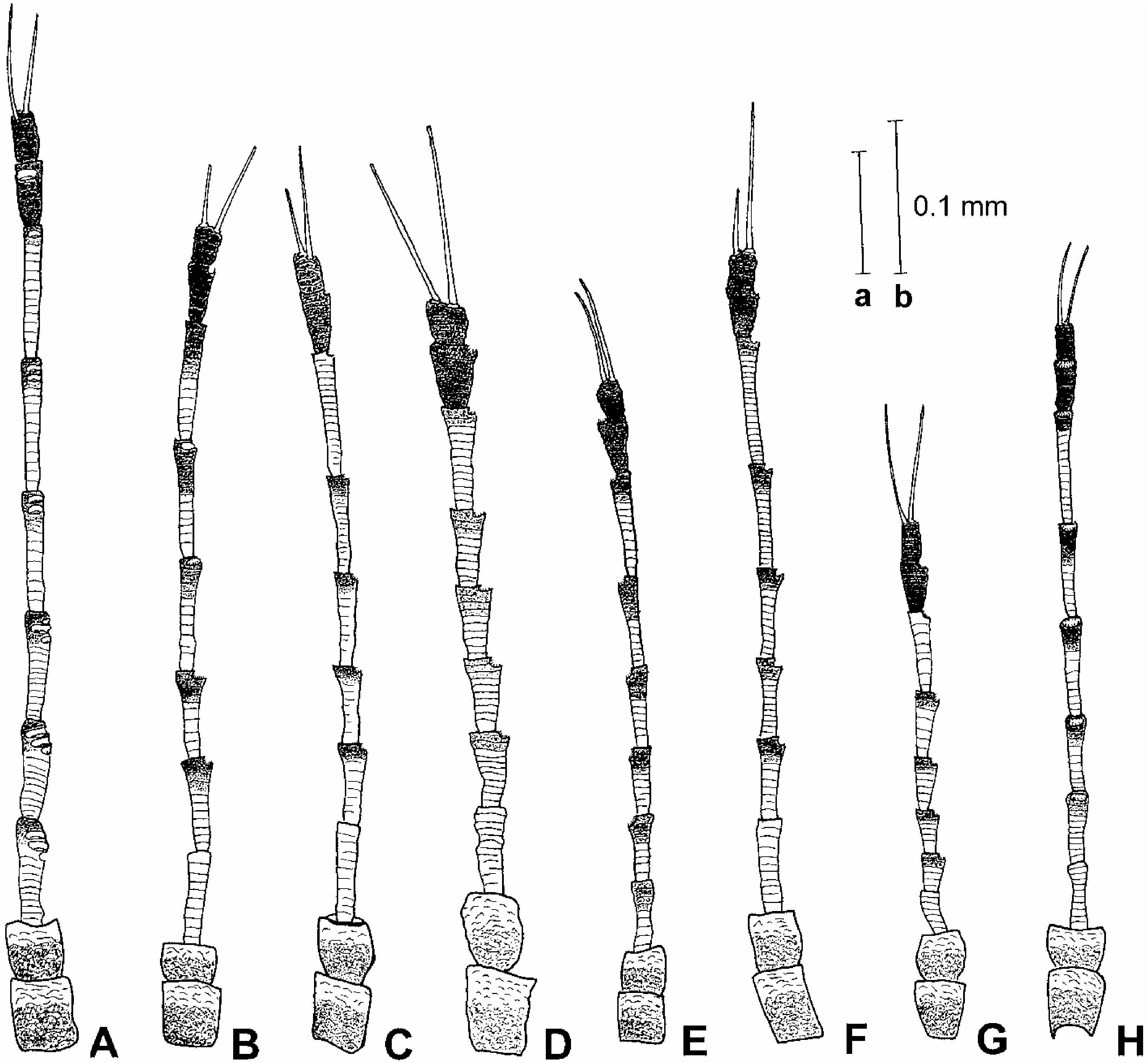
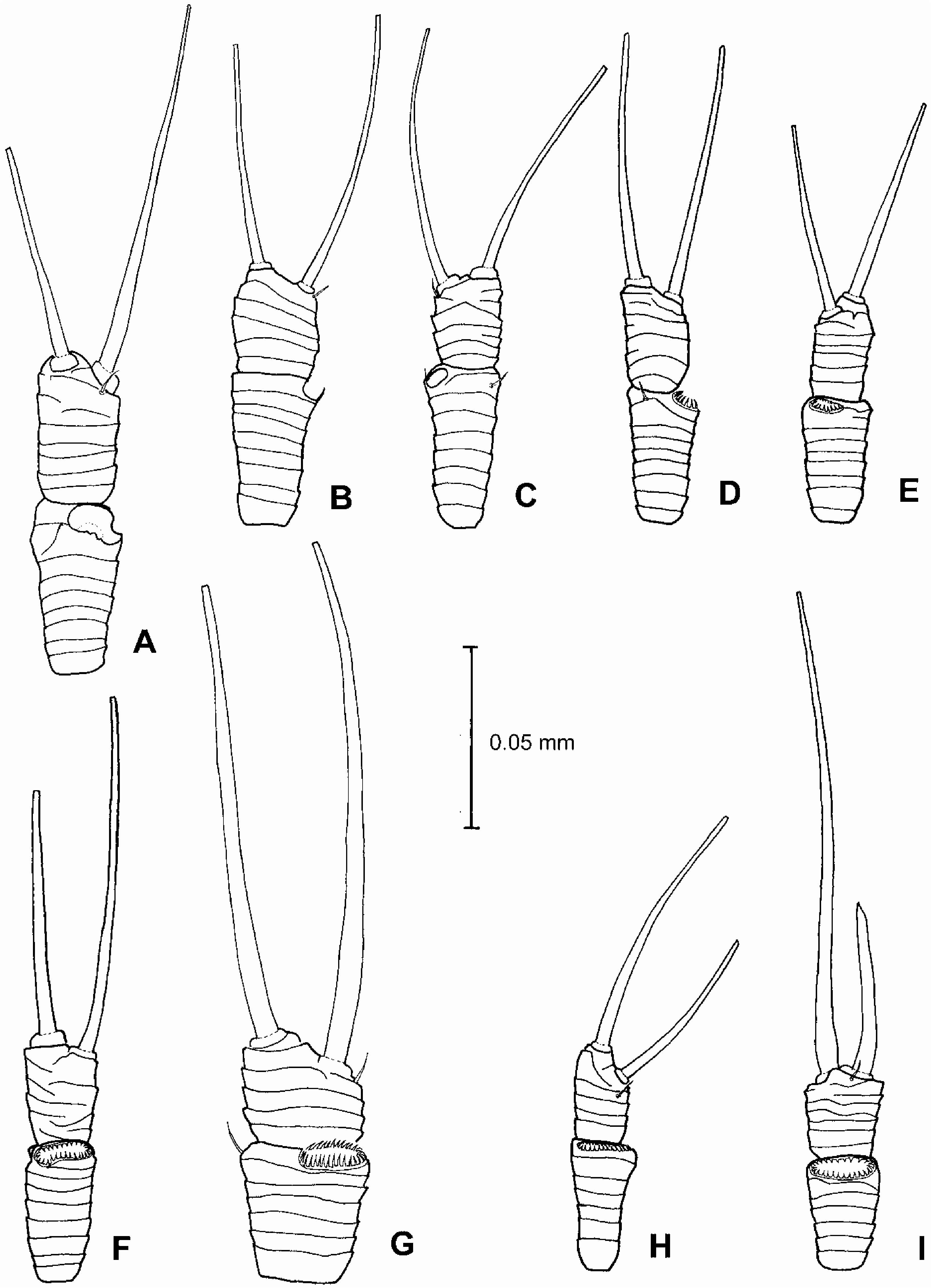
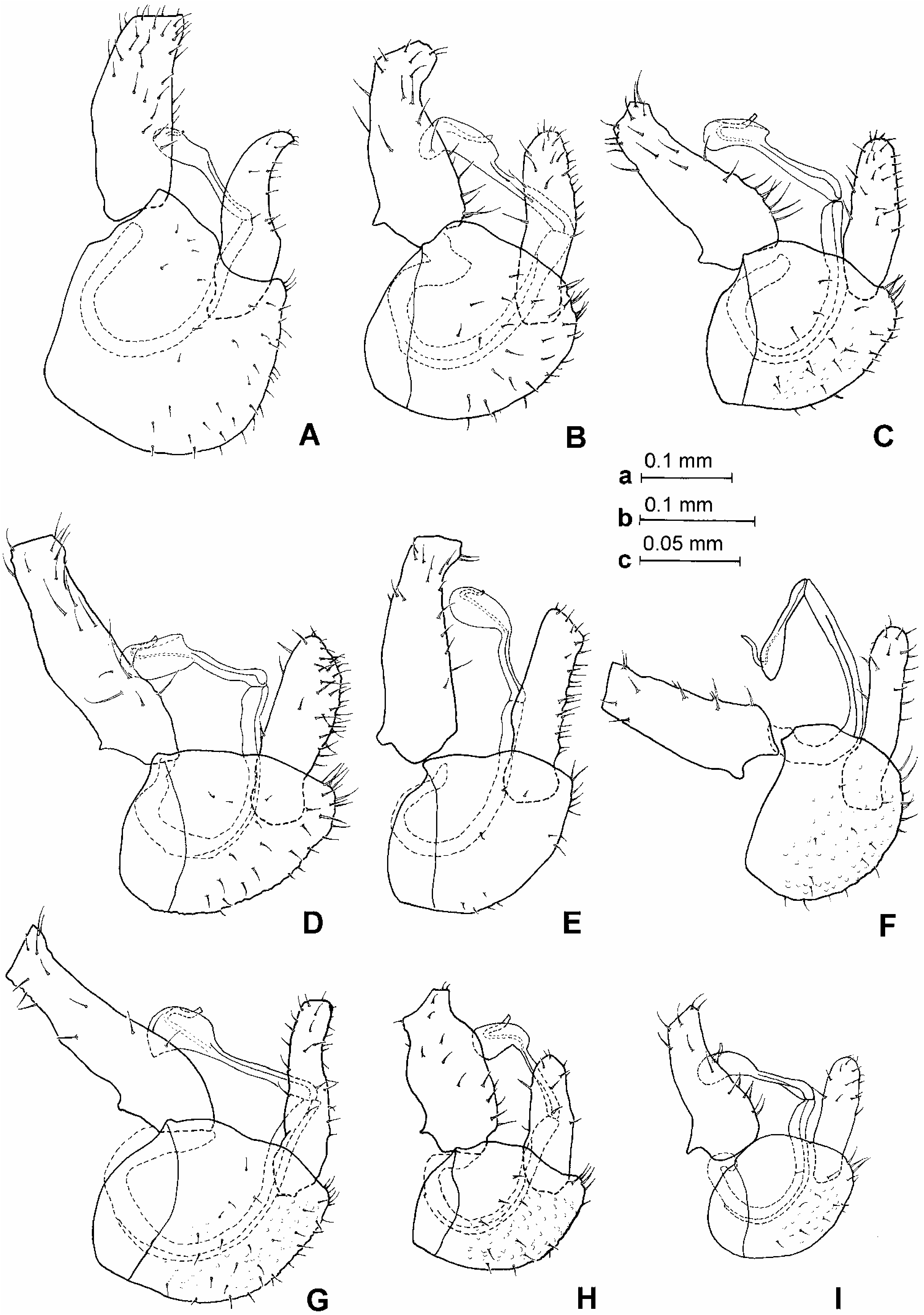
Morphology: head, in dorsal view, slightly wider than mesonotum, subglobular. Vertex with microsculpture, matt, about twice as wide as long along midline, rounded down in front (Figure 1G). Coronal suture reduced throughout. Median ridge on vertex raised, distinct. Vertex on either side of the median ridge convex, distinctly bulging. Lateral ocelli lying on small tubercles slightly above the plane of vertex. Occiput in dorsal view narrowly triangular. Preoccipital sclerite narrow, eyes not stalked, in frontal view subglobular. Genae slightly swollen (Figure 1H). Tubercle below torulus pointed, forming an acute angle. Frons narrow, parallel-sided. Clypeus broadly pyriform. Antenna moderately elongate, slender, segments cylindrical, weakly widening to apex ( Figure 4C View Figure 4 ); a single subapical rhinarium on each of segments 4–9; rhinaria elliptic with a wreath of cuticular spines; terminal setae subequal, the longer seta distinctly longer than segments 9 and 10 together ( Figure 5F View Figure 5 ). Fore wing moderately elongate, almost parallel-sided in apical half, anterior margin in outer half more or less straight; apex unevenly rounded. Vein Rs relatively long. Surface spinules present in all cells except for basal part of r2, densely arranged especially in the apical part, only a few spinules present in cells c+sc and r1 ( Figure 7D View Figure 7 ). Costal break in apical sixth or seventh of C+Sc. Hind legs relatively short and robust. Meracanthus short, acute, pointed. Metafemur constricted medially. Metatibia bearing an open crown of 10–12 unsclerotized apical spurs and additional two rows of five to seven similar spurs laterally. Metabasitarsus bearing two black sclerotized spurs. Dorsal margin of abdomen, in profile, serrate; posterior margin of tergites 3 and especially 4 and 5 medially swollen into a prominent tubercular procession. Male terminalia as in Figure 8D View Figure 8 . Proctiger relatively long, slightly narrowing to apex. Subgenital plate, in profile, longer than high, dorsal margin straight. Paramere, in profile, relatively short, robust, anterior margin straight, posterior margin convex, apex blunt; in posterior view, narrowing to apex; inner surface covered in many more or less regularly arranged setae, subapical setae slightly thicker and longer ( Figure 9E View Figure 9 ). Basal segment of aedeagus stout; apical segment relatively short, with a more or less parallel-sided, apically broadly rounded head, its dorsal margin angular basally; sclerotized end tube of ductus ejaculatorius relatively short, sinuate ( Figure 10D View Figure 10 ). Female terminalia as in Figure 12A View Figure 12 . Proctiger relatively short, with dorsal margin weakly sinuate, almost straight; apical process short; circumanal ring with two rows of pores, pores of outer row contiguous. Subgenital plate, in profile, relatively short, dorsal margin more or less straight in apical half, ventral margin strongly convex, abruptly narrowing, apex forming a small pointed tooth; in ventral view, broadly pointed apically ( Figure 13D View Figure 13 ). Dorsal and ventral valvulae with a few (two or three) distinct lateral teeth at apex. Measurements and ratios in Tables I–III.
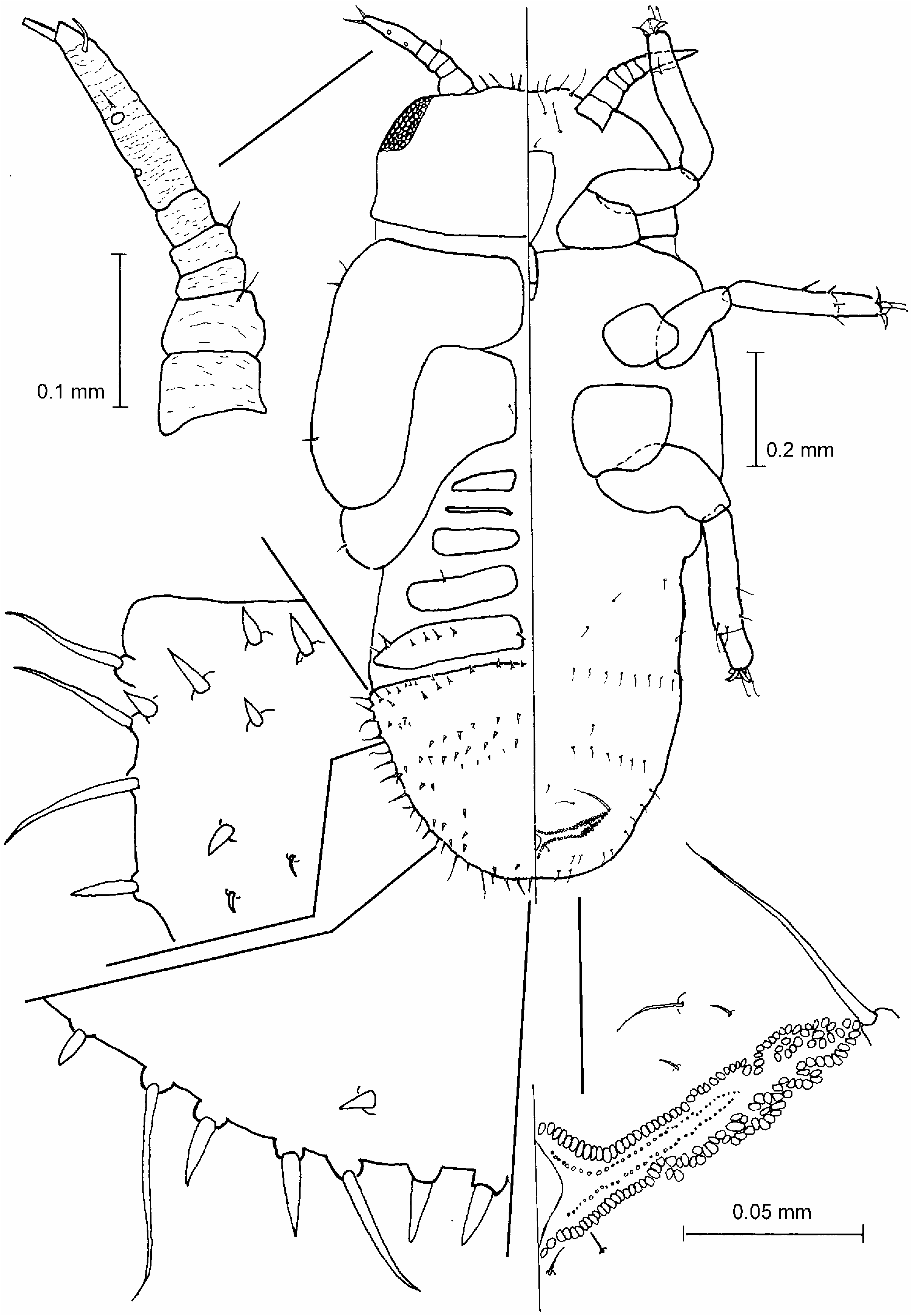
Fifth instar larva ( Figure 16 View Figure 16 ). Uniformly pale yellow. Body narrow, more or less parallelsided. Body margin with simple setae on head in front of antennal insertion, cephalothorax behind eyes, humeral region of fore wing pad, hind wing pad, and abdomen. Many small pointed lanceolate setae present near caudal plate margin and in irregular rows over entire width of disc of caudal plate and preceding free tergal sclerite. A simple seta present in ocular region. Eyes with ca 50 distinct ommatidia. Antenna straight, with six distinct segments; two rhinaria on segment 6. Tarsal arolium large relative to claws, with a broad pad and indistinct petiole ( Figure 21C View Figure 21 ). Abdomen dorsally on each side with five free sclerites and a completely fused caudal plate; apex of caudal plate slightly truncate. Anus in ventral position. Outer circumanal ring wide, anterior and posterior margin close together, composed of a single row of pores, laterally narrowly sinuate. Measurements and ratios in Table IV.
Host plant
Deinbollia sp. (Sapindaceae) .
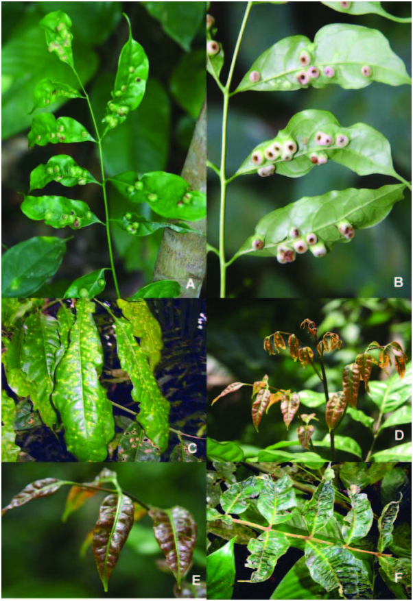
Biology
Inducing nut galls on the leaves ( Figure 22A, B View Figure 22 ). Eggs are laid on the young leaves. After eclosion, the larva establishes itself along a secondary vein on the lower leaf surface and gradually sinks into the leaf with gall tissue growing around it. With the leaf development the size of the gall increases, becoming subglobular and closed, except for a minute aperture on the lower leaf surface. At the adult moult, the gall opens as the apical aperture widens becoming a round hole letting the adult escape. Often several galls are present on a single leaf which becomes severely distorted.
Distribution
Cameroon.
Material examined
Holotype: „, Cameroon: Centre Province, Mbalmayo , 40 km S Yaounde´, 3 ° 309N, 11 ° 309E, 660 m, 15 November 2005, Deinbollia sp. ( D. Burckhardt) . Dry-mounted ( NHMB) . Paratypes: Cameroon: 3 „, 8♀, 25 larvae, same data as holotype; 5 „ , 5♀, six larvae, South Province, Nkouemvone, Ebolowa , 2 ° 559N, 11 ° 099E; 583 m, 22 May 2004, Deinbollia sp. , degraded primary forest ( J. L. Tamesse). Dry- and slide-mounted, and preserved in alcohol ( BMNH, LZUY, MHNG, MMB, NHMB) .
Etymology
From the Latin adjective fuscus 5 dark-coloured and the noun vena 5 the vein, referring to the darkened veins on the fore wing.
Comments
P. fuscivenosum View in CoL sp. n. is close to Pseudophacopteron zimmermanni ( Aulmann, 1912) View in CoL in sharing a similar size, the body colour, a clear fore wing membrane near apices of veins R1, Rs, M1+2, M3+4, and Cu1a, the head shape, the robust paramere with convex posterior margin and the inner face with many stout setae, the apical basally angular dilation of the distal segment of aedeagus, the relatively short female proctiger and subgenital plate, the latter, in profile, with strongly convex ventral margin, abruptly narrowing to apex and forming a small pointed tooth, and the dorsal and ventral valvulae with a few distinct lateral teeth at apex. P. fuscivenosum View in CoL differs from P. zimmermanni View in CoL as follows: fore wing membrane along veins R, basal half of M, entire Cu1b, and basal half of Cu1a infuscated; terminal setae on antenna slightly shorter; paramere longer and narrower, posterior margin of paramere, in profile, more evenly curved; ventral margin of female subgenital plate, in profile, less convex; in the host plant association.
P. zimmermanni is characterized by the following characters: veins R, basal half of M, basal half of Cu1a and base and apex of Cu1b are dark but the infuscation is not extended to the membrane; T 150.11–0.13; PL 50.12–0.14; paramere, in profile, broader and posterior margin more angular; ventral margin of female subgenital plate, in profile, more convex; the host plant is Khaya senegalensis (Meliaceae) . Material of P. zimmermanni examined: syntypes, 3 „, 1♀, Tanzania: Amani, Khaya senegalensis (Marshall) . 102 „, 130♀, Uganda: District Masindi, Budongo Forest North Sonso, 1 ° 459N, 31 ° 359E, 19 June to 30 July 1995 and 15–25 January 1997, canopy fogging ( T. Wagner). Dry- and slide-mounted, and preserved in alcohol ( BMNH, MAKB, MHNG, MMB, NHMB, ZMB).


No known copyright restrictions apply. See Agosti, D., Egloff, W., 2009. Taxonomic information exchange and copyright: the Plazi approach. BMC Research Notes 2009, 2:53 for further explanation.
|
Kingdom |
|
|
Phylum |
|
|
Class |
|
|
Order |
|
|
Family |
|
|
Genus |
Pseudophacopteron fuscivenosum
| Malenovský, Igor, Burckhardt, Daniel & Tamesse, Joseph L. 2007 |
P. fuscivenosum
| Malenovský & Burckhardt & Tamesse 2007 |
P. fuscivenosum
| Malenovský & Burckhardt & Tamesse 2007 |