
Plectranthias takasei, Gill, Anthony C., Tea, Yi-Kai & Senou, Hiroshi, 2016
|
publication ID |
https://doi.org/ 10.11646/zootaxa.4205.4.3 |
|
publication LSID |
lsid:zoobank.org:pub:645E1862-14A5-45A7-B94B-C93353B7081B |
|
DOI |
https://doi.org/10.5281/zenodo.5687661 |
|
persistent identifier |
https://treatment.plazi.org/id/038D87EA-FFC6-FFB7-FF1D-1976FD445B1E |
|
treatment provided by |
Plazi |
|
scientific name |
Plectranthias takasei |
| status |
sp. nov. |
Plectranthias takasei new species
New standard Japanese name: Hinomaru-hanadai English common name: Hinomaru perchlet Figures 1–5 View FIGURE 1 View FIGURE 2 View FIGURE 3 View FIGURE 4 View FIGURE 5
Holotype. KPM-NI 21068 About KPM-NI , 40.0 mm SL, Japan, Honshu , Sagami Bay , E of Izu Peninsula, Izu Oceanic Park, 52 m, collected by W. Takase, 18 March 2008.
Paratype. KPM-NI 21 About KPM-NI 286, 36.0 mm SL, Japan, Honshu , Sagami Bay , E of Izu Peninsula, Izu Oceanic Park, 55 m, donated by K. Endoh, April 2008.
Diagnosis. The following combination of characters distinguishes P. hinomaru from congeners: dorsal rays X,15; no fleshy flaps on dorsal-fin spines; pectoral rays 13, all unbranched; branched caudal-fin rays 8 + 7; lateral line scales 28 (including intermittent and terminal pitted scales); circumpeduncular scales 12; fourth dorsal-fin spine longest; and preopercle without antrorse spines or serrations ventrally, with 2–3 weak serrations or crenulations posteriorly.
Description. (Data given first for holotype, followed where different by data for paratype in parentheses.) Dorsal rays X,15, all segmented rays branched; anal rays III,7, all segmented rays branched; pectoral rays 13/13, all rays unbranched; pelvic fin I, 5, all segmented rays branched; upper procurrent caudal-fin rays 4; lower procurrent caudal-fin rays 4; principal caudal-fin rays 9 + 8; branched caudal fin rays? + 7 (8 + 7); total caudal-fin rays 25; lateral line complete with 28 tubed scales on the left side, and 20 tubed scales followed by a pitted scale and 7 tubed scales on the right side (23 tubed scales followed by a pitted scale, 3 tubed scales then a pitted scale on the left side; scales damaged on right side); scales above lateral line to origin of dorsal fin 2/2; scales above lateral line to base of middle dorsal spine 3/2 (2/?); scales below lateral line to origin of anal fin 9/9 (9/?); diagonal rows of scales on cheek 5; predorsal scales 18 (17), extending to just short of posterior nostrils; circumpeduncular scales 12; gill rakers 4 + 12, the upper 3 and lower 5 rudiments (4 + 11, the upper 3 and lower 5 rudiments); pseudobranchial filaments 11 (10); branchiostegal rays 7.
Vertebrae 9 + 17; supraneurals 3, the third reduced in size; predorsal formula 0/0+0/2/1+1; no trisegmental pterygiophores associated with dorsal and anal fins; ribs present on vertebrae 3 through 9; epineurals present on vertebrae 1 through 13 (1 through 12); parhypural and hypurals autogenous; well-developed hypurapophysis on parhypural; epurals 3; single uroneural (posterior uroneural absent); ventral tip of cleithrum with well-developed posteroventral process ( Figure 5 View FIGURE 5 ).
Dorsal-fin spines without fleshy tabs on their distal tips; fourth dorsal spine longest; dorsal fin deeply incised before first segmented-fin ray; second anal-fin spine longest and stoutest; anal fin rounded with third segmented ray longest; caudal fin truncate to slightly rounded, with some ray branches slightly elongated past fin margin; lower 7 pectoral fin rays somewhat thickened, with membranes between thickened rays deeply incised; eighth (counting from dorsal-most) pectoral ray longest, reaching vertical through posterior edge of anal-fin base; pelvic fins short, not reaching anus, second segmented ray longest.
Morphometric values are summarised in Table 1 View TABLE 1 .
Mouth large, slightly oblique, posterior margin of maxilla reaching almost to vertical through posterior edge of eye; maxilla expanded posteriorly, with long, low, lateral ridge running parallel to dorsal margin; mouth terminal; upper jaw with several fixed, short stout outer canines on either side of symphysis flanked internally by villiform band with about 5–8 rows of depressible, smaller, sharp-tipped teeth, with inner rows becoming progressively longer, band reducing to 3 rows posteriorly; lower jaw with villiform band of about 4–5 rows of small depressible teeth at symphysis, teeth becoming progressively longer on inner rows, an enlarged, curved canine on middle of jaw, band narrows to single row posteriorly; vomer with roughly V-shaped band of 2–4 rows of sharp-tipped conical teeth; palatine with a band of 2–3 rows of small, sharp-tipped conical teeth; ectopterygoid and mesopterygoid edentate; tongue narrow, pointed and edentate.
Opercle with 3 flat spines, middle spine longest, upper spine concealed by scales; preopercle with 2 (3) weak serrations or crenulations on lower part of posterior margin, ventral margin smooth; interopercle and subopercle smooth. Anterior nostril positioned at middle of snout, tubular with small flap on posterior rim; posterior nostril at anterior border of orbit, with slightly raised rim but no flap.
Scales ctenoid with peripheral cteni ( Roberts 1993); lateral line broadly arched over pectoral fin following body contour to caudal-fin base; no scales on chin, branchiostegal membranes, maxilla or snout; no auxiliary scales on head or body; dorsal fin with intermittent row of scales along base of fin; anal fin with low scaly sheath basally, with some small scales extending on to fin membranes; caudal fin with scaly basal sheath, with small scales extending on to basal third to half of fin membranes; pectoral fins with basal sheath and small scales extending on to fin membranes.
Colour in life (based on colour photos of the holotype and paratype when freshly dead, and underwater photos of individuals at Izu Oceanic Park; Figures 1–4 View FIGURE 1 View FIGURE 2 View FIGURE 3 View FIGURE 4 ): Head and body pale pink to white; nape, top of head and front of lips orange to reddish brown; maxilla and lips sometimes bright yellow; iris pale pink with two orange oblique stripes (one above and one below pupil), bluish grey spots anteriorly and posteriorly, and sometimes small yellow spots ventrally; large (up to twice orbit diameter) greyish red to bright red spot on upper sides below middle of dorsal fin; pink to orange-red oblique bar extending below large red spot to small (pupil-sized) greyish red to red spot near anal-fin origin; small (pupil- to orbit-sized) greyish red to red spot below lateral line and vertical through middle of soft dorsal, connected to smaller reddish grey to red spot at termination of anal fin by pink to orange-red oblique bar; pink to orange-red bar from termination of dorsal fin to small reddish grey to red spot ventrally at middle of caudal peduncle; a dorsal and a ventral reddish grey to red spot at posterior end of caudal peduncle, these connected by pink to orange red vertical bar; reddish grey to red and pink markings on head and body overlain with a series of oblique to horizontal stripes composed of closely spaced, orange-edged, bright yellow to gold spots (one per scale), stripes usually broken into segments and occurring only when passing directly over reddish grey to red or pink markings; first stripe short and indistinct, from orbital rim at about 1 o’clock position to middle of nape; second stripe from above tip of preopercle to lateral-line origin, tracking lateral line to just behind vertical through dorsal-fin origin, curving ventrally along lower edge of large red spot, and ending at middle of caudal-fin base; third stripe extending obliquely from behind and below lateral-line origin to just above anal-fin origin, then horizontally to ventral edge of caudal-fin base; fourth stripe from about 3 o’clock position on orbital rim, above pectoral-fin base, to lower part of abdomen (just above and in front of anus); fifth stripe from about 5 o’clock position on orbital rim to pectoral-fin base; sixth stripe very short (2–3 scales long) and horizontal, near anterior tip of preopercle; additional yellow or gold spots sometimes present beneath dorsal-fin base and dorsal edge of caudal peduncle; silvery white markings sometimes present on upper snout, on nape, below dorsal-fin origin, below end of spinous part of dorsal fin, at termination of dorsal fin, on upper part of caudal-fin base, on operculum between fourth and fifth gold stripes, and along ventral edge of fifth gold stripe; dorsal fin pinkish or reddish to yellowish hyaline, sometimes with silvery white markings from body encroaching on to fin base; anal fin pinkish to yellowish hyaline, pale pink anteriorly, with reddish grey to red markings at origin and termination of fin encroaching on to fin base; caudal fin pinkish to reddish hyaline; pectoral fins pinkish hyaline; pelvic fins pinkish hyaline, pale blue to pale pink anteriorly.
Colour in preservative: Pale tan, dusky grey-brown on upper part of head and nape, with intermittent dusky grey-brown markings along dorsal part of body and caudal peduncle; large greyish red to red spot on midside and smaller greyish red to red spots (at anal-fin origin, below middle of soft dorsal, at termination of dorsal fin, at termination of anal fin, and dorsally and ventrally at end of caudal peduncle) become dusky grey-brown; all other markings obsolete.
Habitat and distribution. Plectranthias takasei is known only from Izu Ocean Park, where it has been collected and photographed on rocky reefs at depths of 48–60 metres.
Comparisons. Plectranthias takasei is distinct from most members of the genus in having in combination: a complete lateral line; no antrorse spines ventrally on the preopercle; 15 segmented dorsal-fin rays; and pectoral fins with 13 unbranched rays. Five congeners have a similar combination of characters: P. maugei Randall (1980) , P. morgansi ( Smith 1961) , P. cirrhitoides Randall (1980) , P. foresti Fourmanoir (1977) and P. megalepis ( Günther 1880) . Plectranthias maugei , known only from three type specimens from off Madagascar, differs in having a lunate caudal fin with 14 branched rays (versus weakly rounded to truncate with 15 branched rays in P. takasei ) and short fleshy flaps (cirri) on the tips of the dorsal-fin spines. Plectranthias morgansi , from the Western Indian Ocean, has the third (versus fourth) dorsal spine longest, bearing a long fleshy tab (versus no tab), 14 (versus 12) circumpeduncular scales, fine serrations on the preopercle (versus 2–3 weak serrations or crenulations on the posterior margin of the preopercle) and a distinctly serrated (versus smooth) interopercle. Plectranthias cirrhitoides , from Rapa, differs in having 14 circumpeduncular scales, the first segmented dorsal-fin ray unbranched (versus branched) and a shallower body (greatest body depth 31.0–32.4 versus 35.0–38.3 % SL). Plectranthias foresti , from the West Pacific, differs in having an emarginate caudal fin with 14 branched caudal-fin rays (versus weakly rounded to truncate with 15 branched rays), 18–29 serrae on the hind margin of the preopercle, and the second to fourth segmented dorsal-fin rays filamentous (versus not filamentous). Plectranthias megalepis , known only from the lectotype from the Kai Islands, Indonesia, differs in having 14 circumpeduncular scales, 18– 21 serrae on the posterior margin of the preopercle, and the top of the head scaled anteriorly only to about the mid interorbital area (versus almost to posterior nostrils).
The live coloration of P. takasei is distinctive, and will serve to distinguish the species from the above species and all other congeners. Plectranthias takasei may be confused with several species that have yellow to red spots on a pale body — such as P. sagamiensis ( Katayama 1964) , P. elongatus Wu et al. (2011) , P. xanthomaculatus Wu et al. (2011) , P. inermis Randall (1980) and P. flammeus Williams et al. (2013) — but details of the colour pattern are distinctive for P. takasei . In particular, the combination of a large red spot on the mid-side and oblique stripes of closely spaced, orange-edged, bright yellow to gold spots are diagnostic.
Etymology. The species is named for Mr Wataru Takase, who collected the holotype. The Japanese standard name and English common names refer to the prominent red spot on the side, which is reminiscent of the red disk (rising sun) in the Japanese flag (commonly called Hinomaru , ‘the sun disk’).
Remarks. Placement of the new species in Plectranthias should be regarded as provisional. The generic classification of the Anthiadinae is unsatisfactory, with few genera cladistically diagnosed by synapomorphies. The two largest genera, Plectranthias and the Indo-Pacific genus Pseudanthias Bleeker (1871) are particularly problematic. Not only is neither genus defined on the basis of synapomorphies, but both show considerable variation in characters that have been used to diagnose other anthiadine genera. For example, Plectranthias varies in the number of supraneural bones, degree of squamation on the head, presence or absence of antrorse spines on the ventral edge of the preopercle, number of lateral-line scales, and number of branched caudal-fin rays. It is likely that further studies will result in the reassignment of some species to new genera or to nominal genera placed in the synonymy of Plectranthias by Randall (1980).
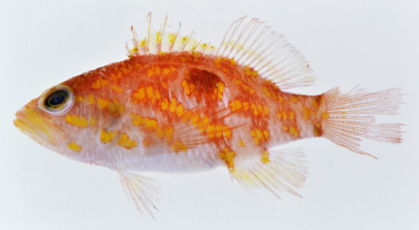


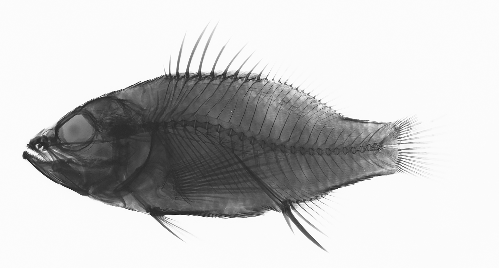
TABLE 1. Morphometric values for Plectranthias takasei, expressed as percentage SL.
| SL (mm) | KPM-NI 21068 (holotype) 40.0 | KPM-NI 21286 (paratype) 36.0 |
|---|---|---|
| Greatest body depth Body depth at anal-fin origin Body width | 38.3 35.5 21.0 | 35.0 32.5 22.2 |
| Head length Snout length Orbit diameter | 42.5 9.3 10.8 | 42.8 8.9 10.8 |
| Bony interorbital width Upper jaw length Maxilla width | 4.0 18.0 5.5 | 3.9 19.2 6.4 |
| Caudal peduncle length Caudal peduncle depth Predorsal length | 20.5 14.8 40.0 | 19.7 15.0 40.8 |
| Preanal length Prepelvic length Dorsal fin base length | 68.0 35.3 48.8 | 66.9 36.7 51.4 |
| First dorsal spine Longest dorsal spine (number) First segmented dorsal ray Longest segmented dorsal ray (number) Anal fin base length | 4.0 17.0 (4th) 14.0 17.8 (7th) 15.0 | 4.2 15.3 (4th) 11.7 18.3 (5th) 15.0 |
| First anal spine Second anal spine Third anal spine First segmented anal ray Longest segmented anal ray (number) Caudal fin length | 8.0 17.3 14.3 19.5 21.8 (3rd) broken | 8.6 16.9 14.2 broken 21.4 (3rd) 26.7 |
| Pectoral fin length Pelvic fin spine | 39.0 14.3 | 38.6 15.0 |
| Pelvic fin length | 24.0 | 25.0 |
| KPM-NI |
Kanagawa Prefectural Museum of Natural History |
No known copyright restrictions apply. See Agosti, D., Egloff, W., 2009. Taxonomic information exchange and copyright: the Plazi approach. BMC Research Notes 2009, 2:53 for further explanation.
|
Kingdom |
|
|
Phylum |
|
|
Class |
|
|
Order |
|
|
Family |
|
|
Genus |