
Macgregoromyia
|
publication ID |
https://doi.org/10.5281/zenodo.206902 |
|
DOI |
https://doi.org/10.5281/zenodo.6188595 |
|
persistent identifier |
https://treatment.plazi.org/id/038C87C0-F67C-D21E-FF76-05E73BC8F95E |
|
treatment provided by |
Plazi |
|
scientific name |
Macgregoromyia |
| status |
|
Key to world species of genus Macgregoromyia View in CoL
Except for the new species, the key was compiled based on the literature as indicated. For brevicula , celestia , shikokuana and szechwanensis include reference to the very detailed figures in Savchenko 1983 (wing and male genitalia). For fohkienensis includes a female specimen from Chinese province Zhejiang.
1. Distal cells of wing with macrotrichia..................................................................... 2
- Distal cells of wing bare................................................................................ 4
2. Wing with r-m strongly arcuated, entering R4+5, R4+5 basally bent basad ( Alexander, 1955, p 367, fig. 2)................................................................................................. M. syusiro Alexander, 1955 View in CoL
- Wing with r-m slightly arcuated, entering Rs, R4+5 straight basally............................................... 3
3. Wing with R1+2 absent; posterior margin of tergite 9 with a V-shaped incision; apex of lobe of gonostylus narrowed; dorsal crest of clasper of gonostylus greatly convex ( Alexander 1932, Pl. I, fig. 3, Pl. II, fig. 32)..... M. szechwanensis Alexander, 1932 View in CoL
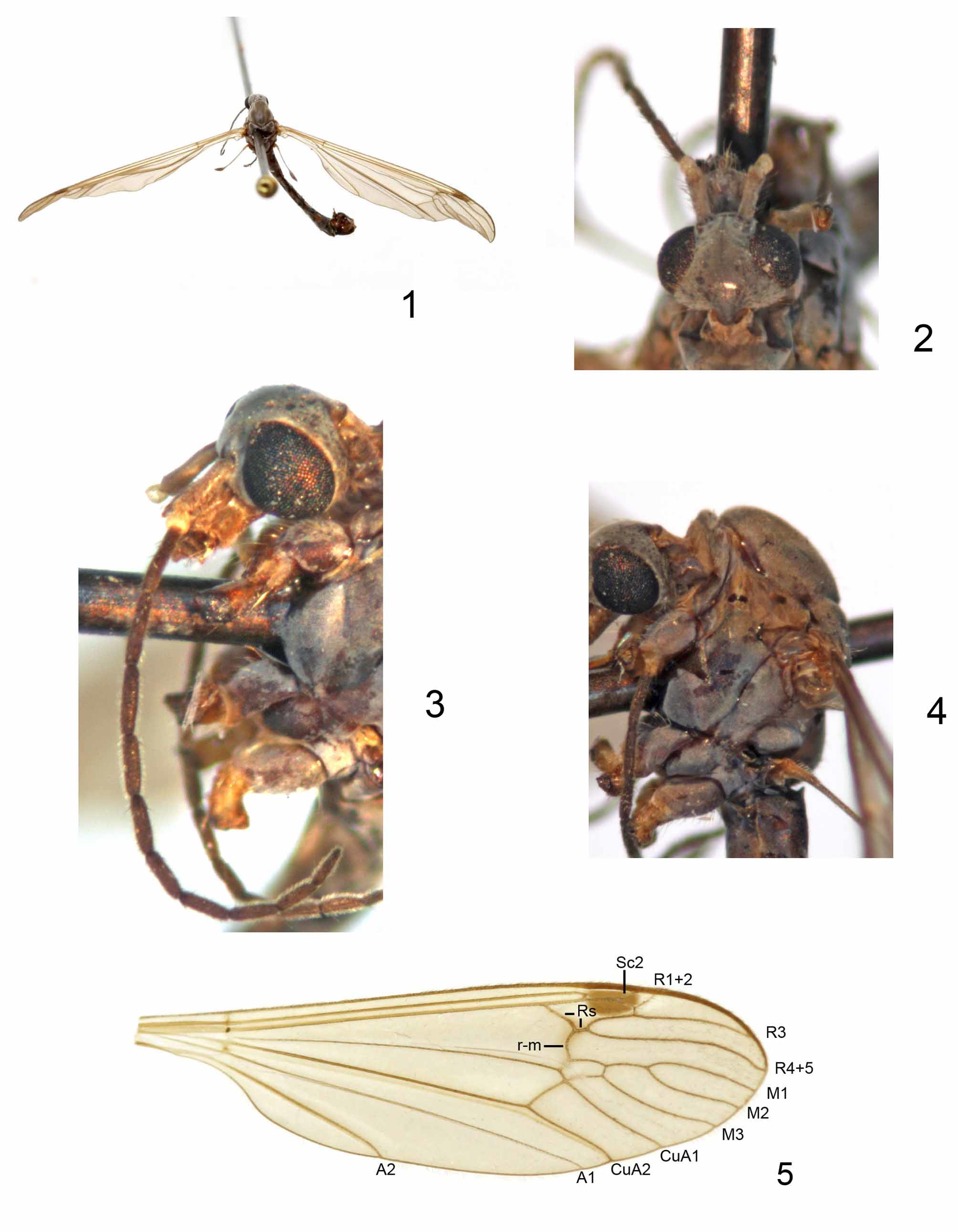
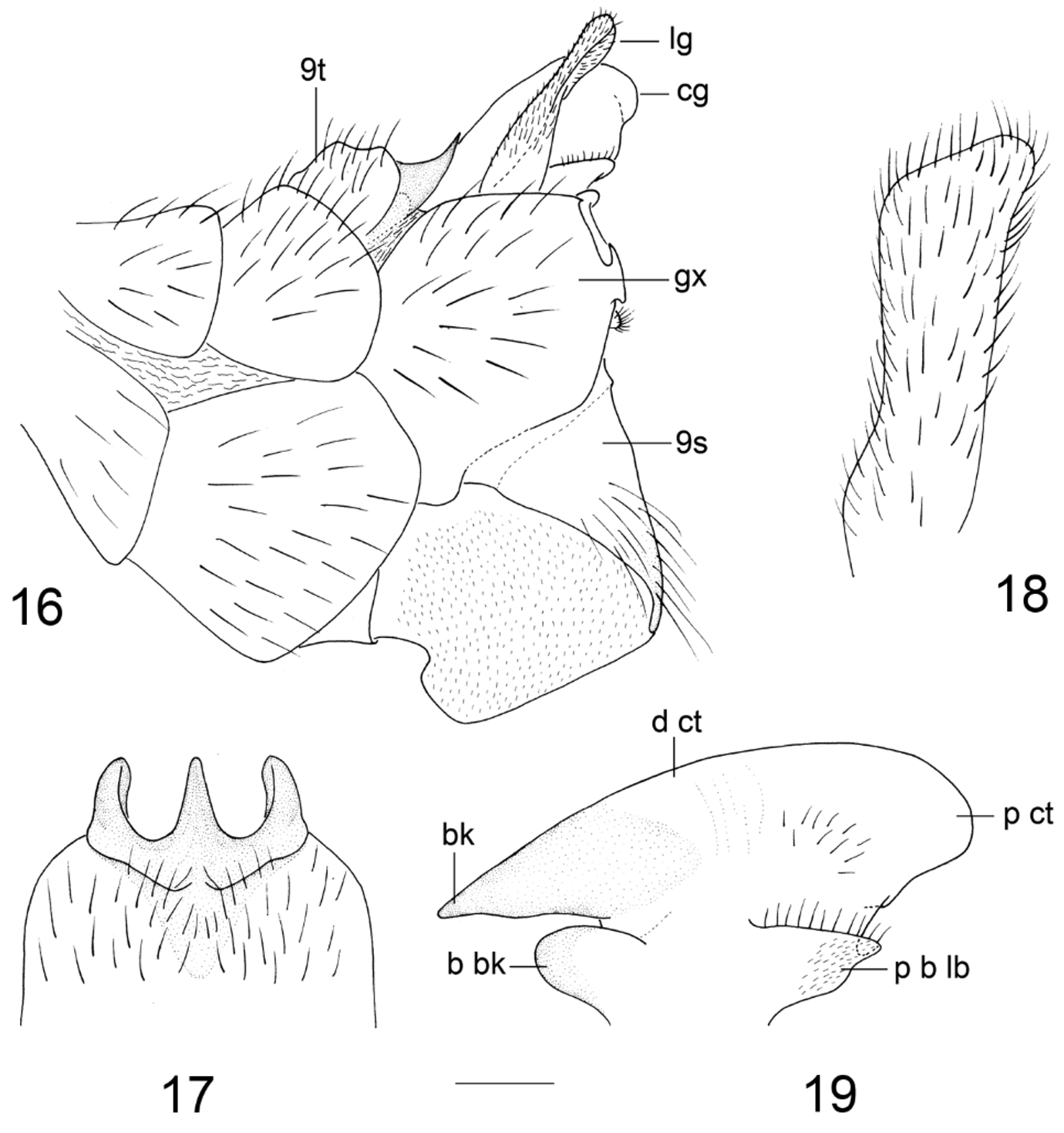
- Wing with R1+2 entire ( Fig. 5 View FIGURES 1 – 5 ); tergite 9 ( Fig. 17 View FIGURES 16 – 19 ) with posterior margin bearing three processes; lobe of gonostylus ( Fig. 18 View FIGURES 16 – 19 ) with tip broad; clasper of gonostylus ( Fig. 19 View FIGURES 16 – 19 ) with dorsal crest smooth........................ M. ternifoliusa View in CoL sp. nov.
4. Wing with r-m entering Rs at fork of Rs.................................................................... 5
- Wing with r-m either entering Rs before fork of Rs or entering R4+5 beyond fork of Rs............................... 6
5. Femora brown with tips blackened; posterior margin of tergite 9 narrowly emarginated; basal beak of clasper of gonostylus short and obtuse, dorsal crest of clasper of gonostylus with a spine-like process, posterior crest laterally with a collar-like flange ( Alexander 1969, p 34, figs. 1, 2)............................................. M. babana Alexander, 1969 View in CoL
- Femora dark brown with bases broadly more yellowed; median lobe of tergite 9 very reduced; basal beak of clasper of gonos- tylus long and pointed, dorsal crest of clasper of gonostylus smooth, posterior crest small, without such flange ( Alexander 1954, Pl. I, figs. 1, 8).......................................................... M. shikokuana Alexander, 1954 View in CoL
6. Wing with r-m entering R4+5 beyond fork of Rs.............................................................. 7
- Wing with r-m entering Rs before fork of Rs................................................................ 8
7. Tergite 9 medially notched; posterior crest of clasper of gonostylus produced into an obtuse lobe ( Alexander 1933, Pl. II, fig. 26).......................................................................... M. brevicula Alexander, 1933 View in CoL
- Tergite 9 medially produced into a semicircular blade; posterior crest of clasper of gonostylus convex with apex produced backwards ( Alexander 1955, p 365)..................................................... M. itoi Alexander, 1955 View in CoL
8. R1+2 entire............................................................................................ 9
- R1+2 spur-like or absent................................................................................ 12
9. Rs original long before line of r-m ( Alexander 1929, Pl. I, fig. 3)..................... M. benguetensis Alexander, 1929 View in CoL
- Rs original nearly in line with r-m....................................................................... 10
10. CuA1 meeting M3 near middle of cell dm ( Edwards 1932, p 54)...................... M. perpendicularis Edwards, 1932 View in CoL
- CuA1 meeting M3 near fork of M........................................................................ 11
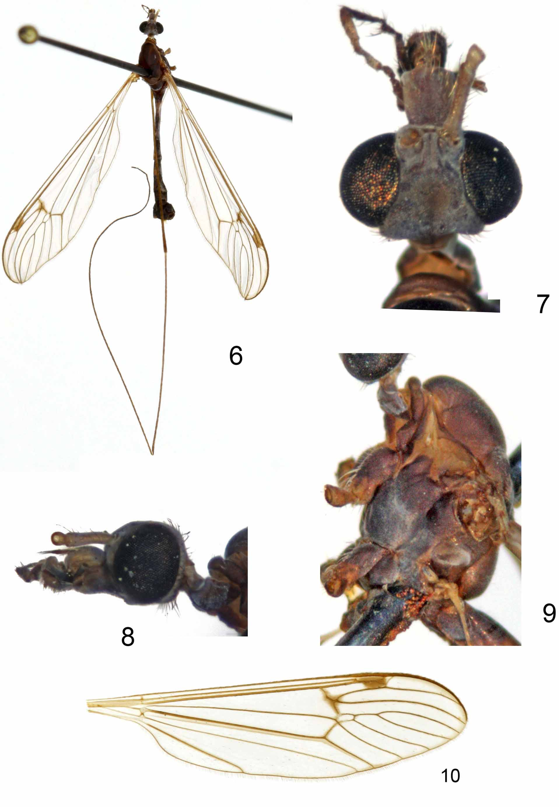
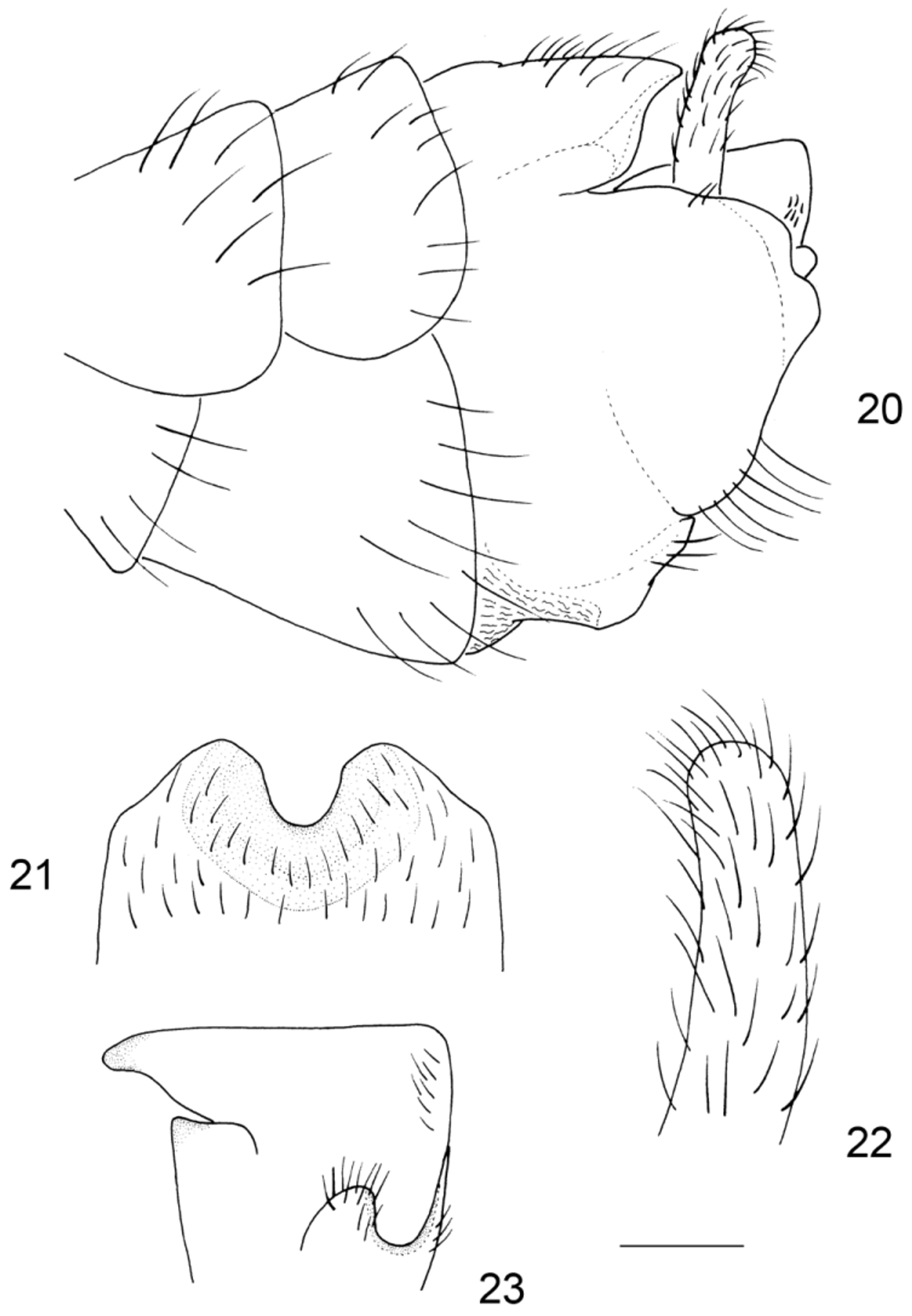
11. CuA1 meeting M3 at a point ( Fig. 10 View FIGURES 6 – 10 ); tergite 9 with posterior margin bearing a shallow U-shaped incision ( Fig. 21 View FIGURES 20 – 23 ); dorsal crest of clasper of gonostylus flat ( Fig. 23 View FIGURES 20 – 23 )................................................... M. flatusa View in CoL sp. nov.
- CuA1 fused with M3 for a short distance; tergite 9 with posterior margin bearing a deep U-shaped notch; dorsal crest of clasper of gonostylus convex ( Alexander 1931, Pl. I, fig. 5, Pl. III, figs. 44–46)................. M. brevisector Alexander, 1931 View in CoL
12. First flagellomere black or brown with base yellow.......................................................... 13
- First flagellomere entirely yellow....................................................................... 14
13. Rs arcuated before joint with r-m; R1+2 spur-like, apex touching Sc2 and closing cell sc2 ( Fig. 15)................................................................................................... M. fohkienensis Alexander, 1949 View in CoL
- Rs straight before joint with r-m; R1+2 represented by a basal spur, paralleled with Sc2 ( Fig. 14 View FIGURES 11 – 14 ).... M. rectangularis View in CoL sp. nov.
14. r-m meeting Rs near fork of Rs; beak of clasper of gonostylus produced ( Alexander 1962, Pl. I, fig. 1, Pl. III, fig. 39).................................................................................. M. sikkimensis Alexander, 1962 View in CoL
- r-m meeting Rs nearly at middle of Rs; beak of clasper of gonostylus small and bent downwards ( Alexander 1932, Pl. I, fig. 4, Pl. II, fig. 34).................................................................. M. celestia Alexander, 1932 View in CoL

No known copyright restrictions apply. See Agosti, D., Egloff, W., 2009. Taxonomic information exchange and copyright: the Plazi approach. BMC Research Notes 2009, 2:53 for further explanation.
|
Kingdom |
|
|
Phylum |
|
|
Class |
|
|
Order |
|
|
Family |
|
|
Genus |