
Sparassocynus maimarai, Abello, María Alejandra, Reyes, Martín De Los, Candela, Adriana Magdalena, Pujos, Francois, Voglino, Damián & Quispe, Bernardino Mamani, 2015
|
publication ID |
https://doi.org/10.11646/zootaxa.3937.1.7 |
|
publication LSID |
lsid:zoobank.org:pub:4147764B-7FA0-475A-909C-E9FCA58E131C |
|
DOI |
https://doi.org/10.5281/zenodo.5613777 |
|
persistent identifier |
https://treatment.plazi.org/id/038987DA-5360-FFCE-FF5A-F951FA88F977 |
|
treatment provided by |
Plazi |
|
scientific name |
Sparassocynus maimarai |
| status |
sp. nov. |
Sparassocynus maimarai new species
Fig. 5 View FIGURE 5 , Tables 1–2 View TABLE 1 View TABLE 2
Etymology. “ maimarai ” in reference to the locality of Maimará where the new species was discovered. Holotype. JUY-P-48, left mandibular fragment with a complete p2–m4 series.
Locality and Horizon. The type specimen was collected in the Maimará Formation (late Miocene-early Pliocene), near the Locality of Maimará, Jujuy Province, Argentina ( Fig. 1 View FIGURE 1 C).
Diagnosis. Sparassocynus maimarai differs from S. bahiai and S. derivatus by its smaller size, the relatively longer m1 with respect to the m4, its more robust and conical entoconids, and the presence of a lingual cingulum extended between para- and metaconid on the m1–3.
Comparative description. Considering the mean of m1–3 length as a proxy of species size, S. maimarai is 9% smaller than S. bahiai and S. derivatus ( Tables 2 View TABLE 2 and 3 View TABLE 3 ). The horizontal ramus of the dentary is short and deep as in S. bahiai and S. derivatus , and about a 40% deeper than Hesperocynus ( Tables 2 View TABLE 2 and 3 View TABLE 3 ). The mandibular symphysis is long, ending posteriorly behind to the p3–m1 boundary ( Fig. 5 View FIGURE 5 A–C). The ventral border of the horizontal ramus between p2 and m4 is not preserved ( Fig. 5 View FIGURE 5 A–B) and the possible presence of a well-developed notch mark for the geniohyoid muscle, a diagnostic character of sparassocynids ( Goin, 1991), could not be evaluated. On the labial side of the dentary two mental foramina are present, one located below the p2 and the other below the posterior root of m1. The anterior end of the mandible is broken in front of the p2 ( Fig. 5 View FIGURE 5 A–B). Behind the root of the canine is the posterior alveolus of p1, which was oblique to the dentary row ( Fig. 5 View FIGURE 5 C). The p2 is labiolingualy compressed, and has a small posterior cusp; the p3 is narrow posteriorly with a small lingual talon (Fig. C, E). Premolars are slightly imbricated, without diastema between them such as some specimens of S. derivatus (e.g., MMP 1088).
Molars increase in length, and have a proportionally longer and wider trigonids, from m1 to m4. First lower molar is relatively longer with respect to the m4 than it is in S. bahiai and S. derivatus ( Tables 2 View TABLE 2 and 3 View TABLE 3 ). Talonids of S. maimarai are shorter and narrower than the trigonids as occur in the remaining Sparassocynus species ( Fig. 5 View FIGURE 5 C; Tables 2 View TABLE 2 and 3 View TABLE 3 ). The talonid of m4 is, in the contact with the trigonid, as wide as that of S. derivatus and S. bahiai ( Figs. 5 View FIGURE 5 C and 3A–B), and wider than in H. dolgopolae ( Fig 3 View FIGURE 3 D; Tables 2 View TABLE 2 and 3 View TABLE 3 ). Similarly to the remaining Sparassocynidae , trigonids are higher than talonids ( Reig & Simpson, 1972; Goin, 1991). The paraconid is lower than proto- and metaconid; metaconid is reduced compared to the protoconid and partially fused to this cusp ( Fig. 5 View FIGURE 5 D–E, G). The paraconid is slightly larger than that of S. bahiai and S. derivatus . Lingually, a basal cingulum is located between para- and metaconid ( Fig. 5 View FIGURE 5 D, F–G). Anterobasal cinguli are narrow and become anteriorly wider along the paraconid where they form deep hypoconulid notchs. The hypoconids are lingual to the protoconids and do not project labially ( Fig. 5 View FIGURE 5 C, F). The entoconids are robust and rounded in section (i.e., are not labiolingually compressed; Fig. 5 View FIGURE 5 C, F) compared to that of other Sparassocynus species. The hypoconulids are well developed, especially on m2–3, and locked in the deep hypoconulid notchs of the respective posterior molars.
‘ Sparassocynus ’ heterotopicus MNHN-BOL- 011896 0.9
continued.
Taxon Lm3 Wtrigm3 Ltrigm3 Wtalm3 Ltalm3 Lm4
Sparassocynus maimarai 3.4 2.2 2.4 2 1 3.5 Sparassocynus derivatus 3.7 2.3 2.7 2.2 1 4.2 Sparassocynus derivatus 3.8 2.6 2.8 2.4 1 4.3 Sparassocynus derivatus 3.4 2.2 2.5 2.2 0.9 4 Sparassocynus derivatus 3.5 2.4 2.4 2.3 1.1 3.9 Sparassocynus derivatus 3.7 2.6 2.7 2.3 1 4.4 Hesperocynus dolgopolae 3.4 2.2 2.3 2.1 1.1
Hesperocynus dolgopolae 3.6 2.25 2.4 2 1.2 3.8 Sparassocynus bahiai 4 2.5 2.9 2.3 1.1 4.4 Sparassocynus bahiai 3.7 2.5 2.8 2.35 0.9 4.2 Sparassocynus bahiai 4 2.3 2.9 2.1 1.1 4.4 ‘ Sparassocynus ’ heterotopicus 3.6 2 2.5 17.5 1.1 3.6 LM2 WM2 LM3 WM3 LM4 AM4 ‘ Sparassocynus ’ heterotopicus 3.25 2.8 3.5 3.2 3.7 3.4 continued.
Taxon Lm1/Ltal Lm2/Ltal Lm3/Ltal Wtrigm4/wtal The assignation of the specimen JUY-P-48 to the Sparassocynidae is based on the presence of the following diagnostic characters of the family (according to Reig & Simpson, 1972; Goin, 1991): (1) high dentary, (2) long symphysis, (3) premolars without diastema, (4) lower molars with high trigonids and low talonids, (5) short and narrow talonids, and (6) metaconid small and partly fused with protoconid.
In the context of the Sparassocynidae specimen JUY-P-48 shared several characters with Sparassocynus species. Like them it has a deeper dentary, talonids of m1–3 relatively shorter, and talonid of m4 proportionally wider than that of Hesperocynus dolgopolae ( Figs. 3 View FIGURE 3 and 5 View FIGURE 5 A; Tables 2 View TABLE 2 and 3 View TABLE 3 ). Forasiepi et al. (2009) have considered that two characters mentioned previously as diagnostic of the Sparassocynidae ( Reig & Simpson, 1972; Goin, 1991) are actually diagnostic of Sparassocynus : an extremely reduced metaconid fused with the protoconid and slightly imbricate premolar tooth row. Unfortunately, the first of these traits could not be evaluated in the specimen JUY-P-48. It is hard to establish if the size difference between metaconid and protoconid was similar to that in S. bahiai and S. derivatus because of worn degree of molars ( Figs. 3 View FIGURE 3 A–B and 5A). In relation to the second character, we observed that the imbrication degree of the premolars shows intraspecific variability, at least in one species of this genus; therefore, we considered that this trait is not diagnostic of Sparassocynus . All studied specimens of S. bahiai have slightly imbricate premolars, but in S. derivatus specimens premolars could be not imbricated (e.g., MMP 1379) to slightly imbricated (e.g., MMP 412).
Compared with S. derivatus and S. bahiai , specimen JUY-P-48 is characterized by its smaller size, the m1 relatively longer with respect to the m4, the proportionally robust and conical entoconids, and the presence of a lingual cingulum extended between para- and metaconid on the m1–3 ( Tables 2 View TABLE 2 and 3 View TABLE 3 ). These characters indicate that the new specimen from Maimará locality represents a new species of Sparassocynus : S. maimarai sp. nov.
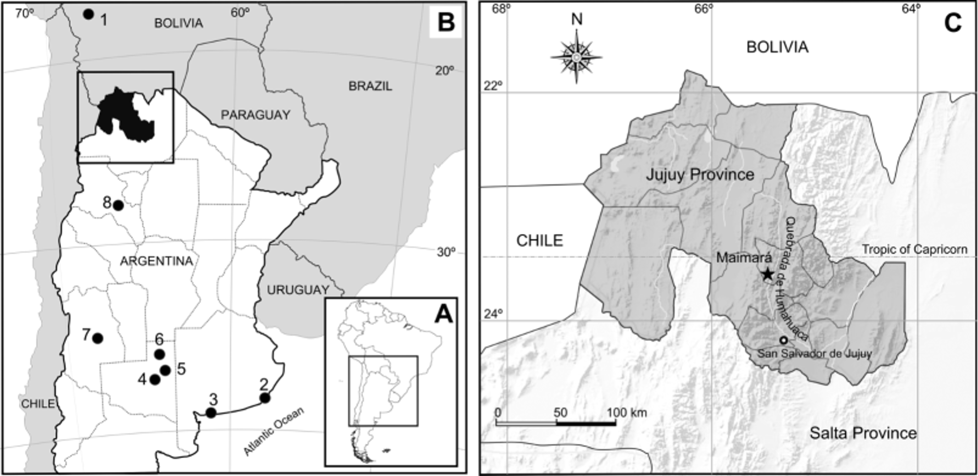
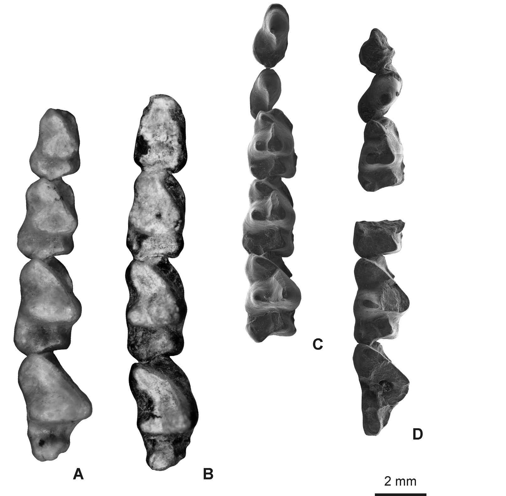
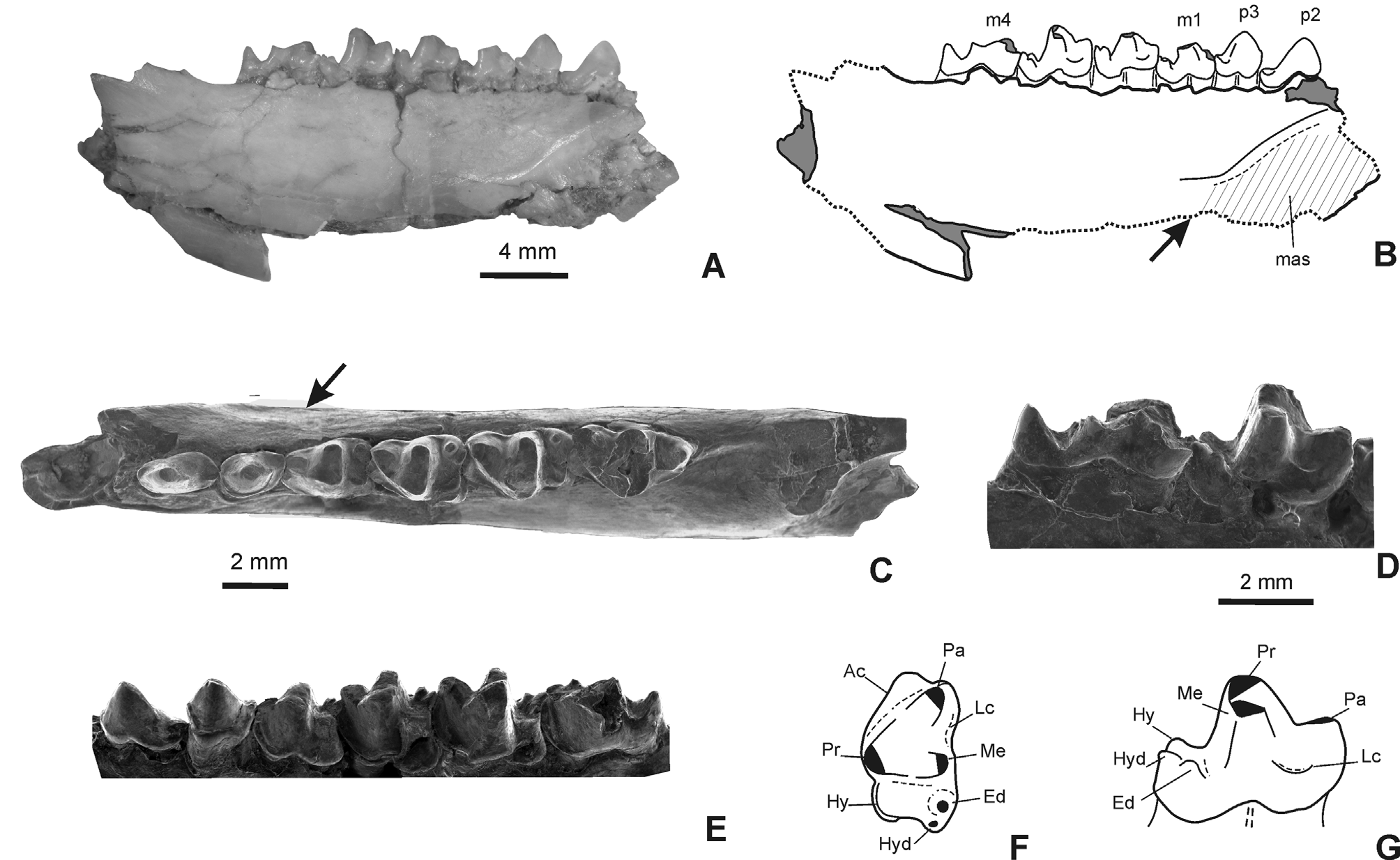
TABLE 2. Mandibular and dental ratios of Sparassocynidae species and ‘ Sparassocynus ’ heterotopicus estimated from measurements in Table 1. Upper values are means and lower values are ranges; sample size (n) is given in taxon column.
| Taxon | Wtrigm4 | Ltrigm4 | Wtalm4 | Ltalm4 | Lm1-3 | Lm2-3 |
|---|---|---|---|---|---|---|
| Sparassocynus maimarai | 2.2 | 2.5 | 1.5 | 1 | 9 | 6.4 |
| Sparassocynus derivatus | 2.5 | 3.1 | 1.5 | 1.1 | 9.7 | 6.9 |
| Sparassocynus derivatus | 2.6 | 3 | 1.8 | 1.3 | 9.8 | 7 |
| Sparassocynus derivatus | 2.2 | 2.6 | 1.5 | 1.4 | 9 | 6.4 |
| Sparassocynus derivatus | 2.5 | 2.9 | 1.7 | 1 | 9.2 | 6.5 |
| Sparassocynus derivatus | 2.5 | 3 | 1.9 | 1.4 | 10.1 | 7.1 |
| Hesperocynus dolgopolae | 9 | 6.3 | ||||
| Hesperocynus dolgopolae | 2 | 2.8 | 0.6 | 1 | 9 | 6.2* |
| Sparassocynus bahiai | 2.5 | 3 | 1.8 | 1.4 | 10.2 | 7.5 |
| Sparassocynus bahiai | 2.5 | 3 | 1.7 | 1.2 | 9.5 | 7 |
| Sparassocynus bahiai | 2.4 | 3 | 1.6 | 1.4 | 10 | 7.4 |
| ‘ Sparassocynus ’ heterotopicus | 2 | 2.3 | 1.5 | 1.3 | 6.8 |
TABLE 3. Compared mandibular and dental characters diagnostic of Sparassocynid species.
| Character | Hesperocynus dolgopolae | Sparassocynus bahiai |
|---|---|---|
| horizontal mandibular ramus (Lm1-3/MH) | shallow | deep |
| m1-3 talonid relative length (Lm1/Ltal; Lm2/Ltal; Lm3/Ltal) | long | short |
| relative width of talonid and trigonid of m4 (Wtrigm4/wtal) | narrow m4 talonid | wide m4 talonid |
| entoconid shape | gracile and labiolingually compressed | gracile and labiolingually compressed |
| lingual cingulum between para- and metaconid | absent | absent |
| relative length of m1 and m4 (Lm1/Lm4) | relatively longer m1 with respect to the m4 | relatively longer m1 with respect to the m4 |
| continued. | ||
| Character | Sparassocynus derivatus | Sparassocynus maimarai sp. nov. |
| horizontal mandibular ramus (Lm1-3/MH) | deep | deep |
| m1-3 talonid relative length (Lm1/Ltal; Lm2/Ltal; Lm3/Ltal) | short | short |
| relative width of talonid and trigonid of m4 (Wtrigm4/wtal) | wide m4 talonid | wide m4 talonid |
| entoconid shape | gracile and labiolingually compressed | robust and conical |
| lingual cingulum between para- and metaconid | absent | present |
| relative length of m1 and m4 (Lm1/Lm4) | relatively longer m1 with respect to the m4 | relatively longer m1 with respect to the m4 |
No known copyright restrictions apply. See Agosti, D., Egloff, W., 2009. Taxonomic information exchange and copyright: the Plazi approach. BMC Research Notes 2009, 2:53 for further explanation.
|
Kingdom |
|
|
Phylum |
|
|
Class |
|
|
SuperOrder |
Marsupialia |
|
Order |
|
|
Family |
|
|
Genus |