
Seborgia sanctensis, Jaume & Sket & Boxshall, 2009
|
publication ID |
https://doi.org/10.5252/z2009n2a3 |
|
persistent identifier |
https://treatment.plazi.org/id/038887C1-FFDC-E854-BDDD-030CFDCCA4F4 |
|
treatment provided by |
Marcus |
|
scientific name |
Seborgia sanctensis |
| status |
sp. nov. |
Seborgia sanctensis View in CoL n. sp.
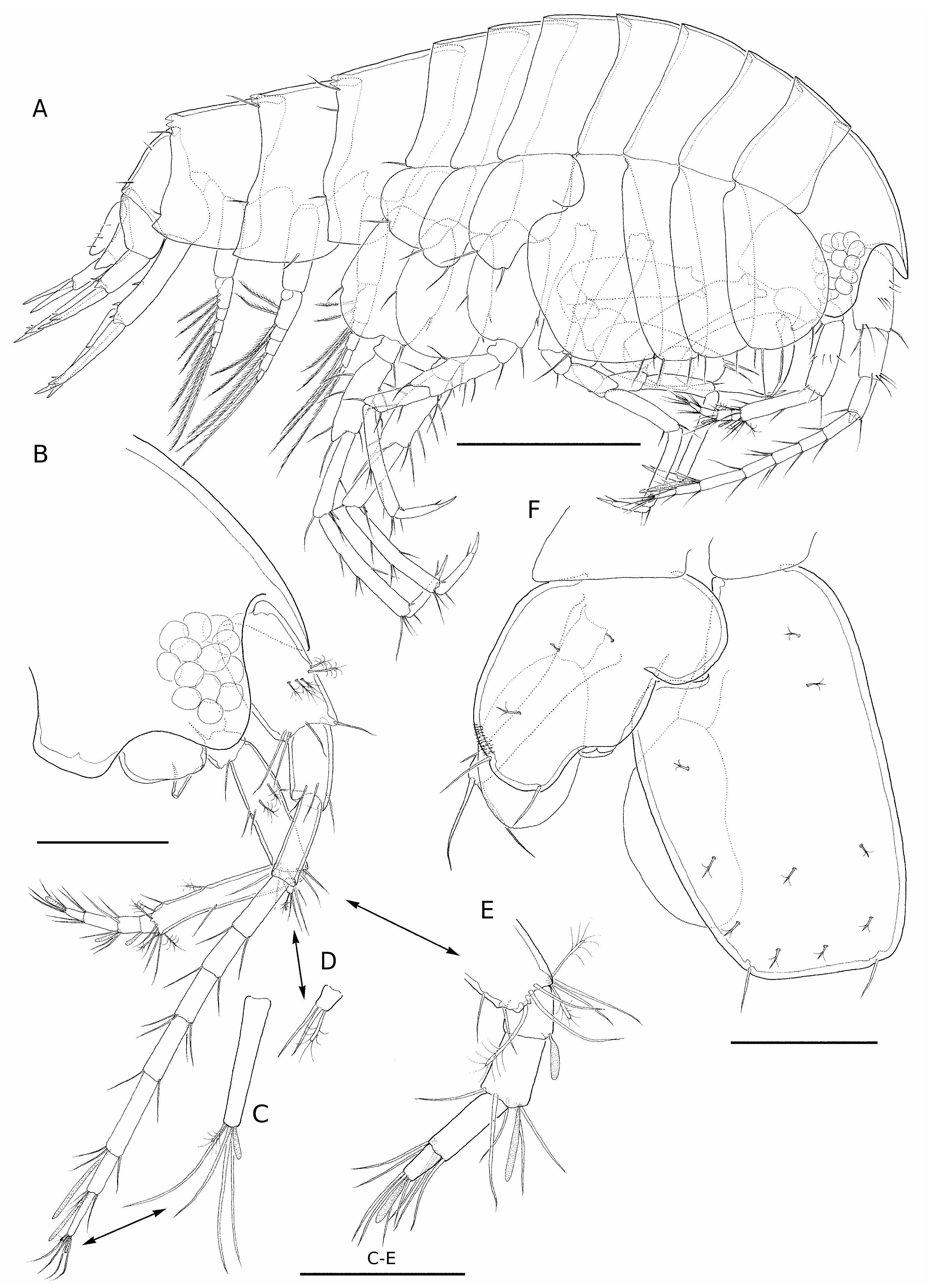
( Figs 1-6 View FIG View FIG View FIG View FIG View FIG View FIG )
TYPE MATERIAL. — Vanuatu. Espíritu Santo, anchialine pool located outside entrance to Loren cave ( 14°58’49.2”S, 167°3’28.08”E), G. A. Boxshall and D. Jaume coll., 8.IX.2006, holotype brooding ♀ 1.90 mm preserved in 70% ethanol in single vial (MNHN-Am7544).
Paratypes: same data as holotype, 7 brooding ♀♀ 1.63, 1.92, 1.71, 1.65, 1.81, 1.83 and 1.68 mm, plus additional 11 brooding ♀♀, 4 specimens of unknown sex, and 19 juveniles, not measured, all preserved in 70% ethanol in single vial (MNHN-Am7545) ; 10 specimens, none measured, preserved in 100% ethanol in single vial ( BMNH 2009.28 - 37 ) .
DIAGNOSIS. — Flagellum of antennule longer than corresponding peduncle. Fifth peduncle segment of antenna elongate, longer than first peduncle segment of antennule. Mandibular palp segment 3 rhomboid, with several D-setae ( sensu Stock 1974). Maxilla bilobed. Unguis of gnathopod I elongate. Uropod III biramous.
ETYMOLOGY. — The species name refers to the type locality on the island of Espíritu Santo ( Vanuatu).
DESCRIPTION OF BROODING FEMALE
(MALE UNKNOWN)
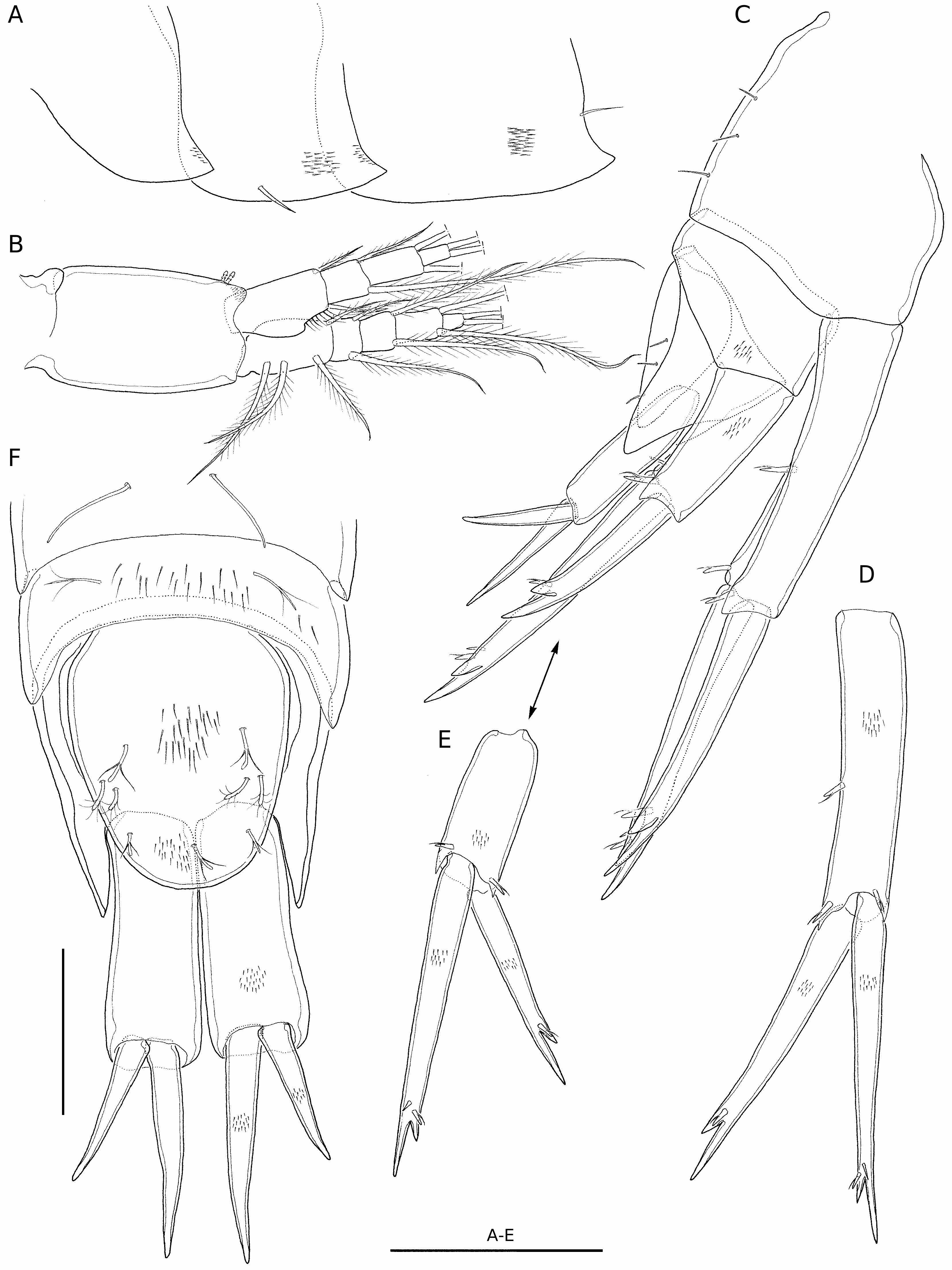
Body ( Fig. 1A View FIG ) compact, with integument covered with densely set microspinules (partially shown in some figures); colourless; eyes lateral, fully developed and pigmented (pigment not shown in figures). Head ( Fig. 1B View FIG ) with triangular rostrum in lateral aspect, not over-reaching distally proximal half of first segment of antennulary peduncle; head lobe well developed, evenly rounded, extending slightly beyond tip of rostrum; antennary sinus broad, shallowly excavated.Hyaline frill along posterodorsal margin of each pleonite with four large triangular serrations ( Fig. 1A View FIG ). Epimeral plates ( Fig. 6A View FIG ) with posterodistal angle weakly produced into pointed process; plates progressively larger towards posterior, with armature reduced to 0 or 1 robust seta on distal margin of second plate only; distal margin of third plate straight. Urosomites II and III strongly telescoped, with third virtually hidden beneath second ( Fig. 6C, F View FIG ).
Antennule ( Fig. 1A, B View FIG ) peduncle segments length ratio 44: 29: 27. Peduncle-to-main flagellum length ratio 45: 55. Main flagellum 7-articulate, article 5 longest; three distal articles each bearing aesthetasc, that on distal article reduced ( Fig. 1C View FIG ). Accessory flagellum bearing two simple slender setae plus one shorter penicillate seta ( Fig. 1D View FIG ).
Antenna ( Fig. 1A, B, E View FIG ) shorter than antennule. Gland cone short, directed posteriorly. Peduncle segments 4 and 5 length ratio 47: 53; fifth peduncle segment longer than first peduncle segment of antennule. Flagellum 4-articulate, shorter than fifth peduncle segment, with short aesthetasc on first, second and fourth articles only ( Fig. 1E View FIG ).
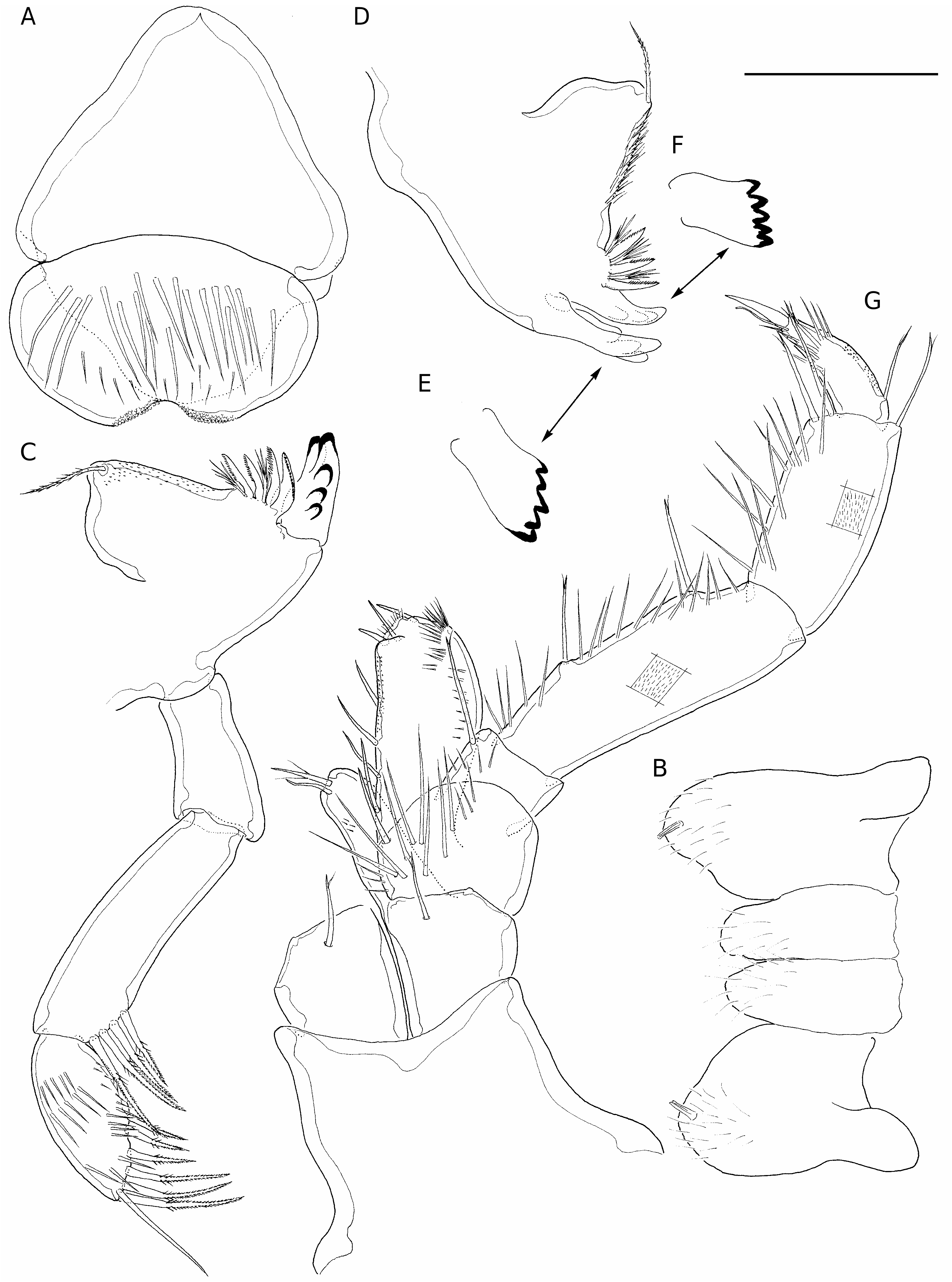
Labrum ( Fig. 2A View FIG ) with slightly produced epistome; anterior margin with sparsely-set, long and stout, simple setules; both lobes of bilobed distal margin microspinulate medially. Paragnaths ( Fig. 2B View FIG ) inner lobes shorter than outer, separate; both inner and outer lobes covered with sparsely-set short setules distally.
Right mandible ( Fig. 2C View FIG ) spine row comprising three elements with brush-like setules between.Molar process distal seta slender, pappose; anterior margin of process covered with sparsely-set microspinules. Palp segments length ratio 28: 43: 29; second segment with cluster of 3 or 4 setae on distomedial margin. Distal segment rhomboidal, sparsely ornamented with long spinules and armed with three D-setae and four E-setae ( sensu Stock 1974). Left mandible ( Fig. 2D, E View FIG ) as right counterpart except for lacinia ( Fig. 2F View FIG ), and with anterior margin of molar process covered with densely-set setules.
Maxillule ( Fig. 3A View FIG ) with smooth conical coxal endite (= inner plate). Endopod with three short stiff simple setae on distal segment; latter longer than proximal segment (relative proportions of segments 20: 80). Basal endite (= outer plate) and endopod sparsely covered with long simple setules.
Maxilla ( Fig. 3B View FIG ) bilobed, outer lobe with four setae, inner lobe with two; outer element on each lobe simple and reduced; one of two terminal elements on outer lobe simple; remaining elements on lobes stout, smooth distally but ornamented with stout pinnules proximally. Long setules sparsely-set on both lobes as figured.
Maxilliped ( Fig. 2G View FIG ) palp relative length of carpus: propodus: dactylus: unguis as 40: 31: 20: 9. Maxillipedal segments from ischium (included) onwards ornamented with long simple spinules as figured; dactylus with conspicuous transverse comb row of long spinules subdistally.
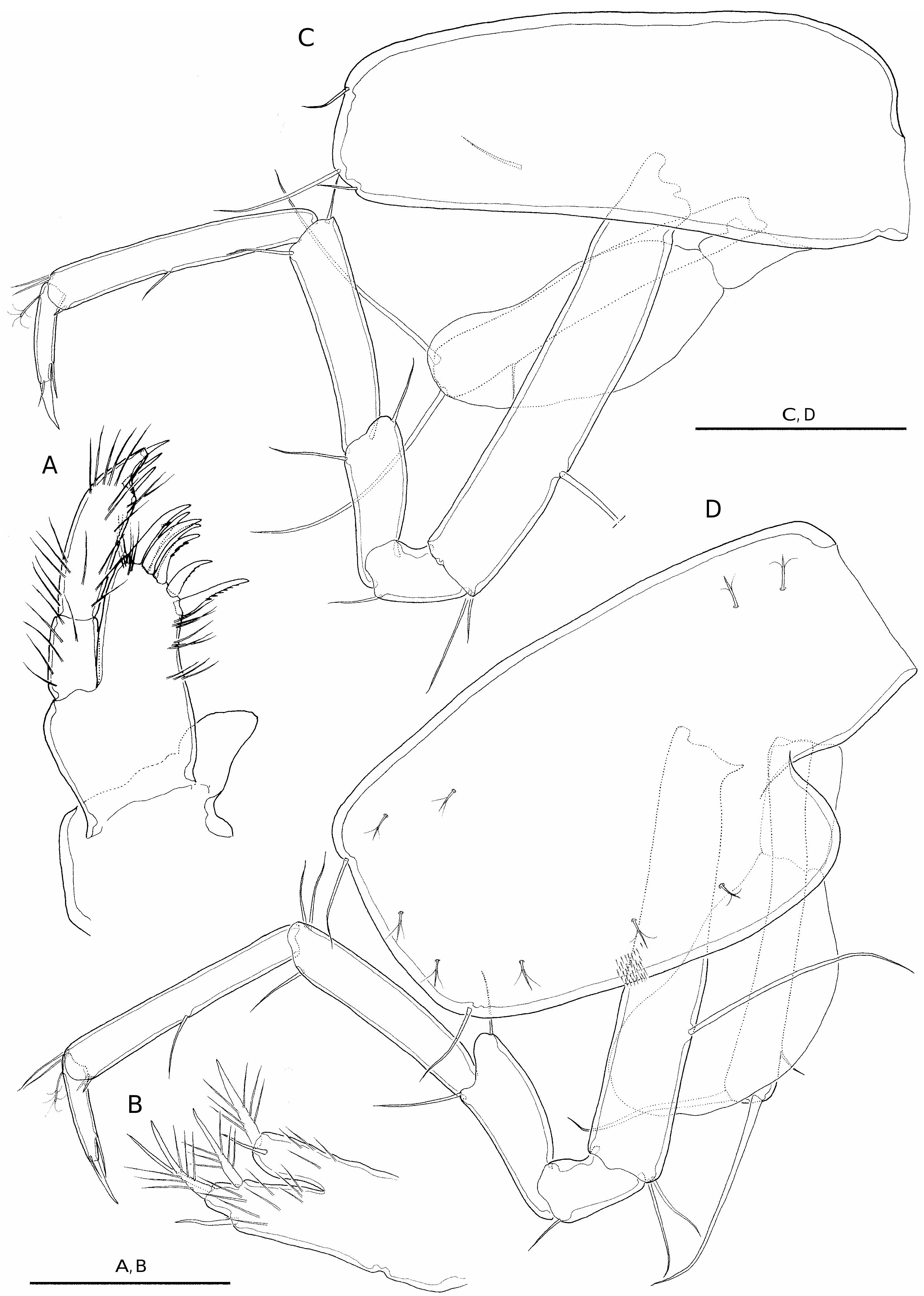
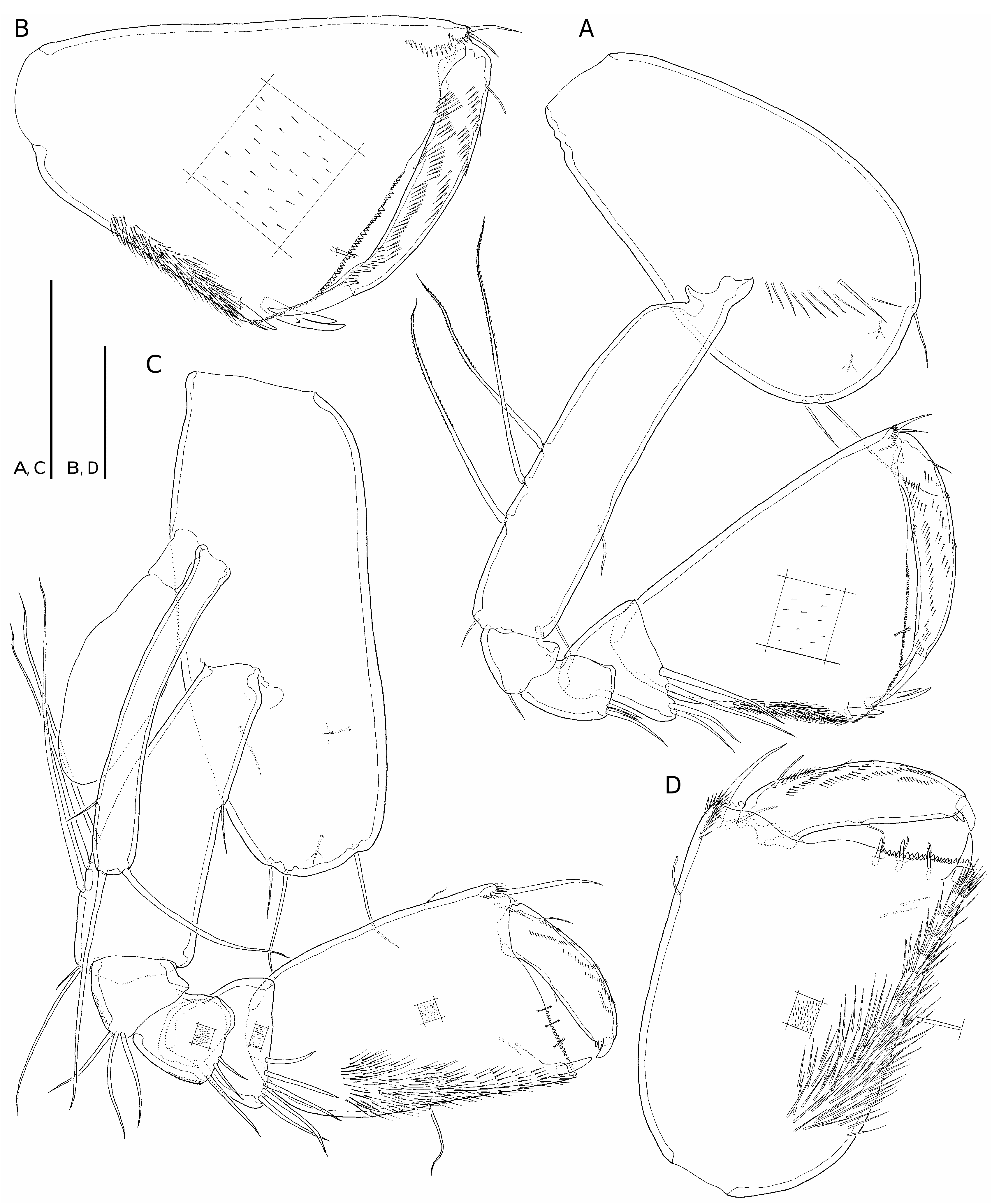
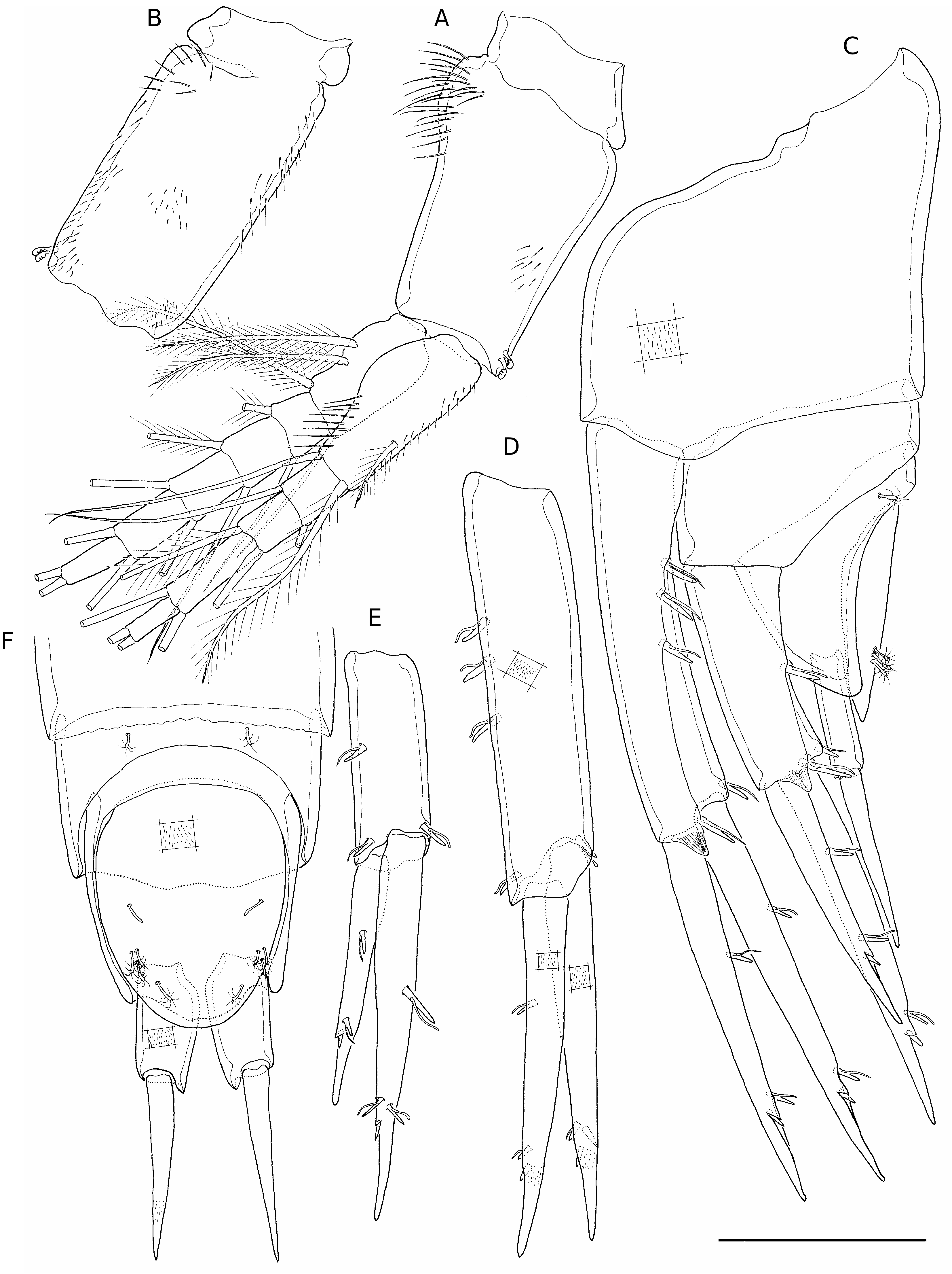
Coxal plate IV ( Figs 1F View FIG ; 3D View FIG ) about 1.6 times longer than broad, widest just below square posterior excavation; anterior and posterior margins of plate slightly convex; excavation occupying proximal 26% of posterior margin. Coxal plate V ( Figs 1F View FIG ; 5A View FIG ) bilobed, smaller anterior lobe covering square posterior excavation of coxa IV.
Gnathopod I ( Fig. 4A View FIG ) subchelate, propodus about 1.5 times longer than broad, palm angle evenly rounded, located at 47% of maximum length of segment. Palm margin slightly convex, covered with short stout triangular denticles and with single flagellate robust seta located submarginally ( Fig. 4B View FIG ); palm angle with two stout robust bifid setae, one either side of angle, that on lateral side longer. Dactylus with terminal spine on medial margin. Unguis long and slender, about 33% length of dactylus.
Gnathopod II ( Fig. 4C View FIG ) propodus subrectangular, about 1.3 times longer than broad; palm angle armed with single long, stout simple robust seta; palm margin ( Fig. 4D View FIG ) covered with stout triangular denticles and with three flagellate robust setae submarginally. Dactylus with two short terminal robust setae on medial margin; unguis short, about 15% length of dactylus.
Pereiopods III and IV ( Fig. 3C, D View FIG ) slender, similar, with unguis not incorporated into dactylus, i. e. articulating basally with it; dactylus with subterminal simple seta on medial margin and shorter simple seta on distal margin.
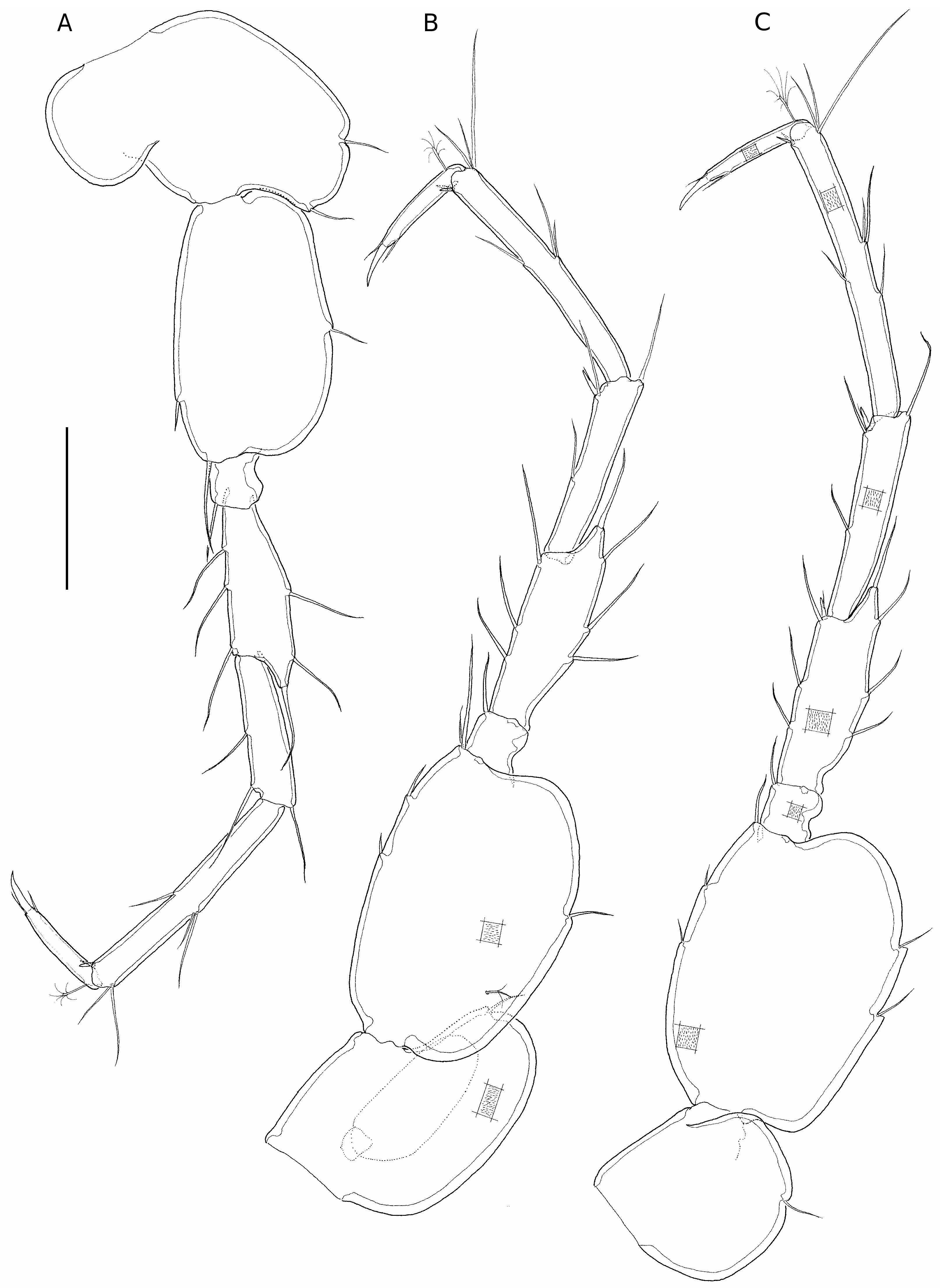
Pereiopods V-VII ( Fig. 5 View FIG ) bases about 1.6, 1.5 and 1.4 times longer than broad, respectively, progressively broader towards posterior, with anterior and posterior margins displaying 1 or 2 simple slender setae at most; posterior margins evenly convex and virtually smooth (displaying 1 or 2 shallow notches at most).
Pleopods ( Fig. 6B View FIG ) with protopod smooth, lacking setulation, each with subparallel margins except pleopod I (not figured; but see Fig. 11A View FIG ), which bears swelling proximally on lateral margin. Protopods each with two retinacles.
Uropods progressively shorter towards posterior ( Figs 1A View FIG ; 6C View FIG ). Uropod I ( Fig. 6D View FIG ) protopod as long as rami, with robust flagellate seta on both posterolateral and posteromedial distal angles, plus another about midway along posterolateral margin. Rami about equal in length; endopod armed with flagellate robust seta; exopod with pair of robust setae, one simple, other flagellate; setae on both rami located around subterminal notch. Uropod II ( Fig. 6E View FIG ) similar to uropod I except for length of protopod, shorter than rami and lacking seta on lateral margin, and for relative length of rami, with exopod clearly shorter than endopod. Uropod III ( Fig. 6F View FIG ) protopod longer than rami, thick; rami unarmed, endopod longer.
Telson ( Fig.6F View FIG ) with evenly rounded distal margin; armature comprising two pairs of penicillate setae plus five trifid setae distributed as figured.
REMARKS
Seborgia sanctensis View in CoL n. sp. is the only sebid possessing a biramous third uropod, and the bilobed state of its maxilla is also noteworthy since it was previously known to occur only in the Sebinae . Even though these features provide some evidence to support the erection of a separate, new genus for this taxon, its overall similarity with the other Seborgia species is striking, and we prefer here to treat it as the representative of the genus exhibiting the most plesiomorphic character states. The other Seborgia species display a secondarily reduced state for both of these characters. By analogy, the validity of the monotypic Caribseba Shaw, 1989 View in CoL within the Sebinae should be questioned, since the unique traits distinguishing this taxon from the extremely similar Seba View in CoL are also reductions, such as the absence of the accessory flagellum from the antennule and the loss of certain setae on the appendages (see Shaw 1989: 1889), and so Caribseba View in CoL might represent a terminal branch arising within the genus Seba View in CoL .
Additional unique features of the new taxon compared to other Seborgia species ( Table 1) include: the flagellum of the antennule longer than the corresponding peduncle; the elongated antennal peduncle segment 5; the presence of several D-setae on the third segment of the mandibular palp; the outline of the latter segment (rhomboidal vs. slender and subparallel in other species); and the elongated unguis of the gnathopod I.
ECOLOGY, ETHOLOGY
The new species was discovered in a small anchialine pool (about 5 m in diameter and 0.5 m deep) located about 30 m inland from the shore. An active spring discharged into the pool during low tide but at high tide the water flow was greatly reduced and the water became slightly brackish (3.2 ppt). The pool is formed on coarse coral rubble and is locally surrounded by dense forest. This pool may well be connected to the nearby Loren cave system, the entrance of which is located about 50 m away. In addition to the new species, there were many tanaids in this pool, living within a thick blackish mat of algae/bacteria that coated the surface of the submerged stones and boulders.
Specimens of Seborgia sanctensis n. sp. were observed alive under the stereo-microscope in the laboratory. The animals moved ventral side down, not upside-down or on one side. The stance of the animals is quite unusual for an amphipod, vaguely resembling an isopod due to their slightly dorsoventrally depressed body. It maintains the pleo- and urosome strongly reflexed beneath the pereion, having a short and “tail-less” appearance when viewed in dorsal aspect.Two embryos were invariably carried in the brood pouch of brooding females.
No known copyright restrictions apply. See Agosti, D., Egloff, W., 2009. Taxonomic information exchange and copyright: the Plazi approach. BMC Research Notes 2009, 2:53 for further explanation.
|
Kingdom |
|
|
Phylum |
|
|
Class |
|
|
Order |
|
|
Family |
|
|
Genus |
Seborgia sanctensis
| Jaume, Damià, Sket, Boris & Boxshall, Geoff A. 2009 |
Seborgia sanctensis
| Jaume & Sket & Boxshall 2009 |
Seborgia sanctensis
| Jaume & Sket & Boxshall 2009 |
Seborgia sanctensis
| Jaume & Sket & Boxshall 2009 |
Seborgia sanctensis
| Jaume & Sket & Boxshall 2009 |
Caribseba
| Shaw 1989 |
Caribseba
| Shaw 1989 |
Sebinae
| Holsinger & Longley 1980 |
Sebinae
| Holsinger & Longley 1980 |
Seba
| Bate 1862 |
Seba
| Bate 1862 |