
Tokyosoma cornutum, Mikhaljova, Elena V., Golovatch, Sergei I. & Chang, Hseuh-Wen, 2010
|
publication ID |
https://doi.org/ 10.5281/zenodo.197896 |
|
DOI |
https://doi.org/10.5281/zenodo.6207495 |
|
persistent identifier |
https://treatment.plazi.org/id/038687A9-EF3E-FFB5-73AB-B124FA9340A4 |
|
treatment provided by |
Plazi |
|
scientific name |
Tokyosoma cornutum |
| status |
sp. nov. |
Tokyosoma cornutum View in CoL sp. nov.
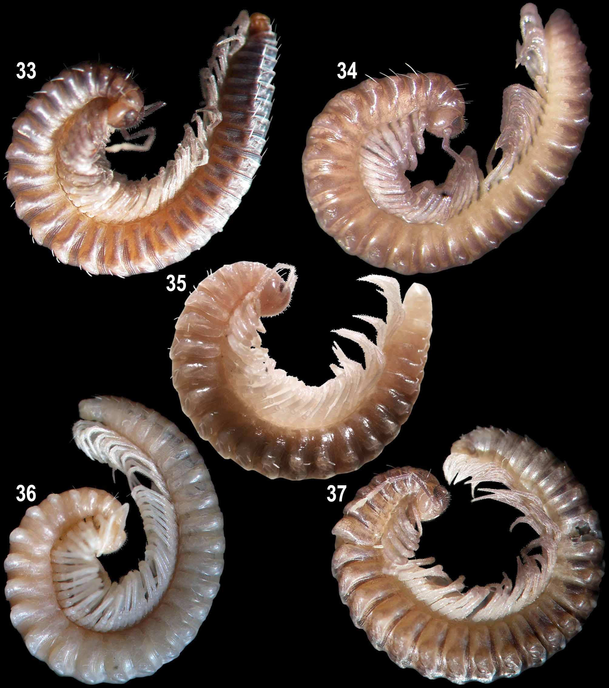
Figs 29–31 View FIGURES 29 – 31 , 36 View FIGURES 33 – 37 .
Material examined: Holotype: 1 male ( NMNS –6345–016), Taiwan, Nantou County, Ren-ai Township, Meifeng, forest, 24°05'49" N, 121°10'51" E, 2158 m, 21 May 2005, leg. Z. Korsós & Ch.-H. Chang.
Diagnosis: Differs from congeners mainly by the shape of the posterior gonopod lateral coxal branch divided into three strong processes distally, as well as in the shape of the posterior gonopod colpocoxite devoid both of a lateral blade and a distal outgrowth.
Description: Male. Length about 23 mm, width with paraterga about 2.0 mm. Coloration light beige throughout, including antennae, legs and ocellaria.
Body with 32 segments. Head covered with long and short setae. Each eye patch composed of at least 28 ocelli. Collum semi-circular. Body width gradually increasing until somite 7, body parallel-sided on somites 8–15(16), thereafter gradually tapering. Beginning from somite 4, paraterga normally developed, rounded, growing increasingly less distinct towards hind part of body, strongly reduced on somites 24–26, absent from somites 27–31. Paraterga 2 and 3 small. In anterior and posterior parts of body, anterolateral and median macrochaetae subequal in length, posterolateral macrochaetae longest, pointed, but not very sharply so. In middle part of body, macrochaetae very short, pointed, but not very sharply so.
Leg pairs 3–7 somewhat enlarged. Leg pair 7 with a small group of funnel-shaped tarsal papillae apically near claw. Claw of leg pair 7 at base without both additional claw dorsally and filament ventrally.
Postgonopodal legs (including leg pairs 10 and 11) without tarsal papillae. Claw of leg pairs 10 and 11 at base with one small additional claw dorsally and a very short setiform filament ventrally. Claw of midbody legs at base with one small additional claw dorsally and a short setiform filament ventrally. Claw of hindmost legs at base only with one small additional claw dorsally.
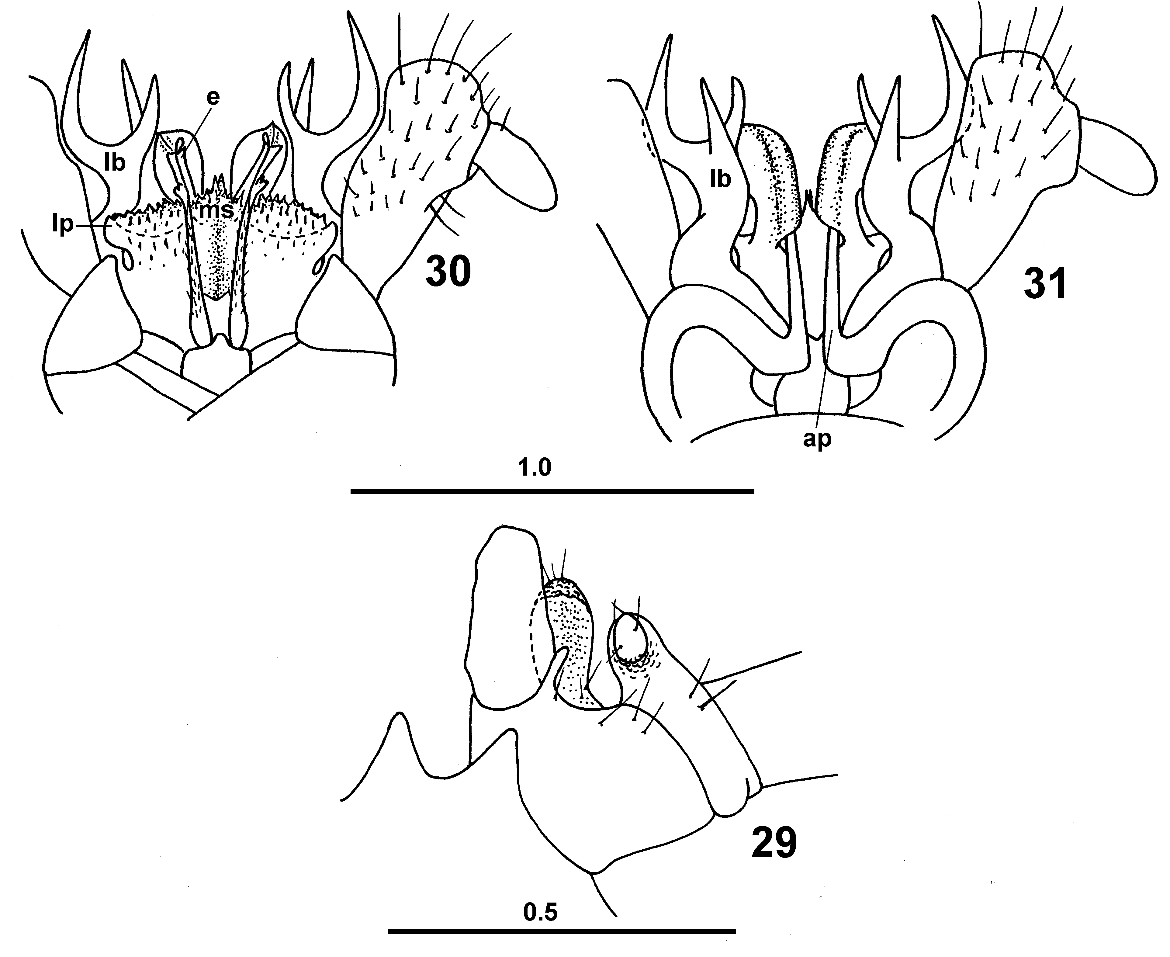
Legs 10 and 11 with coxal glands. Coxa 10 covered with papillae on caudal face, with an oblong caudoventral process concave frontally; lateral edge of orifice of coxal gland extended into a process ( Fig. 29 View FIGURES 29 – 31 ). Trochanter 10 with an anteriad curved, finger-shaped, papillate, ventral, setiferous outgrowth. Coxa 11 papillate on caudal face, with a tiny, papillate, ventral prominence. Trochanter 11 with a caudal finger-shaped process papillate mesally and rounded apically.
Anterior gonopod telopodite 1-segmented, flagelliform, beset with cuticular spinules, its distal part positioned inside sheaths with very strongly elevated and tightly closed edges (e) ( Fig. 30 View FIGURES 29 – 31 ). These sheath edges like high plates in lateral view. Telopodite base and distal part of coxosternum tightly attached to adjacent mesal portion of posterior gonopod. Posterior gonopod colpocoxites fused sub-basally, somewhat curved caudad. Mesal sheath processes of posterior gonopod colpocoxites fused medially into a single cupshaped structure (ms) with a middle outgrowth covered with very short pointed spinules. Lateral sheath processes of colpocoxites (lp) cup-shaped, carrying such spinules caudally.
Posterior gonopod angiocoxite with a subtriangle globule, but without process in posterior view. Posterior gonopod coxal part with a long lateral branch (lb) divided into three strong processes distally, of which lateral process longest and mesal ones subequal in length. Basal part of this branch fused with both colpocoxite and anterior angiocoxite.
Angiocoxite depressed centrally in anterior view ( Fig. 31 View FIGURES 29 – 31 ), supplied with a long process (ap); distal portion of this process penetrating colpocoxite. Posterior gonopod telopodite 2-segmented; trochanteroprefemur hollow for accommodation of lateral process of coxal branch (lb) mediocaudally; femur short and thin.
Female unknown.
Name: The specific epithet refers to the antler-shaped lateral coxal branch of the posterior gonopod.
| NMNS |
National Museum of Natural Science |
No known copyright restrictions apply. See Agosti, D., Egloff, W., 2009. Taxonomic information exchange and copyright: the Plazi approach. BMC Research Notes 2009, 2:53 for further explanation.