
Tokyosoma distinctum, Mikhaljova, Elena V., Golovatch, Sergei I. & Chang, Hseuh-Wen, 2010
|
publication ID |
https://doi.org/ 10.5281/zenodo.197896 |
|
DOI |
https://doi.org/10.5281/zenodo.6207486 |
|
persistent identifier |
https://treatment.plazi.org/id/038687A9-EF3B-FFA9-73AB-B761FEDE451F |
|
treatment provided by |
Plazi |
|
scientific name |
Tokyosoma distinctum |
| status |
sp. nov. |
Tokyosoma distinctum View in CoL sp. nov.
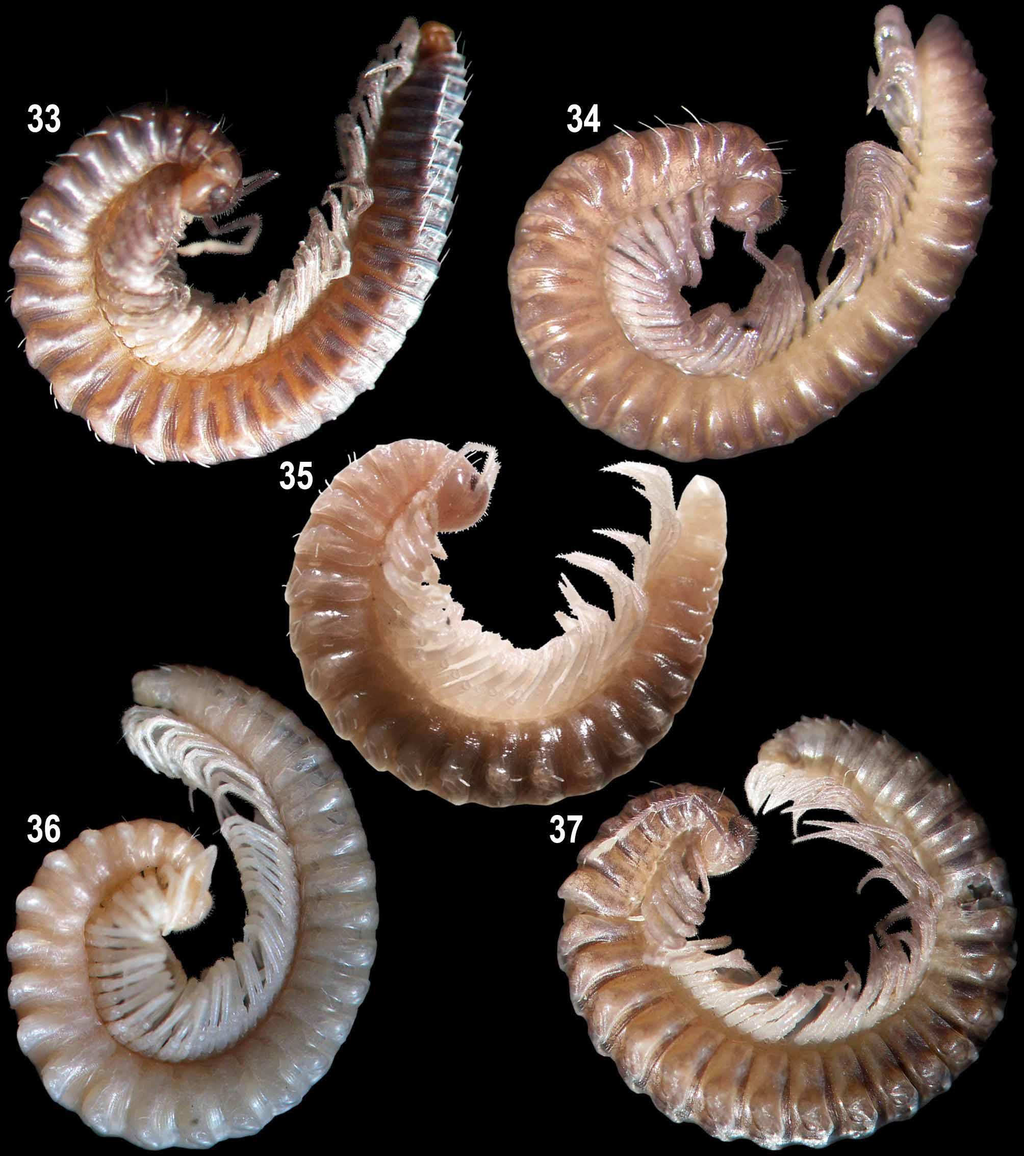
Figs 23–25 View FIGURES 23 – 25 , 35 View FIGURES 33 – 37 .
Material examined: Holotype: 1 male ( NMNS –6345–012, D–0498), Taiwan, Nantou County, Ren-ai Township, Meifeng, 19 February 2002, leg. S.H. Wu. Paratypes: 1 male ( NMNS –6345–013, D–0207), Taiwan, Nantou County, Ren-ai Township, Meifeng, 22 October 2001, leg. S.H. Wu; 1 male ( ZMUM), D– 0 504, same locality as in holotype, 19 February 2002, leg. S.H. Wu; 1 male ( IBSS), D–0502, Taiwan, Nantou County, Ren-ai Township, Meifeng, 15 April 2002, leg. S.H. Wu.
Diagnosis: Differs from congeners mainly by the shape of the posterior gonopod colpocoxite, which carries a lateral horn-shaped process, and in the form of the posterior gonopod lateral coxal branch with a narrow lateral blade and small subapical outgrowths, as well as in a subconical, anteriorly curved, coxal process of male leg 10.
Description: Male. Length about 13 mm, width with paraterga 1.0– 1.1 mm. Coloration in alcohol beige, light brown to brown with a pattern of marbled brown bands and pale spots on dorsum, legs with marbled brown distal parts, ocellaria brown (possibly faded) or black.
Body with 29 segments. Head covered with sparse, relatively long and short setae. Each eye patch composed of at least 20 ocelli. Collum semi-circular. Body width gradually increasing until somite 7, body parallel-sided on somites 8–15(16), thereafter gradually tapering. Beginning from somite 4, paraterga normally developed, rounded, growing increasingly less distinct towards hind part of body, strongly reduced on somites 22–24, absent from somites 25–28. Paraterga 2 and 3 small. Metazonital macrochaetae in a transverse row on somite 28, like an elongate (to different degrees) triangle on preceding somites. Macrochaetae in anterior part of body relatively long, pointed apically, but in middle and posterior parts of body short and blunt. Metazonites with two very low knobs placed near axial suture on each side and with an oblong bolster on paraterga.
Leg pairs 3–7 somewhat enlarged. Leg pairs 5–7 with a group of funnel-shaped tarsal papillae apically near claw. Claw of leg pairs 5–7 at base without additional claw dorsally. Claw of leg pairs 5 and 7 at base with a long setoid filament ventrally, but claw of leg pair 6 without such outgrowth. Postgonopodal legs (including leg pairs 10 and 11) without tarsal papillae. Claw of legs 10 and 11 at base with one small additional claw dorsally and a long setoid filament ventrally. Claw of midbody legs at base with one small additional claw dorsally, with or without long setoid filament ventrally. Claw of hindmost legs at base with neither an additional claw dorsally nor a filament ventrally.
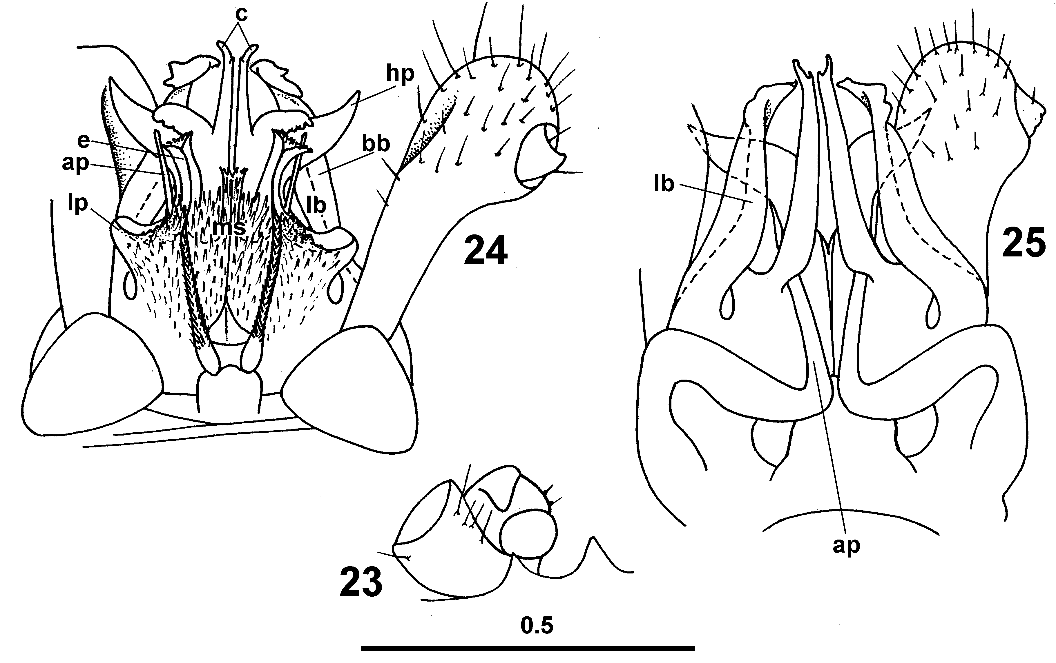
Legs 10 and 11 with coxal glands. Coxa 10 papillate caudally, with a caudoventral subconical process curved anteriad ( Fig. 23 View FIGURES 23 – 25 ). Trochanter 10 with a small ventral outgrowth setose apically. Coxa 11 without processes. Trochanter 11 with a caudal finger-shaped process papillate mesally and rounded apically.
Anterior gonopod telopodite 1-segmented, flagelliform, densely beset with cuticular spinules, its distal part positioned inside sheaths with very strongly elevated and tightly closed edges (e) ( Figs. 24 View FIGURES 23 – 25 ). These sheath edges like high plates in lateral view. Telopodite base and distal part of coxosternum tightly attached to adjacent mesal portion of posterior gonopod. Posterior gonopod colpocoxites fused sub-basally, somewhat curved caudad. Colpocoxites distally with slender flat processes (c) and, laterally, with horn-shaped processes (hp). Mesal sheath processes of posterior gonopod colpocoxites fused medially into a single cup-shaped structure (ms) with a middle outgrowth covered with long pointed spinules. Lateral sheath processes of colpocoxites (lp) cup-shaped, carrying pointed spinules caudally.
Posterior gonopod angiocoxite with a globule, but without process in posterior view. Posterior gonopod coxal part with a long lateral branch (lb) carrying a narrow lateral blade (bb) and two small subapical outgrowths. Apex of the branch somewhat twisted, curved anteriad. Basal part of this branch fused with both colpocoxite and anterior angiocoxite.
Angiocoxite depressed centrally in anterior view ( Fig. 25 View FIGURES 23 – 25 ), supplied with a long process (ap); distal portion of this process penetrating colpocoxite and visible outside in lateral view. Posterior gonopod telopodite 2-segmented; trochanteroprefemur hollow for accommodation of lateral colpocoxite process (hp) mediocaudally; femur like a conical knob (part of femur visible through wall of trochanteroprefemur of posterior gonopod telopodite).
Female unknown.
Name: The specific epithet refers to the posterior gonopod colpocoxite, which carries a lateral hornshaped process, this being the main feature making this species distinguished among the remaining congeners in Taiwan.
No known copyright restrictions apply. See Agosti, D., Egloff, W., 2009. Taxonomic information exchange and copyright: the Plazi approach. BMC Research Notes 2009, 2:53 for further explanation.