
Ponsoonops vuena, Bolzern, 2014
|
publication ID |
https://doi.org/10.1206/3803.1 |
|
DOI |
https://doi.org/10.5281/zenodo.5460109 |
|
persistent identifier |
https://treatment.plazi.org/id/FA66A32B-FFDF-4741-FE2C-D5FDA10CF918 |
|
treatment provided by |
Carolina (2021-08-29 17:36:42, last updated 2021-09-05 23:32:49) |
|
scientific name |
Ponsoonops vuena |
| status |
new species |
Ponsoonops vuena , new species
Figures 274–318 View FIGURES 274–288 View FIGURES 289–303 View FIGURES 304–318
NOTE: Males and females have not been collected together. Overall, they are a good match but still may be mismatched.
TYPE: Male holotype from a Berlese sample of floor litter from a deep ravine taken at an elevation of 1494 m at site W of Finca Palo Santo, near Nueva California, Chiriquí, Panama (Mar. 9, 1959, H.S. Dybas), deposited in FMNH (34859 PBI_ OON 10576 View Materials ) .
ETYMOLOGY: The specific name is a noun in apposition, a shortened anagram taken from the type locality.
DIAGNOSIS: Males resemble those of P. boquete , P. lerida , and P. panto in having an embolus with a median protrusion and a narrowed, distinctly elongated embolus tip (figs. 300–303). They differ from P. lerida and P. panto in having a distally pointed conductor that is longer than the embolus (rather than truncated, or with short narrow projections, and shorter), and from P. boquete in having a broad conductor base, a relatively broad embolus, and a mound-shaped median protrusion (rather than a narrow conductor base, a relatively narrow embolus, and a ridge-shaped median protrusion). Females resemble those of P. bollo and P. boquete in having a narrow, long-stalked anterior genitalic process with enlarged apex (fig. 213, 288). They differ from P. bollo in having long, filiform glands at the base of the anterior genitalic process, reaching almost the enlarged part (rather than short ones), and from P. boquete in the presence of distinctly long, protruding glands at the posterior genitalic process (rather than absent)
MALE (PBI_ OON 10576 View Materials , figs. 274–282, 289–318): Total length 1.37. Leg spination: tibiae: I v4-2-2; II v4-2-1p; metatarsi: I v2-2-2; II p1-0-0, v2-2-2. Sperm pore oval. Embolus broad, tube-shaped, longitudinally folded, with ventral slit and prolateral opening, midway with distinct dorsal mound, tip narrowed, elongated, embolus opening retrolaterally rebordered, ventrally with spikelike projection; conductor originates at embolus base, broad, lamelliform elongated, distally narrowed, twisted, tip simple, pointed, moderately longer than embolus.
FEMALE: (PBI_OON 5689, figs. 283–288): Total length 1.80. Leg spination: tibiae: I p1-1-0, v4-2-2, r1-0-0; II p0-1-0, v4-2-1p; IV p0-1-0, v1-0-2, r0-1-0; metatarsi: I, II p1-0-0, v2-2-2; IV p0-1-0. Large genital plate W-shaped, median protruding part wider than long; anterior genitalic process long, narrowly stalked, apex enlarged; cluster of few, distinctly long filiform glands present at base of anterior genitalic process; posterior genitalic process pocketlike, without remarkable duct, weakly sclerotized, distally with distinct, long glands.
OTHER MATERIAL EXAMINED: PANAMA: Chiriquí: W Finca Palo Santos, Mar. 5, 1959, Berlese , chips, leaf mold, etc., at base of cut stump, elev. 1448 m (H. Dybas, FMNH PBI_ OON 10403 View Materials ), 2♂ ; El Volcán , Aug. 9–14, 1950 (A. Chickering, MCZ 72228 View Materials PBI_OON 5689), 2♀ .
DISTRIBUTION: Panama (Chiriquí).
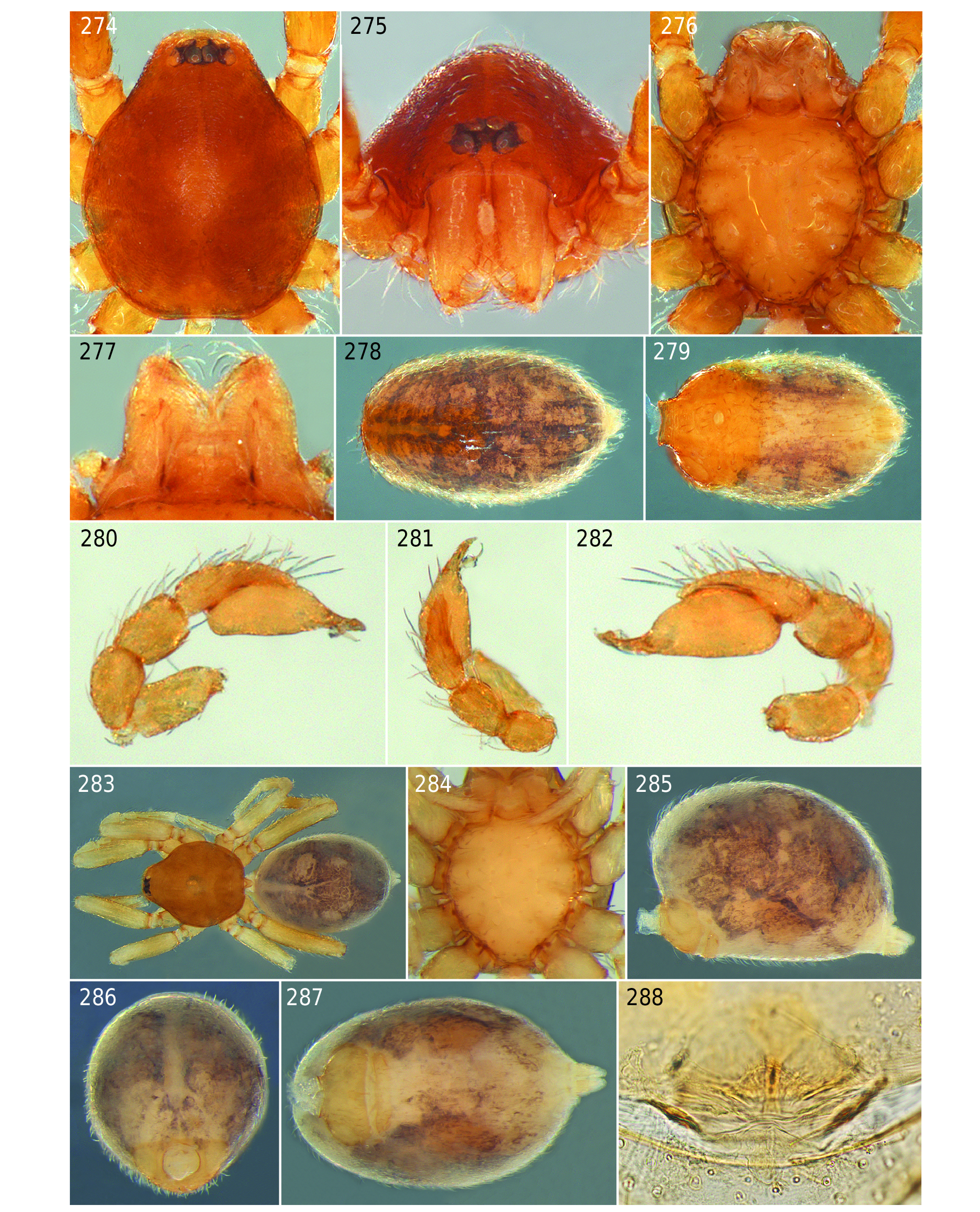
FIGURES 274–288. Ponsoonops vuena, new species, male (274–282) and female (283–288). 274. Carapace, dorsal view. 275. Same, anterior view. 276. Sternum, ventral view. 277. Mouthparts, ventral view. 278. Abdomen, dorsal view. 279. Same, ventral view. 280. Palp, prolateral view. 281. Same, dorsal view. 282. Same, retrolateral view. 283. Habitus, dorsal view. 284. Sternum, ventral view. 285. Abdomen, lateral view. 286. Same, anterior view. 287. Same, ventral view. 288. Genitalia, dorsal view.
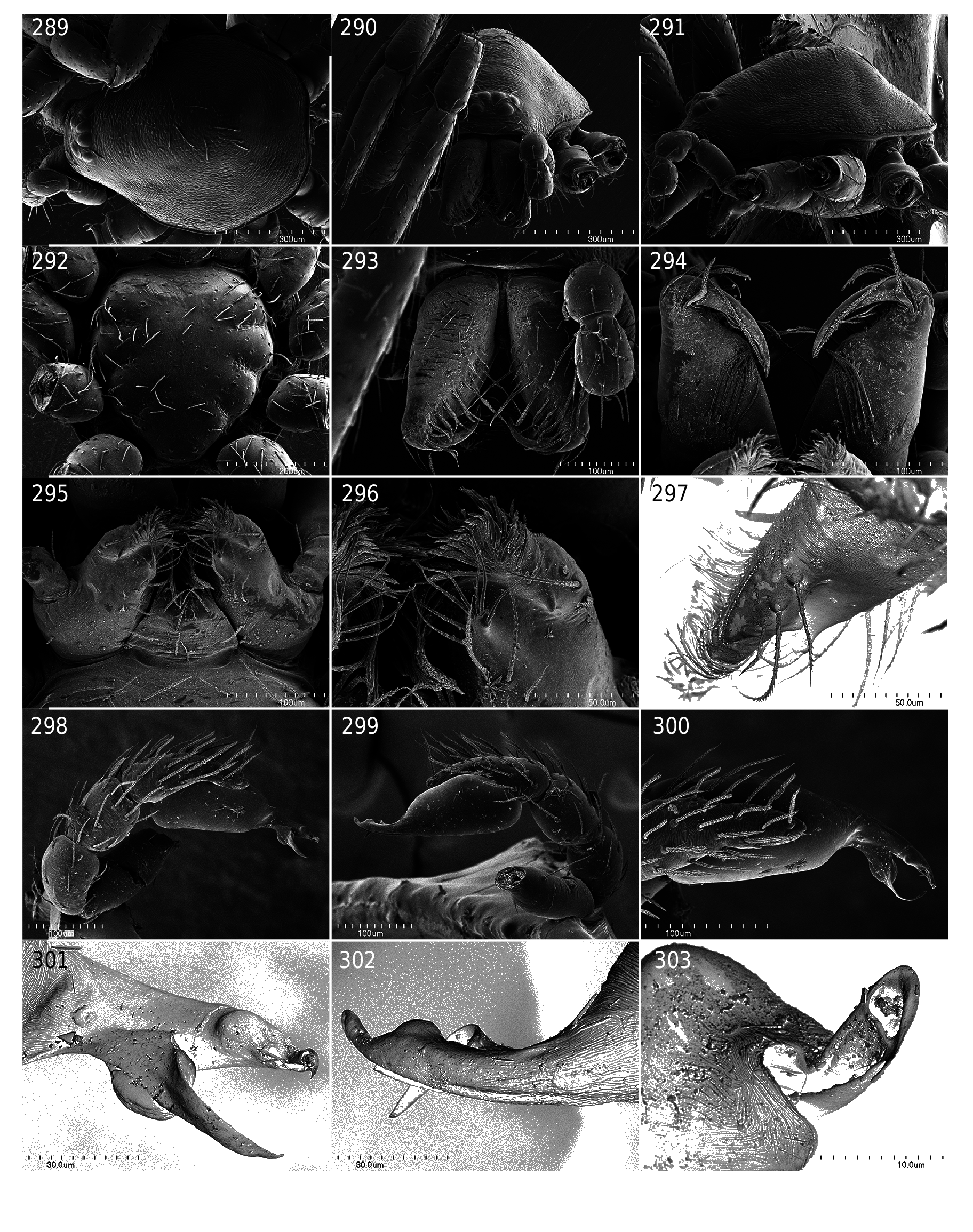
FIGURES 289–303. Ponsoonops vuena, new species, male. 289. Carapace, dorsal view. 290. Same, anterior view. 291. Same, lateral view. 292. Sternum, ventral view. 293. Chelicerae, anterior view. 294. Same, posterior view. 295. Mouthparts, ventral view. 296. Endite tip, ventral view. 297. Same, lateral view. 298. Palp, prolateral view. 299. Same, retrolateral view. 300. Same dorsal view. 301. Embolus and conductor, prolateral view. 302. Same, retrolateral view. 303. Embolus tip, prolateral view.
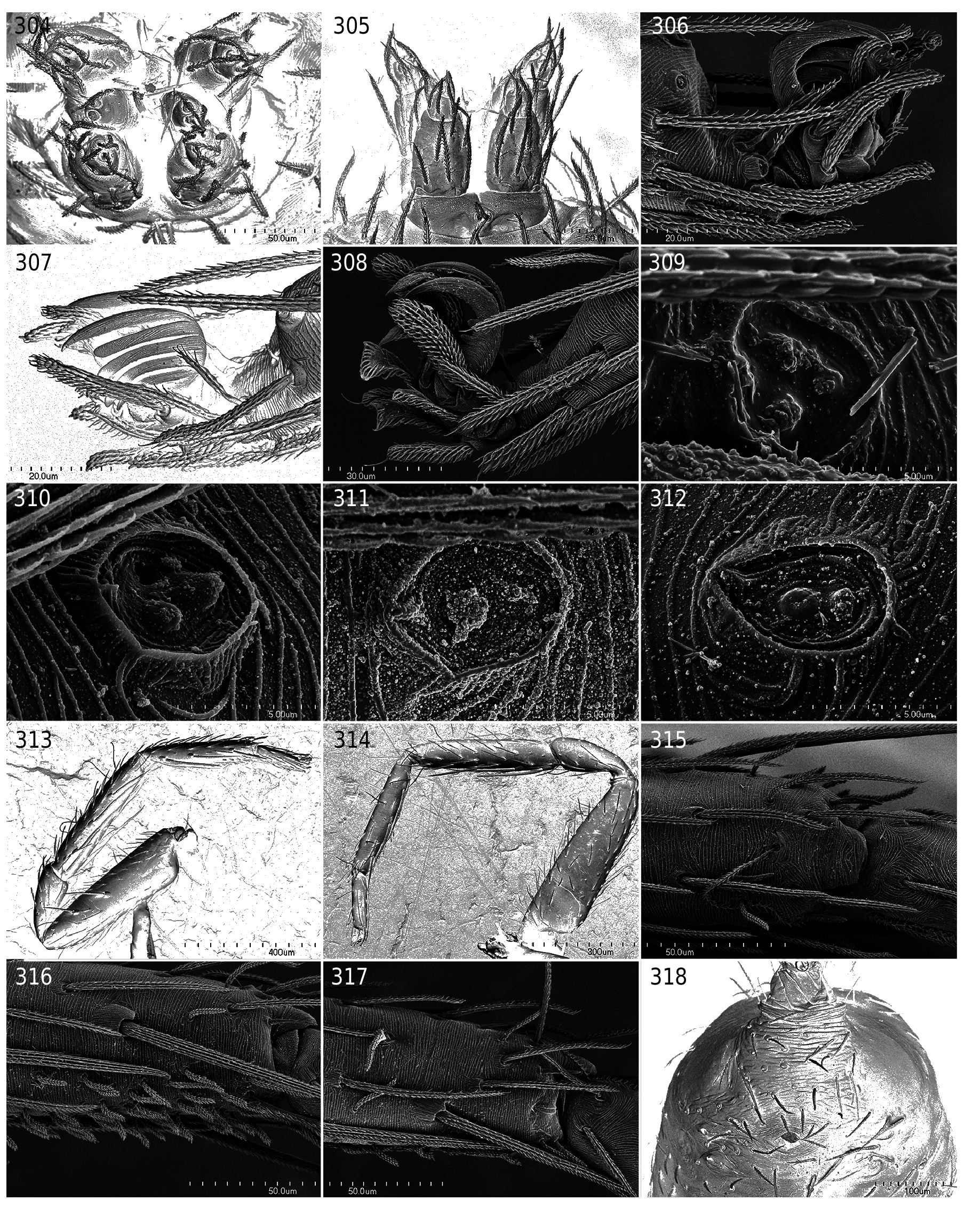
FIGURES 304–318. Ponsoonops vuena, new species, male. 304. Spinnerets, posterior view. 305. Same, ventral view. 306. Claws, leg II, retrolateral view. 307. Same, prolateral view. 308. Same, leg IV. 309. Tarsal organ, leg II, dorsal view. 310. Same, leg III. 311. Same, leg IV. 312. Same, palp. 313. Leg I, prolateral view. 314. Leg II, retrolateral view. 315. Metatarsus tip, leg I, dorsal view. 316. Same, lateral view. 317. Same, leg II. 318. Epigastric area, ventral view.
No known copyright restrictions apply. See Agosti, D., Egloff, W., 2009. Taxonomic information exchange and copyright: the Plazi approach. BMC Research Notes 2009, 2:53 for further explanation.
|
Kingdom |
|
|
Phylum |
|
|
Class |
|
|
Order |
|
|
Family |
|
|
Genus |