
Ceratophila
|
publication ID |
https://doi.org/10.11646/zootaxa.4508.2.1 |
|
publication LSID |
lsid:zoobank.org:pub:5E2BC894-1919-4F63-8EF5-BAAC91913388 |
|
DOI |
https://doi.org/10.5281/zenodo.5957999 |
|
persistent identifier |
https://treatment.plazi.org/id/F95B87D7-FFB1-951D-FF49-F8E7D7FA5E38 |
|
treatment provided by |
Plazi (2019-03-26 07:33:51, last updated 2024-11-26 23:51:32) |
|
scientific name |
Ceratophila |
| status |
|
Key to adult Ceratophila
Because of variable and overlapping morphological character states with some species, it is desirable, and in some cases necessary, to have a series of specimens representing color variations and both sexes to properly identify a
species. In the following key, text in brackets before the species name are the Mexican states of occurrence and the known host species of Ceratozamia .
1. Pronotum not explanate laterally, surface convexly curved to lateral carinae; lateral carinae in lateral view distinctly thicker anteriorly, anterior thickness 2× posterior thickness ( Fig. 1D View FIGURE 1 ); pronotal disc lacking longitudinal groove extending anteriorly from lateral basal pore in margin ( Fig. 1B View FIGURE 1 ); metatibia triangularly dilated toward apex ( Fig. 1F View FIGURE 1 )......................................................................................... [subgenus Ceratophila, new subgenus] ... 2
- Pronotum explanate laterally, surface broadly concave near lateral carinae; lateral carina weakly thickening anteriorly, anterior thickness <1.5× that at base ( Fig. 1E View FIGURE 1 ); pronotal disc with longitudinal groove extending anteriorly from lateral basal pore in margin ( Fig. 6D View FIGURE 6 ); metatibia not triangularly dilated toward apex ( Fig. 1G View FIGURE 1 )............... [ Vovidesa , new subgenus] … 5
2(1). Head with broad medial transverse ridge anterior of a distinct transverse basal groove in males ( Fig. 2D View FIGURE 2 ), ridge and groove weak in females, ridge interrupting supraocular line in both male and female; head broad, width = 0.74–0.75× pronotal width; anterior clypeal margin emarginate; pronotal hypomeron densely punctate ( Fig. 2E View FIGURE 2 ); [Veracruz— C. euryphyllidia View in CoL ].................................................................................... C. (C.) chemnicki , new species
- Base of head at most with indistinct shallow transverse groove, supraocular line complete ( Fig. 3D View FIGURE 3 ); head narrower, width = 0.66–0.68× pronotal width; anterior clypeal margin truncate or weakly convex; pronotal hypomeron with punctures sparse and minute or lacking ( Fig. 3E View FIGURE 3 ).............................................................................. 3
3(2). Dark elytral markings (when present) laterally covering most of disc ( Fig. 4A View FIGURE 4 ); pronotum generally more elongate, pronotal length/pronotal width (PL/PW) = 0.83–0.90; [Chiapas— C. alvarezii View in CoL , C. mirandae View in CoL , C. norstogii View in CoL , C. vovidesii View in CoL ]......................................................................................... C. (C.) picipennis , new species
- Dark elytral markings (when present) only along suture and laterally ( Fig. 5A View FIGURE 5 ); pronotum generally shorter, PL/PW = 0.80– 0.84................................................................................................ 4
4(3). Color of pronotum and elytra similar ( Fig. 5A View FIGURE 5 ); pronotum generally more quadrate; male genitalia with penile struts relatively long, length of penile strut relative to median lobe 2:1; setae at apex of parameres with greatest length <dorsal width of parameres ( Fig. 5F View FIGURE 5 ); [Veracruz— C. tenuis View in CoL ]......................................... C. (C.) sanchezae , new species
- Pronotum reddish brown contrasting with light brown of elytra ( Fig. 3A View FIGURE 3 ); pronotum generally more trapezoidal, narrowing anteriorly; male genitalia with penile struts shorter, length of penile struts relative to median lobe 3:2; setae at apex of parameres with greatest length ± dorsal width of parameres ( Fig. 3H View FIGURE 3 ); [Oaxaca— C. mixeorum ]...................................................................................................... C. (C.) gregoryi , new species
5(1). Male meso- and metatibiae each with medial subapical emargination ( Fig. 1G View FIGURE 1 ); female abdominal segments weakly alutaceous, punctures and setae visible except on lateral quarters.................................................... 6
- Male tibiae lacking medial subapical emargination; female abdominal segments strongly alutaceous ( Fig. 6B View FIGURE 6 ), punctures obscured across middle, setae reduced [Chiapas— C. alvarezii View in CoL , C. mirandae View in CoL , C. norstogi , C. vovidesii View in CoL ]............................................................................................... C. (V.) chiapensis , new species
6(5). Submentum of male and female with sparse patch of setae-bearing punctures, male with long setae projecting anteriorly ( Fig. 7E View FIGURE 7 ); male genitalia more dorsoventrally compressed, median lobe nearly cylindrical; [Veracruz— C. mixeorum ]........................................................................................ C. (V.) mixeorum , new species
- Submentum of male and female with dense patch of setae-bearing punctures, male with long setae projecting laterally ( Figs. 8 View FIGURE 8 E–F); male genitalia laterally compressed, median lobe laterally flattened and distinctly curved in lateral view; [Veracruz— C. tenuis View in CoL ]....................................................................... C. (V.) vazquezi , new species
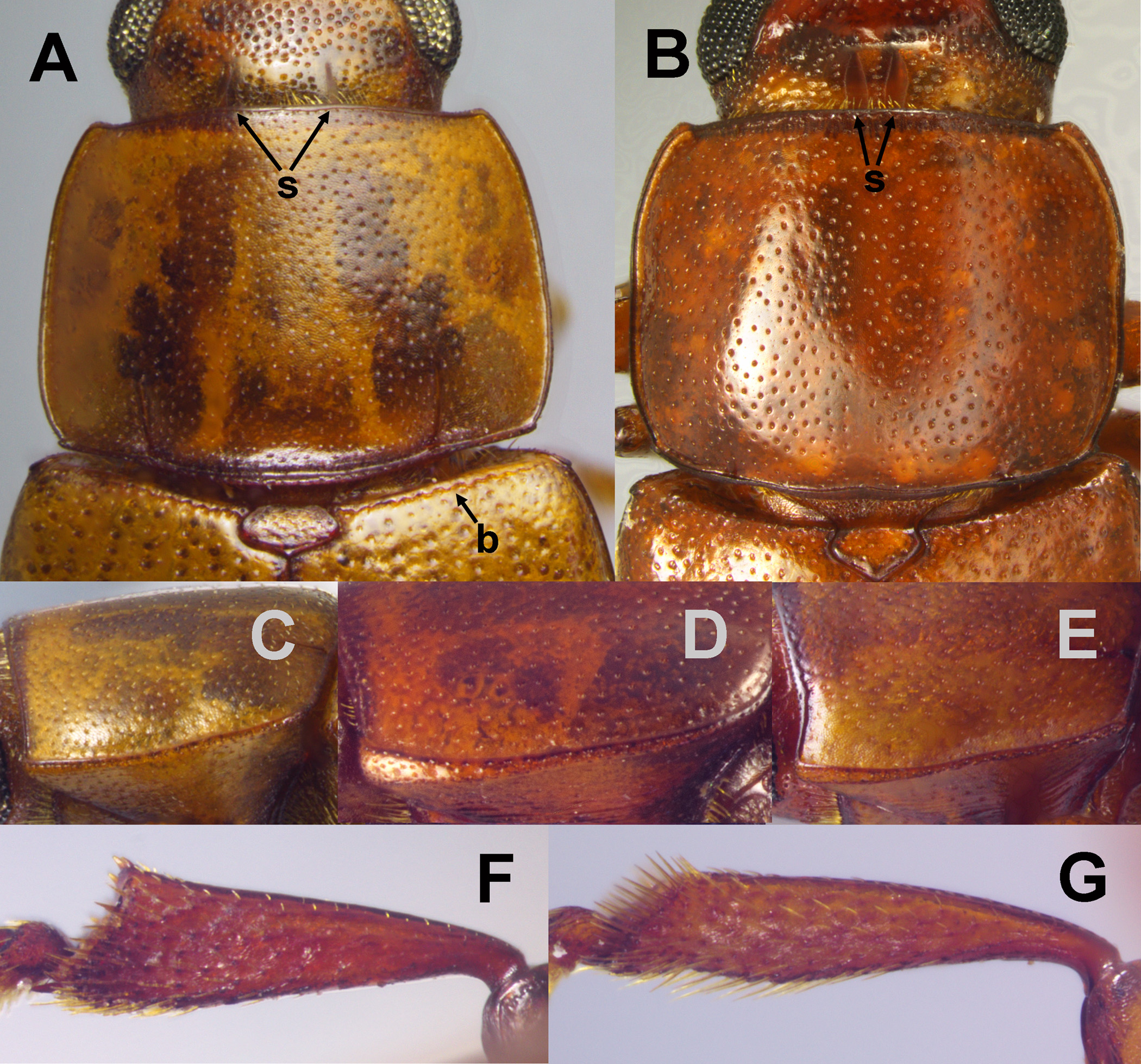
FIGURE 1. Key characters of adults: base of head, pronotum and base of elytra, dorsal view: A) Pharaxonotha sp. collected from Ceratozamia tenuis; B) Ceratophila (C.) chemnicki; pronotal left lateral margins: C) Pharaxonotha sp. from Ceratozamia tenuis; D) Ceratophila (C.) sanchezae; E) Ceratophila (Uovidesa) chiapensis from Ceratozamia vovidesii; right metatibiae of male individuals, posterior view: F) Ceratophila (C.) gregoryi; G) C. (U.) mixeorum; b: basal bead of elytra; s: stridulatory files.
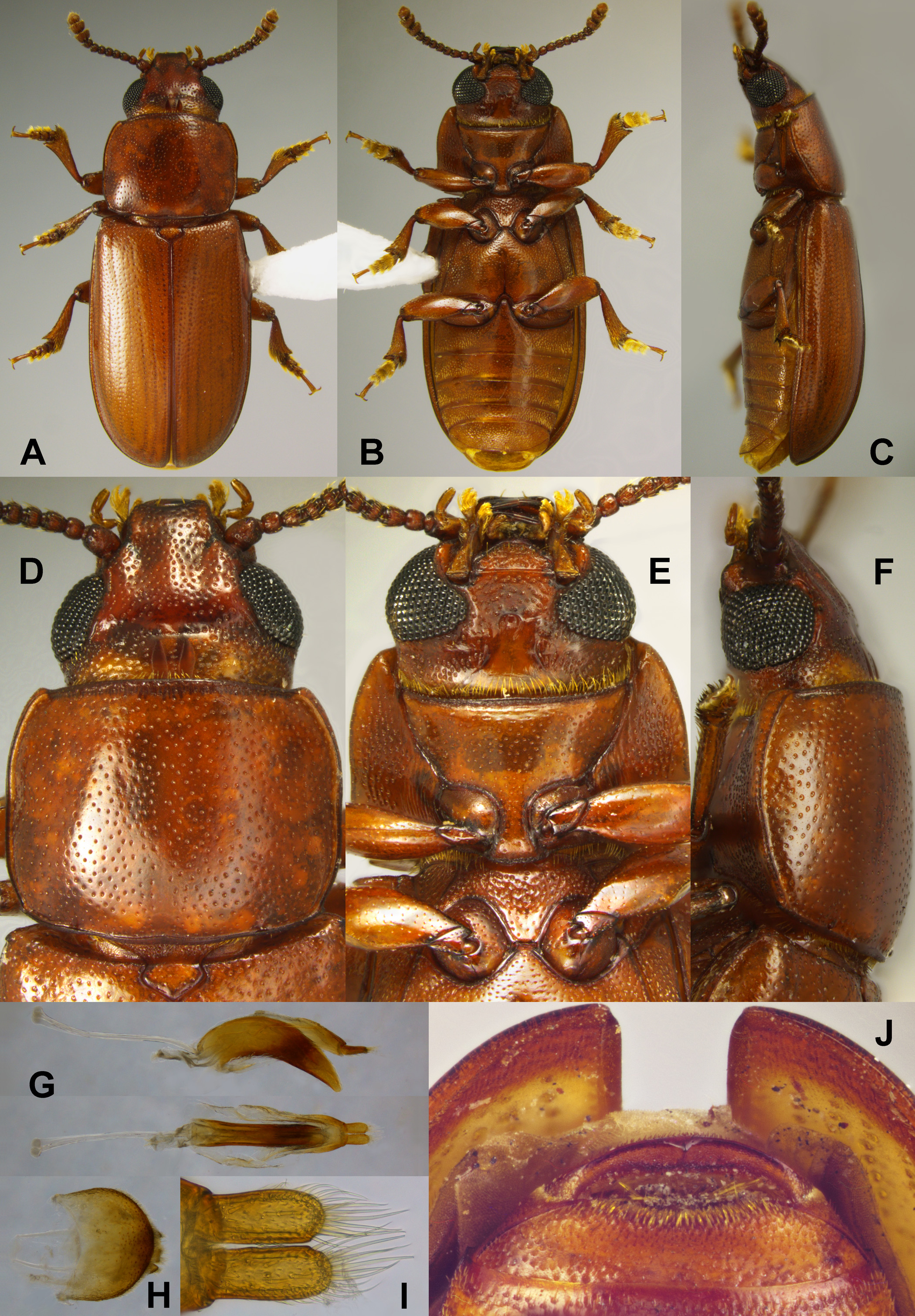
FIGURE 2. Photographs of Ceratophila (Ceratophila) chemnicki (A–I: male holotype): A–C) dorsal, ventral and lateral habitus; D–F) head, dorsal, ventral and lateral views; G) median lobe still attached with tegmen, lateral and ventral views; H) male genital capsule, dorsal view; I) close up of parameres of tegmen, ventral view; J) terminal abdominal ventrite, female.

FIGURE 3. Photographs of Ceratophila (Ceratophila) gregoryi (B–E: female allotype): A) dorsal habitus, female; B–C) ventral and lateral and habitus; D–E) head, dorsal and ventral views; F) male genital capsule, ventral view; G) median lobe still attached with tegmen, lateral and ventral views; H) close up of parameres of tegmen, ventral view.
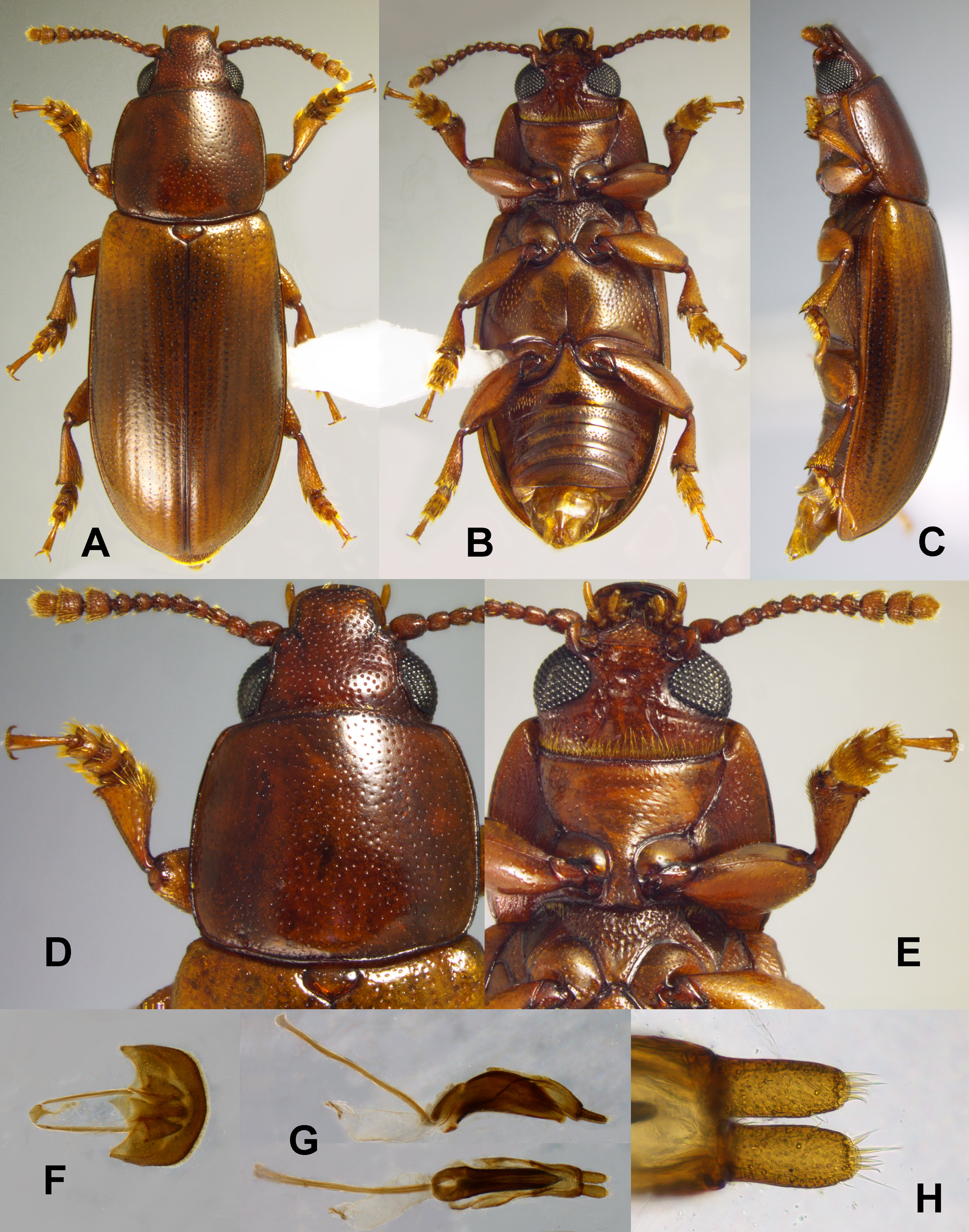
FIGURE 4. Photographs of Ceratophila (Ceratophila) picipennis collected from Ceratozamia mirandae (A–E: male holotype): A–C) dorsal, ventral and lateral and habitus; D–F) head, dorsal and ventral views; G) male genital capsule, ventral view; H) median lobe still attached with tegmen, lateral and ventral views; I) close up of parameres of tegmen, ventral view.
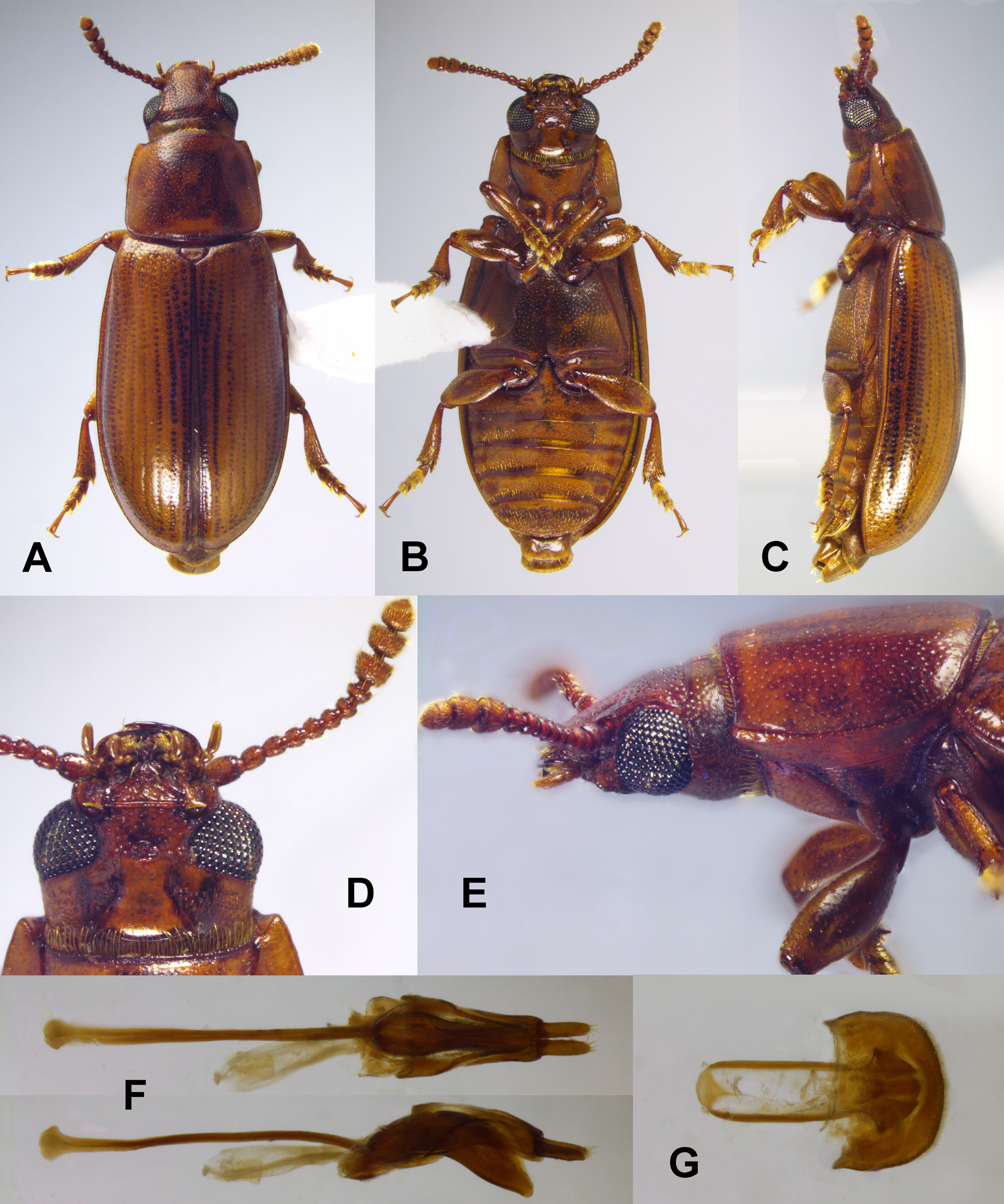
FIGURE 5. Photographs of Ceratophila (Ceratophila) sanchezae (A–E: female allotype): A–C) dorsal, ventral and lateral and habitus; D–E) head, ventral and lateral views; F) median lobe still attached with tegmen, ventral and lateral views; G) male genital capsule, ventral view.

FIGURE 6. Photographs of Ceratophila (Uovidesa) chiapensis collected from Ceratozamia vovidesii (A–F: female allotype): A–C) dorsal, ventral and lateral and habitus; D–F) head, dorsal, ventral and lateral views; G) (left to right) male genital capsule, median lobe and tegmen, ventral views; H) median lobe and tegmen, lateral views; I) close up of parameres of tegmen, ventral view.
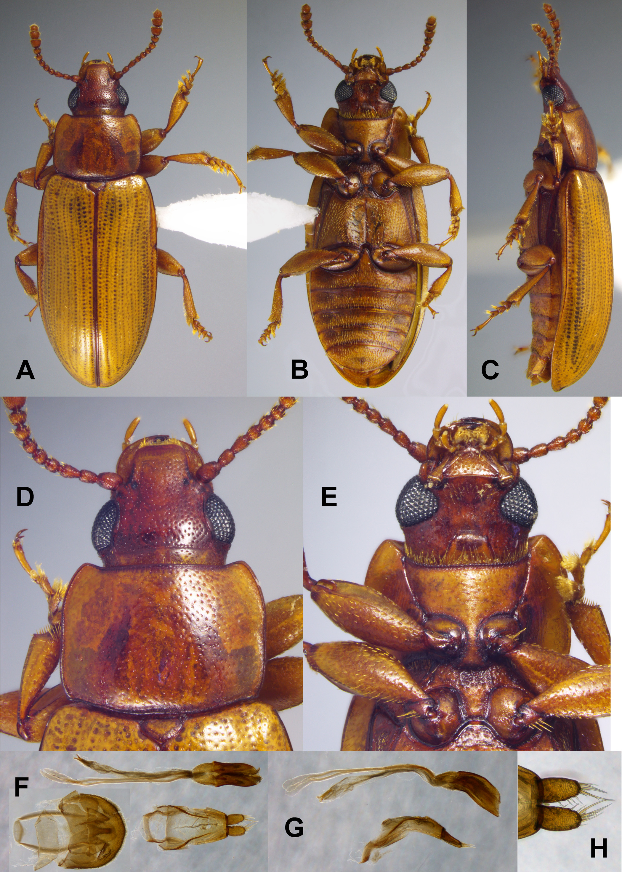
FIGURE 7. Photographs of Ceratophila (Uovidesa) mixeorum (A–E: male holotype): A–C) dorsal, ventral and lateral and habitus; D–E) head, dorsal and ventral views; F) (left to right) male genital capsule, median lobe and tegmen, ventral views; G) (top to bottom) median lobe and tegmen, lateral views; H) close up of parameres of tegmen, ventral view.
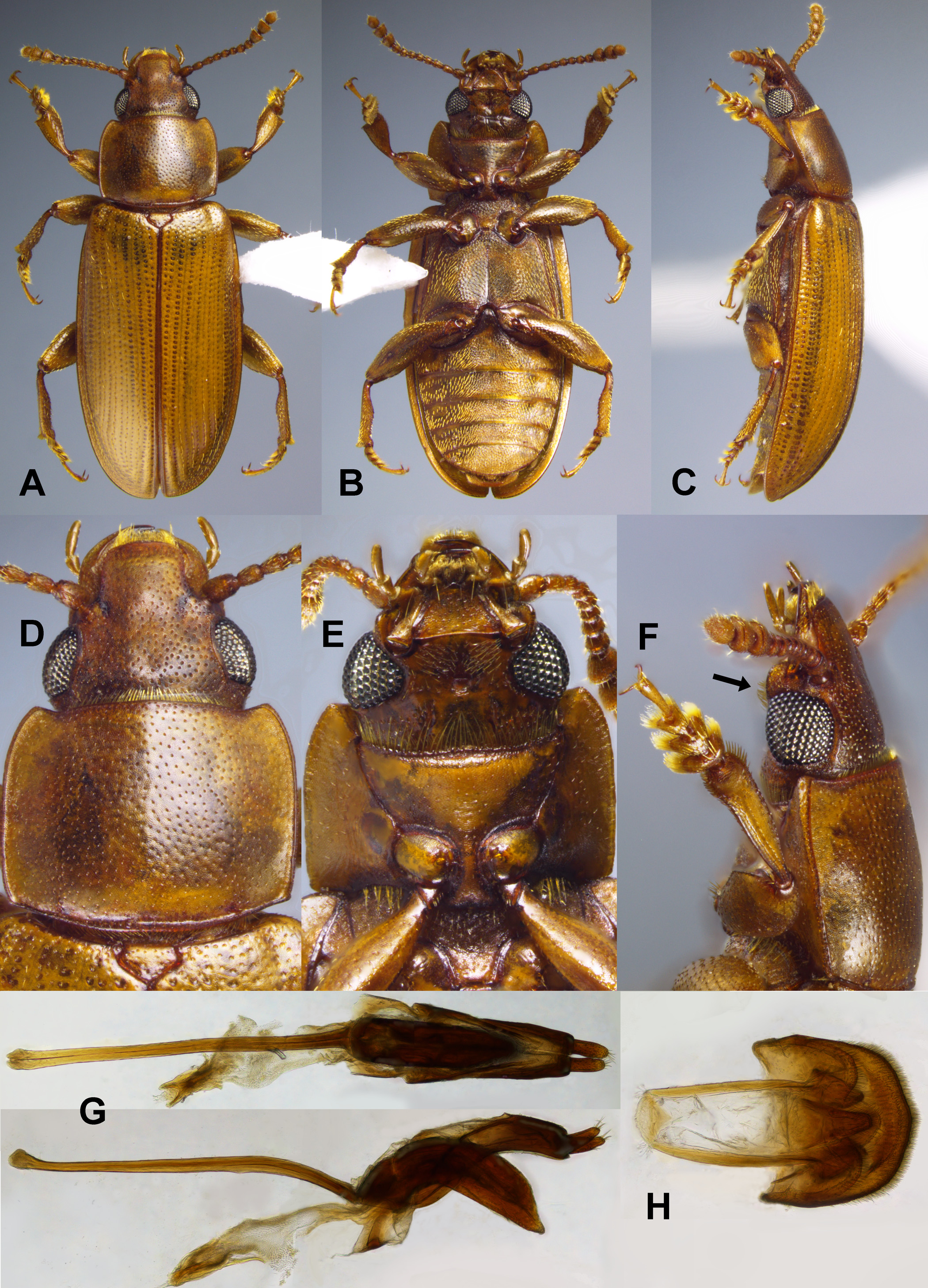
FIGURE 8. Photographs of Ceratophila (Uovidesa) vazquezi (A–D, F: male holotype): A–C) dorsal, ventral and lateral and habitus; D) head, ventral view; E) head, female, ventral view; F) head lateral view; G) median lobe still attached to tegmen, ventral and lateral views; H) male genital capsule, ventral view; arrow indicates tuft of erect setae on mentum and submentum.
No known copyright restrictions apply. See Agosti, D., Egloff, W., 2009. Taxonomic information exchange and copyright: the Plazi approach. BMC Research Notes 2009, 2:53 for further explanation.
|
Kingdom |
|
|
Phylum |
|
|
Class |
|
|
Order |
|
|
Family |
|
|
Genus |