
Baeolidia salaamica ( Rudman, 1982 )
|
publication ID |
https://doi.org/ 10.11646/zootaxa.3802.4.5 |
|
publication LSID |
lsid:zoobank.org:pub:4095CA09-8EA4-4941-8286-32E95F0206AE |
|
DOI |
https://doi.org/10.5281/zenodo.6134217 |
|
persistent identifier |
https://treatment.plazi.org/id/ED36FA51-A033-FF92-FF1B-FE00E12DF868 |
|
treatment provided by |
Plazi |
|
scientific name |
Baeolidia salaamica ( Rudman, 1982 ) |
| status |
|
Baeolidia salaamica ( Rudman, 1982) View in CoL
( Figs. 3 View FIGURE 3 D, 7C–D, 8C–D, 9A)
Spurilla salaamica Rudman, 1982: 173 View in CoL , Figs. 21–23.
Berghia salaamica ( Rudman, 1982) View in CoL : Gosliner 1985, 261.
Type locality. Dar es Salaam, Tanzania.
Type material. According to Rudman (1982), the material was deposited in the Australian Museum, Sydney (C124695).
Material examined. CASIZ 184524, one specimen, dissected, 6 mm in length preserved, Japan, Manadaru, collected by Rie Nakano, 19 April 2006; CASIZ 177397, one specimen, dissected, 3 mm in length preserved, Philippines, Luzon Island, Batangas Province, Balayan Bay, Anilao, Matotonggil, collected by Terrence M. Gosliner, 18 March 2008; CASIZ 177599, one specimen, 2 mm in length preserved, Philippines, Luzon, Batangas Province, Calumpan Peninsula, collected by Terrence M. Gosliner, 16 April 2008.
Geographical distribution. Originally described from Dar es Salaam ( Tanzania) ( Rudman 1982), this species is also known from Papua New Guinea (Gosliner et al. 2008), the Philippines (Gosliner et al. 2008), Japan ( Ono 1999, 2004; Nakano 2004), Korea ( Koh 2006) and Hawaii ( Pittman & Fiene 2012b).
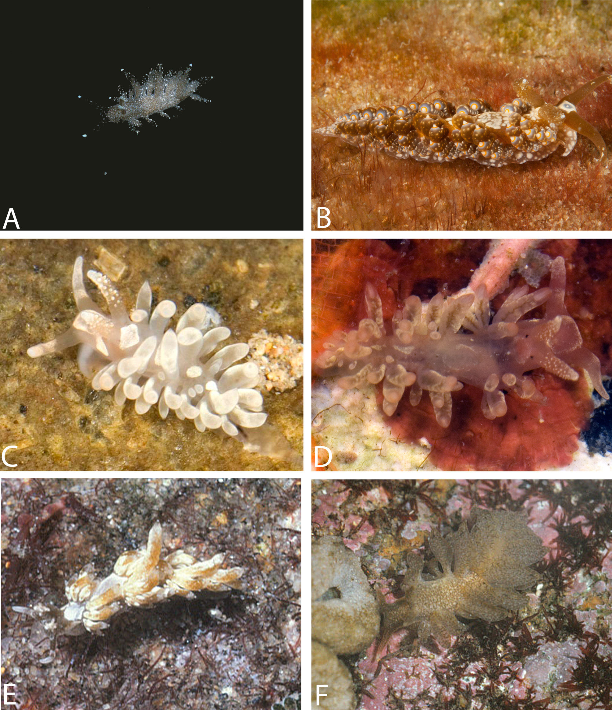
External morphology ( Fig. 3 View FIGURE 3 D, 8C–D): Body short, broad, tapering close towards posterior end of foot. Foot corners tentaculiform. Body colour translucent with white marks scattered over dorsum. White ring, which may have opaque white pigmentation in centre, on head. White diamond-shape mark behind rhinophores. Rhinophores, oral tentacles and foot corners translucent with white marks. Rhinophores shorter than oral tentacles. Rhinophores studded of minute white knobs ( Fig. 3 View FIGURE 3 D). Apex white. Oral tentacles with white tips.
Cerata moderately long, flattened, almost leaf-like. Branches of digestive gland shining through translucent body wall. Cerata may have white pigmentation (as speckles or covering outer side of cerata) on them. Apex translucent white. Cerata forming two arches plus three rows. Each arch or row contains 2–11 cerata, decreasing in size towards foot. Anus cleioproctic located within second right arch. Genital opening among cerata of anteriormost group on right side.
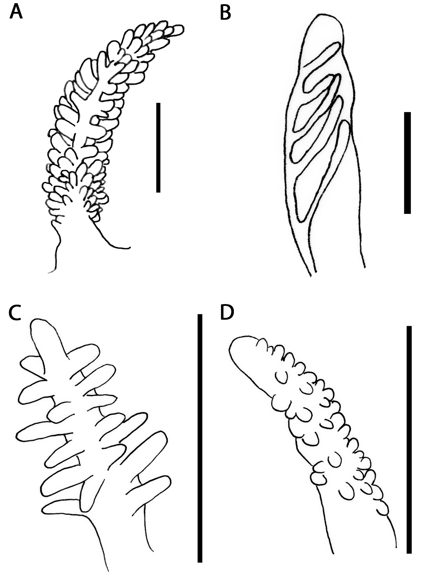
Anatomy. Masticatory border smooth ( Fig. 7 View FIGURE 7 C). Radular formulae 13 x 0.1.0 ( CASIZ 177397, 3mm) and 19 x 0.1.0 ( CASIZ 184524, 6 mm). Radular teeth slightly bi-arched with up to 25 moderately broad, acutely pointed denticles on either side of minute central cusp ( Fig. 7 View FIGURE 7 D). Teeth progressively smaller towards posterior region of radula. Oral glands small, relatively elongate, spongy. Oral glands dorso-laterally to buccal bulb. Salivary glands absent.
Reproductive system diaulic ( Fig. 9 View FIGURE 9 A). Preampullary duct widening into ampulla Postampullary duct dividing into oviduct and vas deferens. Vas deferens elongate, entering into wider proximal portion of penial sac with unarmed penial papilla. Receptaculum seminis bean-shaped, connecting to oviduct, before latter forms female glands. Vagina ventral to penis.
Remarks. Since Rudman (1982) did not consider Berghia and Baeolidia as valid genera he described this species as Spurilla . Gosliner (1985) transferred Spurilla salaamica to Berghia based on its papillate rhinophores and cerata in arches. The molecular phylogeny conducted by Carmona et al. (2013) showed that Spurilla / Berghia salaamica clustered within the Baeolidi a clade, breaking the monophyly of Baeolidia . Thus, this species was transferred to Baeolidia , as the most parsimonious alternative. Baeolidia salaamica also shares some morphological features with other members of this genus such as the knob-like papillae of the rhinophores (similar to those found in B. moebii ), the leaf-like cerata and the cerata arrangement in arches and rows (also in e.g. B. japonica ). Therefore, we can confirm that B. salaamica belongs to this genus.
Concerning the original description, only one of our specimens from the Philippines ( Fig. 8 View FIGURE 8 D) matches completely with Rudman’s (1982) diagnosis. Figure 8 View FIGURE 8 C shows that the other specimen from the Philippines has somewhat large white patches over the outer side of the cerata. Moreover, none of the specimens here examined bear a denticulate masticatory border ( Fig. 7 View FIGURE 7 C) as Rudman (1982) stated in the original description. Based on our results in previous studies ( Carmona et al. 2014a, b, c) we conclude that the latter morphological feature is not significant in Aeolidiidae .
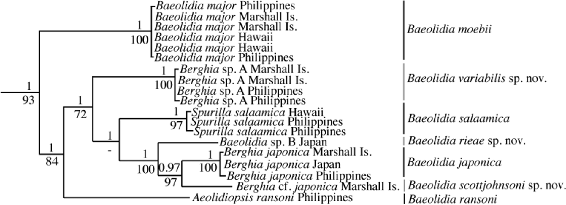
The translucent body, the white ring on the head, the white diamond-shape mark just behind the rhinophores, and mainly the kind of rhinophoral papillae, which is exclusive of Baeolidia but being only present in B. moebii and B. australis , allow us to distinguish B. salaamica from other species of this genus. Concerning the anatomical characters, it seems that B. salaamica has the wider radular teeth denticles of Baeolidia ( Fig. 7 View FIGURE 7 D). Carmona et al. (2013) studied this species from a molecular approach ( Fig. 1 View FIGURE 1 ).


No known copyright restrictions apply. See Agosti, D., Egloff, W., 2009. Taxonomic information exchange and copyright: the Plazi approach. BMC Research Notes 2009, 2:53 for further explanation.
|
Kingdom |
|
|
Phylum |
|
|
Class |
|
|
Order |
|
|
Family |
|
|
Genus |
Baeolidia salaamica ( Rudman, 1982 )
| Carmona, Leila, Pola, Marta, Gosliner, Terrence M. & Cervera, Juan Lucas 2014 |
Spurilla salaamica
| Rudman 1982: 173 |