
Halichoanolaimus ovalis Ditlevsen, 1921
|
publication ID |
https://doi.org/10.5852/ejt.2020.726.1175 |
|
publication LSID |
lsid:zoobank.org:pub:3099C8E5-38D0-4985-90AE-B8AD4CB66D98 |
|
persistent identifier |
https://treatment.plazi.org/id/E71587B1-C76B-8B13-0D4D-FA76CE91F833 |
|
treatment provided by |
Plazi (2020-12-07 10:57:39, last updated 2020-12-07 11:22:27) |
|
scientific name |
Halichoanolaimus ovalis Ditlevsen, 1921 |
| status |
|
Halichoanolaimus ovalis Ditlevsen, 1921
Table 1, Figs 1–3 View Fig View Fig View Fig
Material examined
NEW ZEALAND • 2 ƋƋ, 1 ♀; Kaikōura Canyon , 42.5082° S, 173.6325° E; water depth 1061 m; voyage TAN1006 station 7, site K4; 3 May 2010; NIWA 139245 View Materials GoogleMaps .
Type locality
North Arm of Carnley Harbour, Auckland Islands.
Description
Males
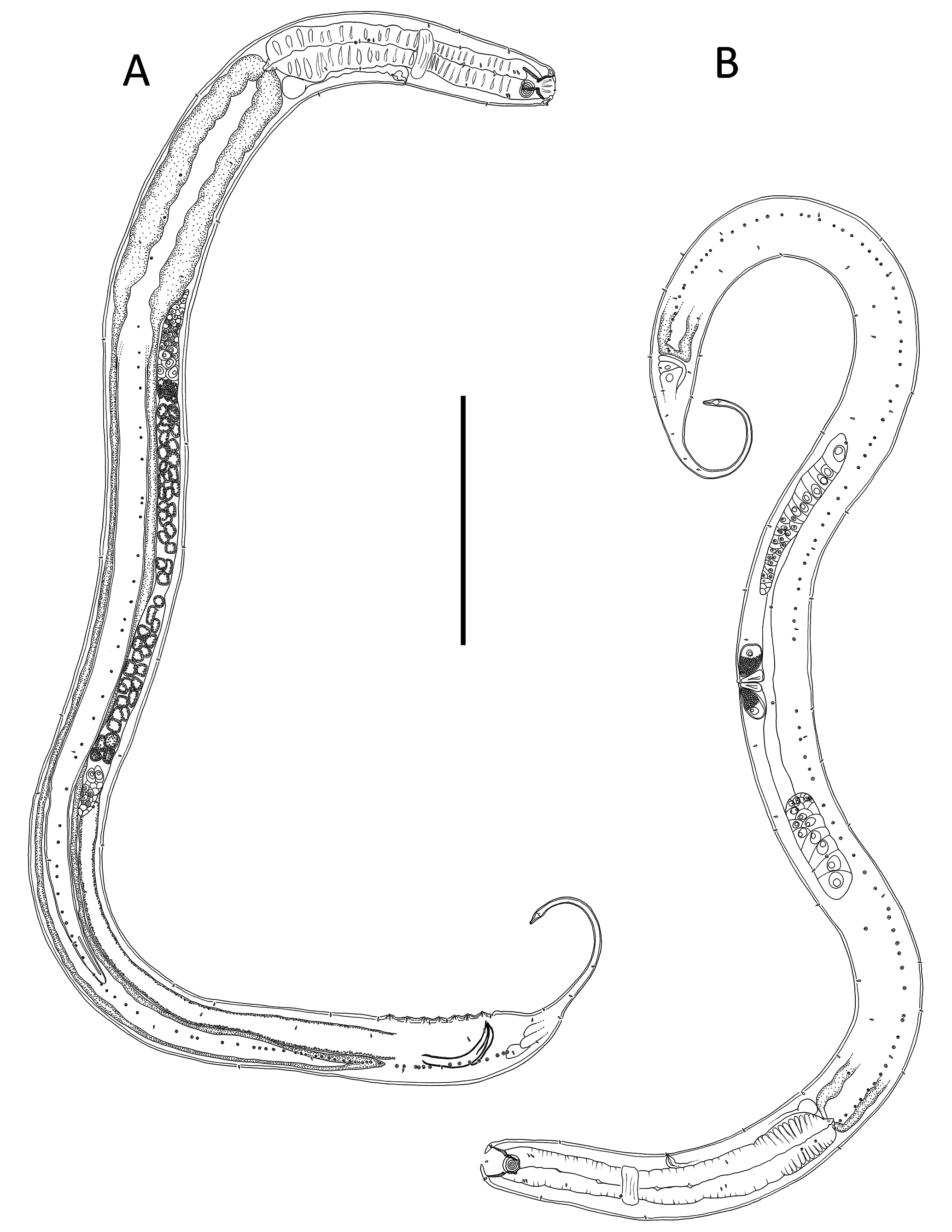
BODY. Cylindrical, tapering slightly towards anterior extremity. Cuticle with transverse rows of punctations; lateral differentiation consisting of larger, more widely spaced punctations. Two dorsosublateral rows of
Halichoanolaimus ovalis Halichoanolaimus ossilagulus sp. nov. Halichoanolaimus funestus Halichoanolaimus Species
Ditlevsen, 1921 sp. nov. pumilus sp. nov.
Males Female Males Females Male Females Male Holotype Paratype Paratype Paratype Holotype Paratype Paratype
Specimen M1 M2 F 1 Holotype M1 M2 F1 F2 M1 F1 F2
L 1461 1453 1356 1068 966 1192 1267 2709 3006 2786 756
a 22222422171817232020 15
b 775 7 6 7 8 7 99 7
c* 89711 913 9 8109 6
cʹ * 4.1 3.7 5.0 2.9 3.0 2.4 3.2 6.3 5.7 5.1 4.7 Head diam. at ceph. setae 29 29 30 20 23 24 23 40 46 44 20 Length of outer labial sensilla 2 2 2 1–2 1–2 1–2 1–2 3–4 4 3 3–4 Length of cephalic sensilla 2 2 2 1–2 1–2 1–2 1–2 3–4 4 3 3–4 Amphid height 12 12 12 8 8 8 9 15 12 13 10 Amphid width 15 16 16 12 11 11 11 17 17 17 15 Amphid width/cbd (%) 38 38 40 43 37 33 33 29 27 30 58 Amphid from anterior end 15 15 16 6 7 8 11 26 24 23 8 Nerve ring from anterior end 103 87 114 83 89 99 92 156 145 154 65 Nerve ring cbd 54 61 50 44 51 54 56 94 117 113 40 Excretory pore from anterior end 117 105 147 95 96 111 101 212 183 184 84 Pharynx length 211 209 256 150 151 162 160 377 348 317 115 Pharyngeal diam. at base 43 46 38 33 39 42 56 81 90 93 30 Pharynx cbd 61 64 53 47 53 56 64 110 125 121 42
Max. body diam. 65 66 56 48 58 68 75 120 148 141 49 Spicule length 68 73 – 56 59 – – 97 – – 49 Gubernacular apophyses length 37 43 – 26 24 – – 47 – – 24 Cloacal/anal body diam.* 45 46 40 33 36 38 46 54 55 62 29
Tail length* 183 170 200 95 109 91 147 341 311 317 136
F 68656654574872778077 61
V – – 639 – – 625 574 – 1341 1288 –
%V –– 47 – – 5245 – 4546 – Vulval body diam. – – 56 – – 68 75 – 130 137 –
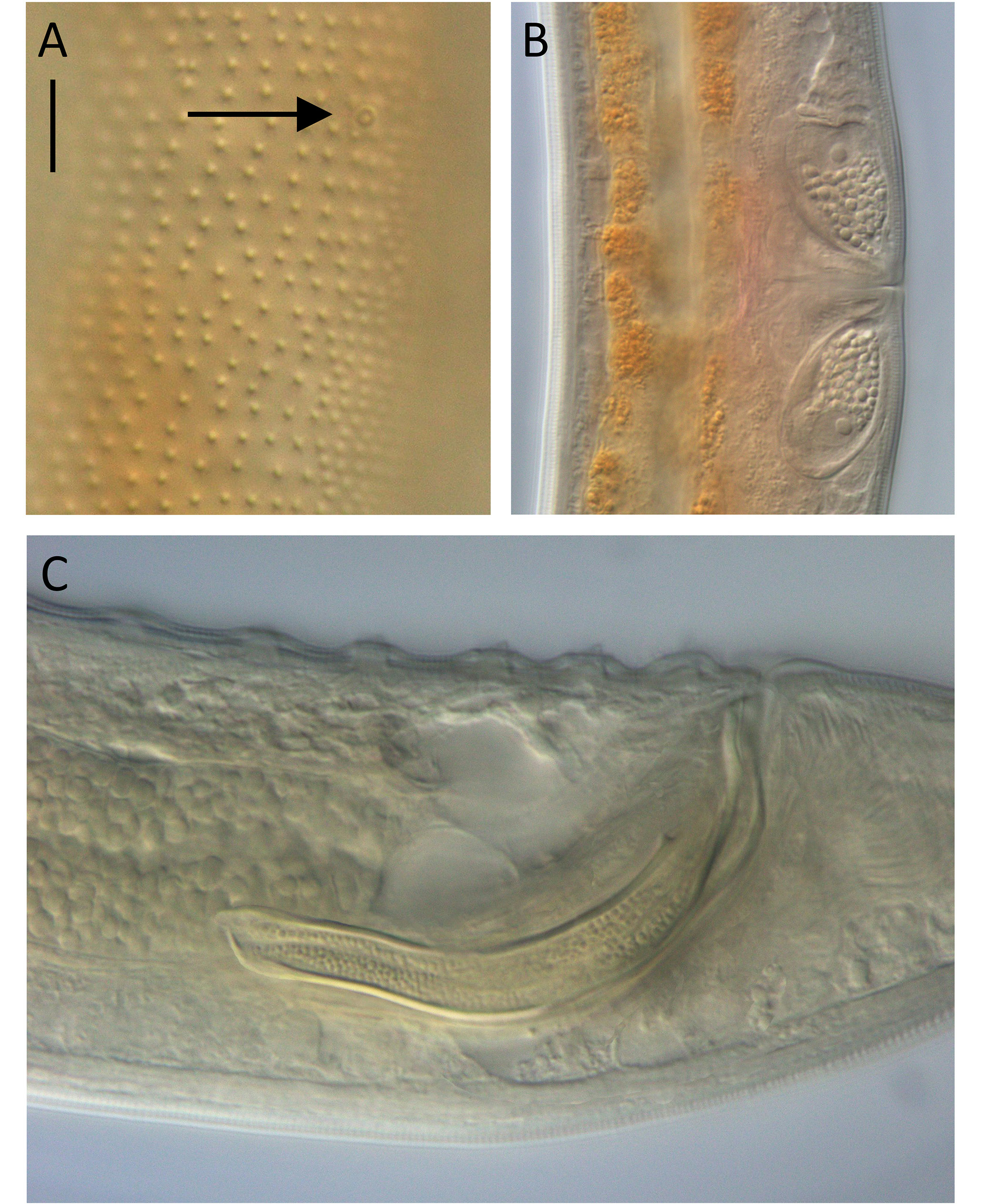
pore complexes extending from posterior to nerve ring to cloacal region, each pore complex ca 1.5 µm in diameter, becoming more closely spaced posteriorly. Up to two or three ventrosublateral pore complexes also present in pharyngeal region or slightly posterior to pharynx. Eight longitudinal rows of short, sparse somatic setae, 2–3 µm long. Cephalic region slightly rounded, with slight indentation immediately posterior to cephalic setae. Lip region not conspicuously differentiated, bearing six inner labial papillae. Six short outer labial papillae, 2 µm long, located at base of labial region and at same level as four cephalic papillae of same length. Amphideal fovea multispiral with 5.0 to 5.25 turns, situated ~0.4 cbd from anterior end. Buccal cavity (pharyngostome) large, 25–30 µm deep, divided into anterior (gymnostome) and posterior portions (stegostome). Anterior portion of buccal cavity cup-shaped, with three sets of six cuticularized rhabdions, 12–14 µm long, terminating in three sets of at least six pairs (one anterior and one posterior) of denticles; posterior portion of buccal cavity narrower, cylindrical, surrounded by three Y-shaped pairs of cuticularized rhabdions with swollen bases, 15–17 µm long. Pharynx cylindrical, muscular, without anterior or posterior bulb. Nerve ring at ca 45–50% of pharynx length from anterior. Secretory-excretory system present; renette cell up to 18 µm wide and 18 µm long, situated at level of cardia. Several nucleated pseudocoelomocytes also present around base of pharynx and either side of secretory-excretory duct; ampulla slightly smaller than renette cell, pore situated posterior to nerve ring. Cardia small, surrounded by intestine; posterior extremity of intestine blind.
REPRODUCTIVE SYSTEM. Diorchic with outstretched testes. Anterior testis to the right or ventrally to intestine, posterior testis to the left side of intestine. Sperm cells globular, 8–12 × 15–17 µm. Spicules paired, curved, tapering distally, length 1.5–1.6 body diameters at level of cloacal opening; minute ventral denticle present at one third of spicule length from distal tip, interior of spicules granular in appearance. Gubernaculum consisting of two detached lateral pieces (crurae) tapering distally, median portion of gubernaculum (corpus and cuneus) not visible. Seven precloacal supplements present, consisting of conical papillae set on cylindrical cuticular elevations each with internal duct, supplements located 10– 14 µm from each other. Tail conicocylindrical with cylindrical portion ca two thirds of total tail length; a few short and sparse somatic setae present subventrally and subdorsally. Three caudal glands located posterior to spicules, spinneret present.
Females
Similar to males but with slightly longer tail. Reproductive system didelphic-amphidelphic, with reflexed ovaries. Anterior ovary to the left of intestine and posterior ovary to the right of intestine. Vulva situated at mid-body. Mature eggs not observed. Proximal portion of vagina surrounded by constrictor muscle, two large and conspicuous vaginal glands with coarsely granulated cytoplasm and large nucleus present. Intestine blind, no rectum or anus.
Remarks
Halichoanolaimus ovalis was originally described by Ditlevsen (1921) based on two females from the Auckland Islands, and males were later described from the littoral zone of Campbell Island by Allgén (1927) (both Auckland and Campbell islands are located in the Southern Ocean directly south of New Zealand’s South Island). The female specimen from Kaikōura Canyon broadly resembles the original description of Ditlevsen (1921), although the Kaikōura Canyon female is shorter (body length 1.4 vs 1.8 mm in H. ovalis ) and has a higher ratio of ‘a’ (24 vs 18). Ditlevsen (1921) states that the amphideal fovea of H. ovalis females has six turns; however, his figure shows only five turns, which is consistent with the Kaikōura Canyon specimen. The present description is also consistent with the observation of conspicuous vaginal glands with coarsely granulated cytoplasm by Ditlevsen (1921). The two male specimens from Kaikōura Canyon agree well with the description of Allgén (1927) in general body dimensions, although like the Auckland Islands specimens, the Campbell Island specimens are somewhat stouter as indicated by a lower value of ‘a’ (17) relative to the Kaikōura Canyon specimens (22). In addition, Allgén counted only four amphideal fovea turns in his male specimens compared to five in the Kaikōura Canyon specimens. However, the structure of the copulatory apparatus, as well as the shape and number of precloacal supplements, are the same.
The intestine of one of the male H. ovalis specimens contained the anterior half of a nematode prey which was identified as belonging to Halalaimus de Man, 1888 based on the structure of the cuticle, amphideal fovea and buccal cavity. The intestine of the other H. ovalis male (from the same sample) contained the posterior half of a nematode, possibly also belonging to the genus Halalaimus .
Allgen C. 1927. Freilebende marine nematoden von den Campbell- und Staten-Inseln. Nyt Magasin for Naturvidenskaberne 66: 249 - 309.
Ditlevsen H. 1921. Papers from Dr. Th. Mortensens Pacific Expedition 1914 - 16. III Marine freeliving Nematodes from the Auckland and Campbell Islands. Videnskabelige Meddelelser fra Dansk naturhistorisk Forening i KJObenhavn 73: 1 - 39.
Man de J. G. 1886. Anatomische Untersuchungen uber freilebende Nordsee-Nematoden. Leipzig, Verlag von Paul Frohberg.
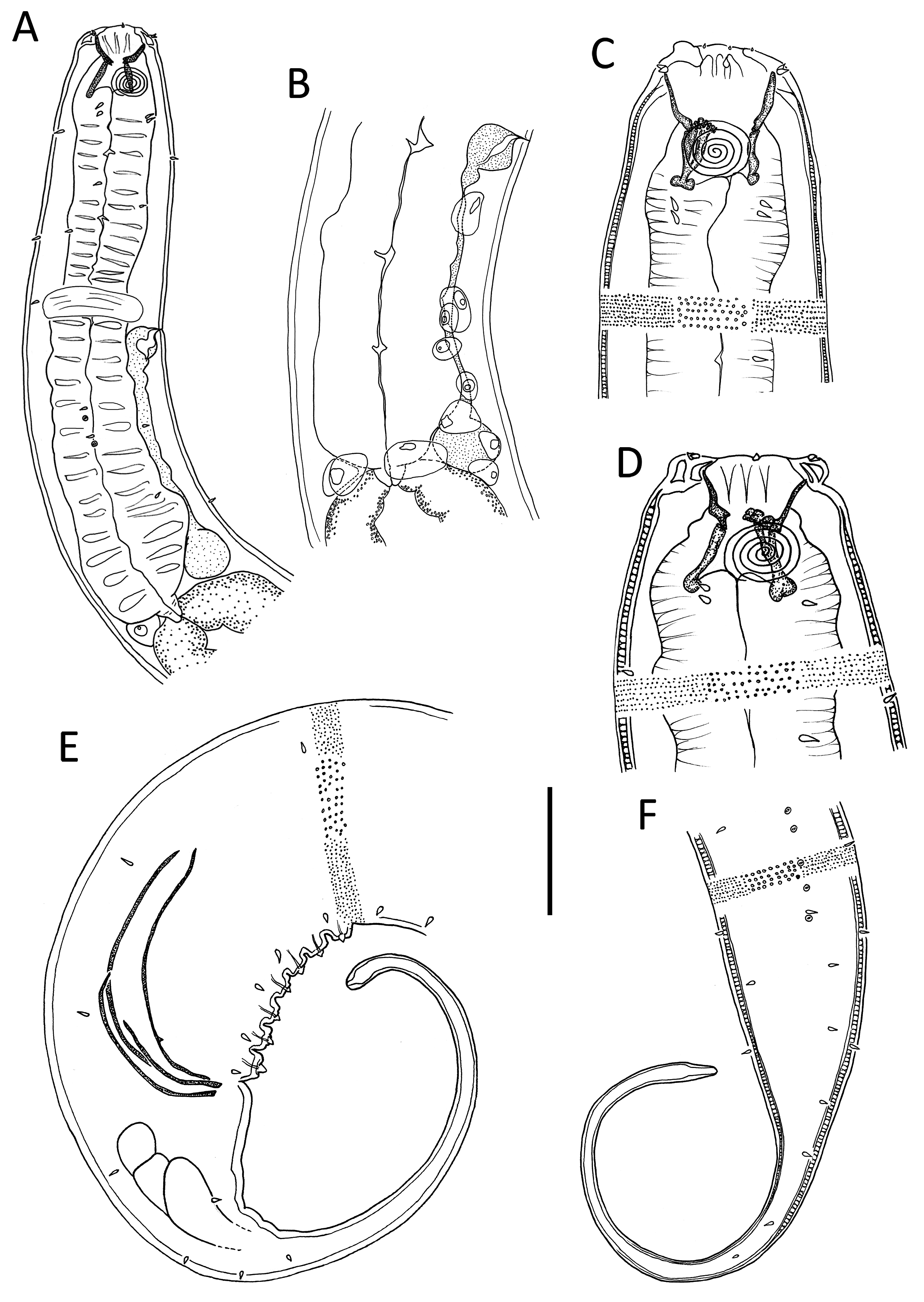
Fig. 1. Halichoanolaimus ovalis Ditlevsen, 1921 A. Male anterior body region. B. Posterior male pharyngeal region and anterior intestinal region showing location of pseudocoelomocytes. C. Female cephalic region. D. Male cephalic region. E. Male posterior body region. F. Female posterior body region. Scale bar: A = 50 µm; B = 37 µm; C, E = 27 µm; D = 25 µm; F = 35 µm.
No known copyright restrictions apply. See Agosti, D., Egloff, W., 2009. Taxonomic information exchange and copyright: the Plazi approach. BMC Research Notes 2009, 2:53 for further explanation.
|
Kingdom |
|
|
Phylum |
|
|
Class |
|
|
Order |
|
|
Family |
|
|
Genus |
1 (by plazi, 2020-12-07 10:57:39)
2 (by ExternalLinkService, 2020-12-07 11:22:27)
3 (by valdenar, 2020-12-07 13:13:11)
4 (by valdenar, 2020-12-07 13:15:28)
5 (by ExternalLinkService, 2020-12-07 13:25:37)
6 (by ExternalLinkService, 2021-11-09 12:10:09)
7 (by ExternalLinkService, 2021-11-09 14:26:09)
8 (by plazi, 2023-11-01 00:06:09)
9 (by ExternalLinkService, 2023-11-01 14:46:02)