
Luciola Laporte
|
publication ID |
https://doi.org/10.11646/zootaxa.3653.1.1 |
|
publication LSID |
lsid:zoobank.org:pub:72A07BC6-AEB0-4EBC-AFA8-F5871065680F |
|
persistent identifier |
https://treatment.plazi.org/id/E46FF216-5E3D-C508-FF6F-B986FC13ECA1 |
|
treatment provided by |
Felipe (2021-08-25 02:01:40, last updated by Plazi 2023-11-04 18:34:06) |
|
scientific name |
Luciola Laporte |
| status |
s. str. |
Luciola Laporte View in CoL s. str.
[ Figs 109–138 View FIGURES 109–119 View FIGURES 120–127 View FIGURES 128–138 ]
Luciola Laporte. Laporte 1833:146 View in CoL . Lacordaire 1857:335. Motschulsky, 1853:52. Gorham 1880:99. Olivier 1902:69; 1907:50. Lea 1909:106.
Type species: Luciola pedemontana Motschulsky designated by Motschulsky 1853.
Luciola (Luciola) Laporte. McDermott, 1966:103 (Partim) . Nec Calder, 1998: 178.
Lampyroidea View in CoL Costa. Costa 1875:clxix. Olivier 1902:69; 1907:49; 1911:102. McDermott 1966:115. Type species: Lampyroidea syriaca Costa monobasic.
Bourgeoisia Olivier. Olivier 1908:17 View in CoL ; 1911b:102. McDermott, 1966:117. Deheyn & Ballantyne 2009:47. Type species: Luciola antipodum Bourgeois View in CoL designated by McDermott 1966.
Diagnosis. Luciola s. str. belongs in a group of genera with aedeagal LL visible beside the ML; distinguished by the separation of the LL dorsally, the often strongly curved/arched ML terminating in a preapical ventral point, and the presence in some species (including italica ) of elongate slender apically pointed lobes arising from the inner ventral margins of the LL. Dorsal colour pattern of orange pronotum and dark brown elytra occurs in several species including the type (which may have a median dark pronotal marking); Pacific Island species have deep grayish brown to black dorsal colour (see Table 9) and several have eye emarginations of varying depth. Females macropterous in L. kagiana , or with varying degrees of fore and hind wing loss in other species. Larvae reliably associated only for kagiana and parvula where they are probably terrestrial and have laterally explanate tergal margins (e.g. Chen 2003:168).
Male. Pronotum: dorsal surface without irregularities in posterolateral areas and longitudinal groove in lateral areas; punctation dense; anterior margin not explanate; lateral margins either diverging posteriorly along their length (C>A, B), or converging in posterior 1/3; width <or subequal to humeral width; anterolateral corners rounded obtuse; lateral margins without indentation at mid-point, or sinuousity in either horizontal or vertical plane; without indentation in lateral margin near posterolateral corner, and irregularities at corner; posterolateral corners usually rounded obtuse, angulate in Pacific Island species where they are 90° approximately and incline obliquely to the median line; posterolateral corners either not projecting, or extending as far as median posterior margin and separated from it by scarce emarginations except in kagiana where the emarginations are well developed.
Hypomera: closed; median area not elevated in vertical direction; posterior area not flat in italica , syriaca and hypocrita , narrowly flat in remainder, where dorsal and ventral surfaces are strongly adpressed; pronotal width/ GHW 1.2–1.6.
Elytron: punctation dense, not linear, not as large as that of pronotum, nor widely and evenly spaced; apices not deflexed; epipleuron and sutural ridge extending beyond mid-point, almost to apex but not extending as a ridge around apex, neither thickened in apical half; no interstitial lines; elytral carina absent; in horizontal specimen viewed from below epipleuron at elytral base wide, covering humerus; viewed from above the anterior margin of the epipleuron arises level with or anterior to posterior margin of MS; epipleuron developed as a lateral ridge along most of length; sutural margins approximate along most of length in closed elytra; lateral margins parallel-sided except for hypocrita where they are slightly convex-sided.
Head: minimally to moderately depressed between eyes; well exposed in front of pronotum, not capable of complete retraction within prothoracic cavity; eyes moderately separated beneath at level of posterior margin of mouthpart complex in L. italica , contiguous ventrally in Pacific Island species ( Fig. 111 View FIGURES 109–119 ); eyes above labrum close, sometimes contiguous; frons-vertex junction rounded, without median elevation; posterolateral eye excavation strongly developed, visible in resting head position in some Pacific Island species only (Figs 110,120,121,122); antennal sockets on head between eyes, contiguous, or separated by <ASW or = ASW; clypeolabral suture present, flexible, not in front of anterior eye margin when head viewed with labrum horizontal; outer edges of labrum reach inner edges of closed mandibles. Mouthparts: probably functional; apical labial palpomere either strongly flattened, shaped like broad triangle (widest at base), with inner edge dentate in L. italica , or ovoid, longer than wide with margins entire; at least half as long as apical maxillary palpomere in the remaining species described below. Antennae 11 segmented; length>GHW up to twice GHW except in Pacific Island species where antennal length is subequal to GHW; no segments flattened, shortened, or expanded; pedicel not produced; FS1 not shorter than pedicel.
Legs: with inner tarsal claw not split; without MFC; no femora or tibiae swollen or curved; no basitarsi expanded or excavated.
Abdomen (Figs114,119,124,125): without cuticular remnants in association with aedeagal sheath; no ventrites with curved posterior margins nor extending anteriorly into emarginated posterior margin of anterior segment; LO absent in L. oculofissa sp. nov. (Figs 124,125); LO absent in V7 and restricted to anterolateral plaques in V 6 in L. hypocrita ( Deheyn & Ballantyne 2009 Fig. 5 a,b View FIGURES 3–5 ); LO in V 7 in remaining species entire, either occupying most of V7, and reaching to sides and almost to posterior margin ( Fig. 119 View FIGURES 109–119 ), or not reaching sides or posterior margin and occupying about half or less of V7 ( Fig. 114 View FIGURES 109–119 ); posterior half of V7 not arched or swollen, muscle impressions not visible in this area; neither anterior nor posterior margin of LO emarginate; if LO present in V6, occupying almost all V6 except in hypocrita . MPP present, symmetrical, apex rounded or truncate, not laterally compressed, short, not inclined dorsally nor engulfed by T8 apex, without dorsal ridge, median longitudinal trough. V7 without median carina, median longitudinal trough, anteromedian depression on face of LO, incurving lobes or pointed projections, median ‘dimple’, or reflexed lobes. T7 without prolonged anterolateral corners. T8 symmetrical, W=L, visible posterior area not narrowing abruptly, median posterior margin shallowly and narrowly emarginate; widest across middle with lateral margins tapering evenly in both an anterior and posterior direction; without prolonged posterolateral corners, median posterior projections, not inclined ventrally nor engulfing posterior margin of V7 nor MPP, not extending conspicuously beyond posterior margin of V7; T8 ventral surface without well developed median longitudinal trough, lateral depressed troughs, asymmetrical projections, median posterior ridge; concealed anterolateral arms of T8 either not as long as visible posterior portion of T8, or shorter, not laterally emarginated before their origins, not expanded dorsoventrally, expanded only in horizontal plane; without bifurcation of inner margin and ventrally directed pieces; lateral margins of T8 not enfolding sides of V7.
Aedeagal sheath: approx. 3 times as long as wide; without bulbous paraprocts; either symmetrical in posterior area where sheath sternite tapers evenly to a narrow rounded apex or slightly emarginated on right side; tergite without lateral arms extending anteriorly at sides of sheath sternite; tergite without projecting pieces along posterior margin of T9, anterior margin without transverse band.
Aedeagus ( Figs 111–113 View FIGURES 109–119 , 122,126) (e.g. Ballantyne 1968 Figs 162–169 View FIGURES 160–167 View FIGURES 168–176 ; Jeng et al. 2003 Figs 21A–C View FIGURES 18–25 ): LL lack lateral appendages; LL visible from beneath beside ML, LL/ML moderate to wide; LL of equal length, slightly longer than ML, either diverging along their length or not diverging basally; separated longitudinally by most of their length; LL base width often narrower than LL apex width which may be wider than that of ML; LL apices often more widely expanded than elsewhere and enfolding the ML at the sides; dorsal base of LL symmetrical, not excavated; LL without lateral hairy appendages along their outer ventral margins; narrowed apices of LL sometimes inturned; without projection on left LL; inner margins often with slender leaf-like projection; ML symmetrical, without paired lateral teeth and tooth to left side, usually strongly arched, preapical ventral area produced and pointed; BP not strongly sclerotised, not hooded, not strongly emarginated along anterior margin, often very narrow.
Female. Macropterous in kagiana ( Chen 2003:168) , or with varying degrees of fore and hind wing loss. Pronotum without irregularities in posterolateral areas; punctation moderate to dense; pronotal width less than, subequal to or greater than humeral width; without indentation of lateral margin, irregularities at posterolateral corner; outline similar to that of male. Elytral punctation not as large as that of pronotum, nor evenly spaced; no interstitial lines; elytral carina absent. No legs or parts thereof swollen and /or curved. LO in V6 only, without any elevations or depressions or ridges on V7; median posterior margin of V7 widely emarginate, median area not broadly rounded; median posterior margin of V8 entire. Bursa plates not observed in dissections of ethanol preserved specimens of italica .
Larva ( Figs 128–138 View FIGURES 128–138 ). Here only L. hypocrita , and New Caledonian larvae associated by label data are described ( Tables 10, 11 indicate rationale for association). Elongate, slender, tapering somewhat in front and behind; with 3 thoracic and 9 abdominal segments; external plates of the dorsal surface very well sclerotised with no obvious dorsal areas of exposed membrane except between segments; all body segments except the last with a median dorsal longitudinal line; lateral margins of tergal plates explanate thickened (except for terminal segment which is parallel-sided), and projecting beyond sides of body and usually covering laterotergites in the abdomen, visible only if they are laterally prolonged. Head: antennal segment 3 surmounted by a ring of hairs and subequal in length to the elongate sense cone. Mouthparts: mandibles without inner teeth; apical palpomeres of maxilla and labium with terminal sense organs. Thorax ( Figs 128, 131, 132, 135, 137, 138 View FIGURES 128–138 ): prothorax longer than wide, always with lateral projections especially at posterolateral corners; meso and metathoracic segments shorter than prothorax and with 2 or 3 lateral projections. Legs: tibiae with an apical brush of fine white hairs reaching over the apical claw (tarsungulus) ( Fu et al. 2012b Fig. 55 View FIGURES 45–55 ). Abdomen ( Figs 129, 130, 133, 134 View FIGURES 128–138 ): median sternal plates of segments 1–6 and laterotergites of segments 1–6 with short posterior projections in Pacific Island species; posterolateral corners of laterotergites angulate, often narrowly prolonged and may be visible at sides of body from above when lateral margins of terga are narrowed and prolonged ( Fig. 134 View FIGURES 128–138 ) ( Fu et al. 2012b Fig. 54 View FIGURES 45–55 ).
Remarks. Luciola s. str. is addressed from scoring a population from Pisa of Luciola italica , the type species ( Ballantyne and Lambkin 2000, 2001, 2006, 2009). Bourgeoisia and Lampyroidea (based only on its type species syriaca ) are submerged under Luciola . Luciola italica exists in a variety of morphological forms across Europe, and such an investigation while necessary is currently beyond our capacity. Suggestions for possible subdivisions below relate to Figs 3, 4 View FIGURES 3–5 and the key to genera and species groups incorporates all these possible subdivisions of Luciola .
1. Fig. 3 View FIGURES 3–5 Node 1 two aquatic Japanese species Luciola cruciata , L. owadai .
2. Fig. 3 View FIGURES 3–5 Node 8 blue number 2 ( Luciola trilucida ).
3. Fig. 3 View FIGURES 3–5 Node 11 blue number 3 ( Luciola indica ).
4. Fig. 4 View FIGURES 3–5 Node 44 blue number 20 Species 8.
5. Fig. 4 View FIGURES 3–5 Node 45 includes species with elytral punctures in lines, an emarginated LO in V7 and sclerites surrounding the aedeagal sheath ( L. carinata – L. aquatilis ).
6. Fig. 4 View FIGURES 3–5 Node 46 ( Luciola dejeani – L. aegrota ).
Footnote: *1 hand collected at night; 2 yellow pan in day
* single larva from Foret du Nord lacks the anterolateral projection; #curved towards rear in 1 larva from Col d’Amieu, 3
Aoupinnie larvae from top camp and sawmill, and in one of two larvae taken on 23.xi.2001; + outer posterior margin of lateral tergal projections with small tooth in Mt Taom, Mt Mou base, one Dzumac Road larva and Foret du Nord larva.
List of species of Luciola s. str.
- aquilaclara sp. nov. *
- italica L.
- oculofissa sp. nov. *
Here we address specifically only the fauna of New Caledonia and Fiji *.
Ballantyne, L. A. (1968) Revisional Studies of Australian and Indomalayan Luciolini (Coleoptera, Lampyridae, Luciolinae). University of Queensland Papers, Department of Entomology, University of Queensland Press, II, no 6, 103 - 139.
Ballantyne, L. A. & Lambkin, C. (2000) Lampyridae of Australia (Coleoptera: Lampyridae: Luciolinae: Luciolini). Memoirs of the Queensland Museum, 46 (1), 15 - 93.
Calder, A. A. (1998) Coleoptera: Elateroidea In Wells, A. (ed.) Zoological Catalogue of Australia, 29.6 (CSIRO Publishing Melbourne).
Chen, T. R. (2003) Fireflies of Taiwan. Field Image Publications, Taipei, Taiwan. 255 pp. (In Chinese)
Costa, A. (1875) The genus Lampyroidea. Bulletin de la Societe Entomologique de France, (5) 1875, clxix.
Deheyn, Dimitri D. & Lesley A. Ballantyne (2009) Optical characterization and redescription of the South Pacific firefly Bourgeoisia hypocrita Olivier (Coleoptera: Lampyridae: Luciolinae). Zootaxa, 2129, 47 - 62.
Fu, X. H., Ballantyne, L. & Lambkin, C. (2012 b) The external larval morphology of aquatic and terrestrial Luciolinae fireflies (Coleoptera: Lampyridae). Zootaxa, 3405, 1 - 34.
Gorham, H. S. (1880) Materials for a revision of the Lampyridae. Transactions of the Royal Entomological Society of London 1880, 1 - 36, 83 - 112. http: // dx. doi. org / 10.1111 / j. 1365 - 2311.1880. tb 02023. x
Jeng, M. L, Lai, J. & Yang, P. S. (2003) Lampyridae: A synopsis of aquatic fireflies with description of a new species (Coleoptera). Pp. 539 - 562 in Jach, MA & Ji, L. Editors. Water Beetles of China. Volume III. Zoologisch-Botanische Gesellschaft in Asterreichand wiener Coleopterologenverein. Vienna, Austria, 572 pp.
Lacordaire, T. (1857) Histoire naturelle des insectes. Genera des Coleopteres. Tome Quatrieme. Roret: Paris, 579 pp.
Laporte, F. L. de (1833) Essai d'une revision du genre Lampyre. Annales de la Societe Entomologique de France, 122 - 153.
Lea, A. M. (1909) Revision of the Australian and Tasmanian Malacodermidae. Transactions of the Entomological Society of London, 1909, 45 - 251. [Lampyridae, 106 - 111].
McDermott, F. A. (1966) Lampyridae Pars. 9. Coleopterorum Catalogus (Junk-Schenkling) (Ed. Sec), 1 - 149 (Luciolini, 98 - 118).
Motschulsky, V. de (1853) Lampyrides. Etudes Entomologiques, I, 33 - 44.
Olivier, E. (1902) Catalogue des especes de ' Luciola' et genres voisins decrits jusqu'a ce jour. Revue Scientifique du Bourbonnais et du centre de la France, 15, 69 - 88.
Olivier, E. (1907) Coleoptera, Lampyridae. Genera Insectorum, 53, 1 - 74 [Luciolinae, 48 - 56].
Olivier, E. (1908) Description d'un nouveau genre de Lampyrides (Col.). Annales de la Societe Entomologique de France, 1908, 17 - 18.
Olivier, E. (1911 b) Revision des Lampyrides. Revue Scientifique du Bourbonnais et du centre de la France, 24, 24 - 27, 39 - 58, 63 - 85, 98 - 112.
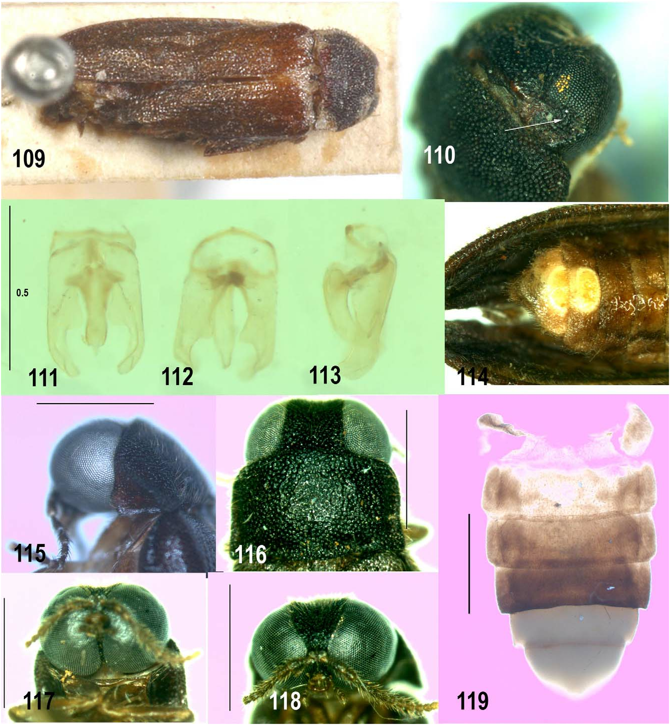
FIGURES 109–119. Luciola spp. Males. 109–114 Luciola antipodum Bourgeois (MNHN). 115–119 Luciola aquilaclara sp. nov. paratype male (QMBA). 109, 116 dorsal (116 head and pronotum only); 110 right posterolateral head (eye excavation arrowed); 111–113 aedeagus ventral (111), dorsal (112) and right lateral; 114 terminal abdomen and elytra ventral; 115–118 head and pronotum left lateral (115), dorsal (116), anteroventral (117) and anterior; 119 abdominal ventrites 3–7 ventral. These figures share scale lines: 111–113.

FIGURES 120–127. Luciola oculofissa sp. nov. paratype males (QMBA). 120, 121 head and pronotum, left lateral (121) and dorsal (Pic du Grand Kaori); 122 head anterior (Foret du Nord); 123, 124, whole body dorsal (123) and ventral (Foret du Nord); 125 V6, 7 ventral (Col de Petchecara); 126, 127 aedeagus in aedeagal sheath ventral (126, anterior end to left of page), ventrolateral (127, anterior end to right of page (Rive Bleu). Figure legends: BP basal piece; LL lateral lobes; ML median lobe. These figures share scale lines: 123, 124.
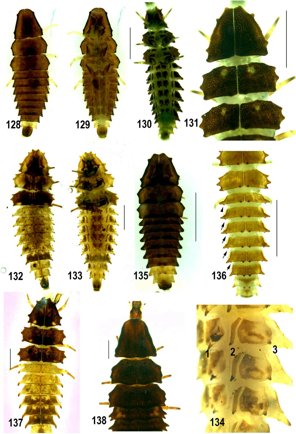
FIGURES 128–138. Luciola spp. Larvae (QMBA). Luciola oculofissa sp. nov. 128, 129 (Foret electrique), 135 (Touhu TV Tower). Luciola aquilaclara sp. nov. 130–134 (130, 131 Col d’Amieu, 132–134 Mandelia summit), 136 (Mt Taom summit), 137 (Aopinnie). 138 Luciola hypocrita. 128, 132, 135 dorsal whole body; 131 dorsal thorax only; 136 dorsal thoracic terga 2, 3, abdominal terga 1–8 (teeth along posterior margins of terga arrowed); 137 dorsal thorax and abdominal segments 1–7; 129, 130, 133 ventral; 134 left ventrolateral 3 abdominal segments. Figure legends: 1 left projection of sternum; 2 posterior projection of laterosternite; 3 posterior projection of laterotergite. These figures share scale lines: 128–130; 132, 133.

FIGURES 3–5. Majority rule tree for the complete analysis of the Luciolinae. Analysis consisted of consensus of 169 MPTs with a length of 5609 from a data set of 143 taxa and 436 morphological characters in (see Table 3). Figure 3, part 1: nodes 1– 34; 4, part 2 Nodes 35–52; 5, part 3 nodes 55–76. Nodes (red). New genera, new species, and still undescribed species in red and bold, with identification numbers blue. Black values above branches show the % of MPTs over 50 %.
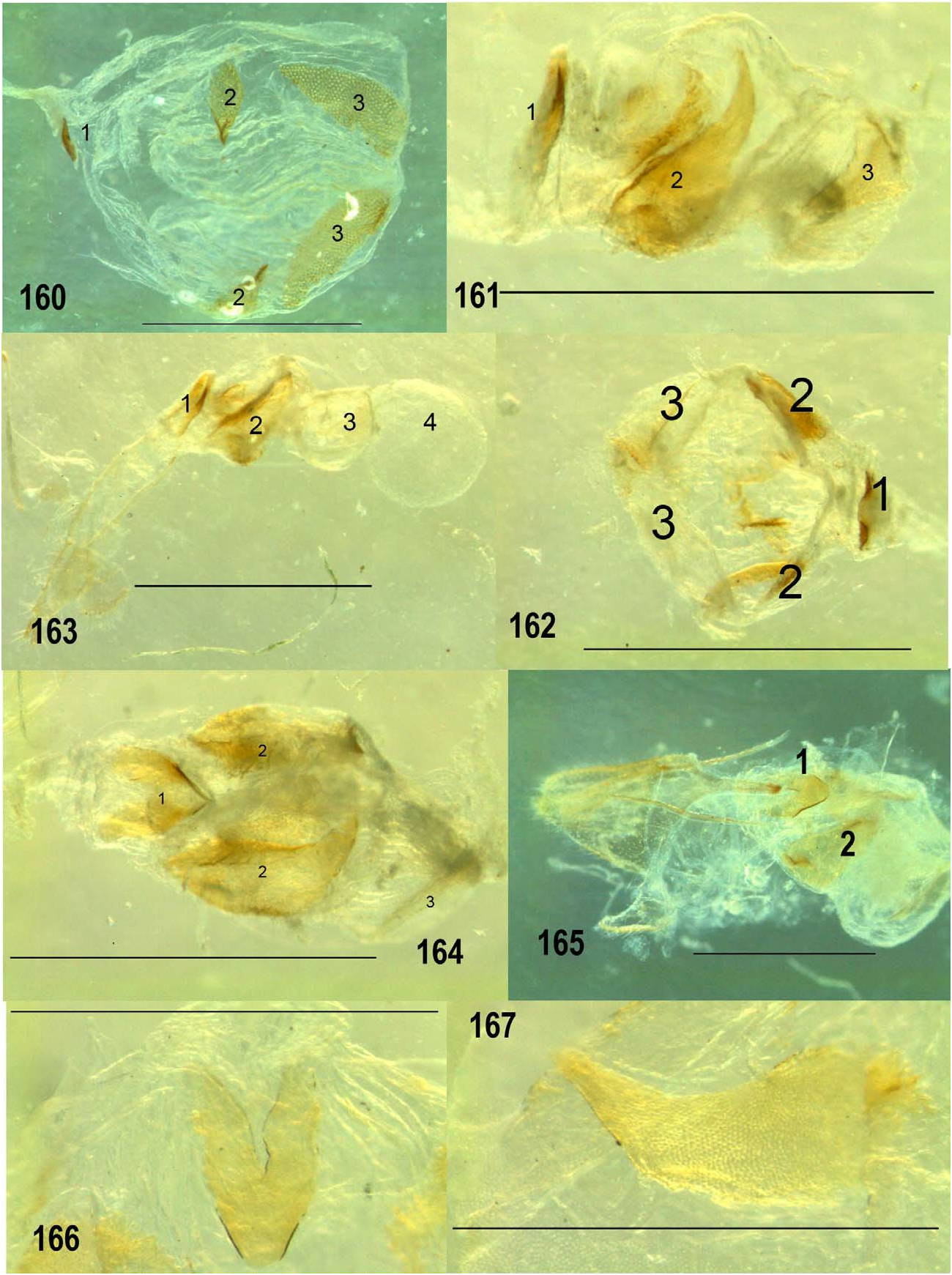
FIGURES 160–167. Female Medeopteryx spp. bursa. 160 Medeopteryx antennata female of mating pair Sek Bridge (ANIC). 161, 162 M. corusca female of mating pair Kondiu (ANIC). 163 M. cribellata Kar Kar Island (ANIC). 164–167 M. similispupillae sp. nov. 164 bursa ventral; 165 reproductive system excluding ovaries lateral; 166 median oviduct plate ventral (anterior end to foot of page); 167 anterior plate lateral. Figure legends: 1 median oviduct plate; 2 posterior bursa plate; 3 anterior bursa plate; 4 spermatophore digesting gland.

FIGURES 168–176. Medeopteryx pupilla male Mt Lamington (AMUS). 168 dorsal; 169, 170 abdomen ventral (169) and dorsal; 171–173 aedeagus left lateral, dorsolateral and ventral; 174, 175 tergite 8 (174 ventral with aedeagal sheath; 175 from right side, dorsal surface uppermost, flanges arrowed); 176 aedeagal sheath dorsal. These figures share scale lines: 171–173.
FIGURES 18–25. Australoluciola aspera, type female (18), Wewak male NHML 19–25. 18, 19 dorsal; 20, 21 ventral (21 abdomen only); 22–24 aedeagus dorsal (22), right lateral (23) and ventral (lateral projections of LL arrowed in 24); 25 pronotum dorsal. These figures share scale lines: 19, 20; 22–24.

FIGURES 45–55. Australoluciola spp. 45, 54, 55 Australoluciola flavicollis, male 45, female 54, 55 (ANIC). 46 Luciola coarcticollis type male (MNHN). 47 Luciola gestroi type male (MNHN). 45–47 dorsal; 48 ventral V4–7 and T8; 49–51 aedeagus left lateral (49), ventral (50) and dorsal; 52, 53 aedeagal sheath with aedeagus ventral (52) and dorsal; 54, 55 female bursa from side (54) and beneath (55). Figure legends: 2 posterior plates; 3 anterior plates. These figures share scale lines:49– 51; 52, 53.
No known copyright restrictions apply. See Agosti, D., Egloff, W., 2009. Taxonomic information exchange and copyright: the Plazi approach. BMC Research Notes 2009, 2:53 for further explanation.
|
Kingdom |
|
|
Phylum |
|
|
Class |
|
|
Order |
|
|
Family |
Luciola Laporte
| Ballantyne, Lesley A. & Lambkin, Christine L. 2013 |
Luciola (Luciola) Laporte. McDermott, 1966:103 (Partim)
| Calder, A. A. 1998: 178 |
| McDermott, F. A. 1966: 103 |
Bourgeoisia Olivier. Olivier 1908:17
| Deheyn & Dimitri D. & Lesley A. & Ballantyne 2009: 47 |
| McDermott, F. A. 1966: 117 |
| Olivier, E. 1911: 102 |
| Olivier, E. 1908: 17 |
Lampyroidea
| McDermott, F. A. 1966: 115 |
| Olivier, E. 1907: 49 |
| Olivier, E. 1902: 69 |
Luciola Laporte. Laporte 1833:146
| Lea, A. M. 1909: 106 |
| Olivier, E. 1907: 50 |
| Olivier, E. 1902: 69 |
| Gorham, H. S. 1880: 99 |
| Lacordaire, T. 1857: 335 |
| Motschulsky, V. de 1853: 52 |
| Laporte, F. L. de 1833: 146 |