
Microporina japonica Canu and Bassler, 1929
|
publication ID |
https://doi.org/10.12782/sd.21.1.009 |
|
publication LSID |
lsid:zoobank.org:pub:61308999-8455-4892-8464-423FFBACF0A1 |
|
DOI |
https://doi.org/10.5281/zenodo.5526917 |
|
persistent identifier |
https://treatment.plazi.org/id/DA5F87F2-467B-FFC2-FCE9-FCE2FD1EFEA0 |
|
treatment provided by |
Felipe (2021-03-04 21:19:55, last updated 2024-11-27 04:26:24) |
|
scientific name |
Microporina japonica Canu and Bassler, 1929 |
| status |
|
Microporina japonica Canu and Bassler, 1929 View in CoL ( Figs 6–8 View Fig View Fig View Fig , 13B View Fig )
Microporina japonica Canu and Bassler, 1929: 139 View in CoL , pl. 14, figs 9–11.
Microporina articulata View in CoL (not of Fabricius, 1821): Sakakura 1936: 259, pl. 15, figs 1–5; Arakawa 1984: 74, pl. 8-1, fig. 2 (only listed and illustrated); Nishizawa 1987: 182, pl. 1, fig. 3; Arakawa 1995: 80 (only listed); Nishizawa 1997: 152, 153 (only listed); Arakawa 1999: 57.
Material examined. NMNS PA 16842 View Materials (eight internodes, A–H), 16844, and 18156 (inner surface of frontal shield), Station 1700, Hakurei-Maru cruise GH80-2; NMNS PA 16843 View Materials (four internodes, A–D), 18152, and 18153, Station 1709, Hakurei-Maru cruise GH80-2; SGBC-0389 (four internodes, A–D), Station 1739, Hakurei-Maru cruise GH80- 2; SGBC-0391, Jizodo Formation , Pleistocene , Jizodo , Chiba Prefecture, Japan; SGBC-0392, Jizodo Formation , Pleistocene , Nanamagari , Chiba Prefecture, Japan; SGBC-0393, Jizodo Formation , Pleistocene , Ichinosawa , Chiba Prefecture, Japan; SGBC-0411 (not coated with metal), Jizodo Formation , Pleistocene, Atebi, Chiba Prefecture, Japan . See Table 1 View Table 1 for coordinates and depths of cruise samples.
Measurements (in milimetres). NMNS PA 16842, 16843, 16844, 18152, and SGBC-0389. Autozooids (n =183, 14): ZL, 0.62–1.18 (0.821±0.094); ZW, 0.25–0.44 (0.338±0.035); OrL, 0.08–0.14 (0.107±0.014); OrW, 0.13– 0.25 (0.201±0.019). Avicularia (n =76, 14): AvL, 0.16–0.25 (0.210±0.020); AvW, 0.13–0.24 (0.180±0.019).
Description. Colony erect, consisting of internodes increasing in width distally. Minimum internode size 2.0 mm long by 0.82 mm in diameter (NMNS PA 16842 A: Fig. 6A View Fig ); maximum size about 7.0 mm long (NMNS PA 16844: Fig. 6D View Fig ) and about 1.5 mm in diameter (NMNS PA 16843 C). Internodes generally circular in transverse section, composed of 8 to 12 columns of zooids; proximal ends of internodes occupied by kenozooids, and mural rim sometimes projected proximally ( Figs 6A View Fig , 7D View Fig ). Zooids elongate, sides nearly parallel, or wide in middle, sometimes tapering proximally. Frontal shield cryptocystal, flat, coarsely granulate, with many conspicuous, evenly distributed pores, surrounded by thick, rounded mural rim. Mural rim salient, finely granulated, and thickened with secondary calcification ( Figs 7 View Fig A–D, 8A). Opesiules originally large, elliptical or rounded triangular, completely occluded by smooth calcification with vein-like sculpturing ( Fig. 8 View Fig A–C). Orifice semielliptical, with proximal border straight or broadly concave. Oral spines lacking. Ovicells absent. Avicularium distal to orifice, oval in outline, longer than wide, directed proximally; rostrum triangular, with complete, M-shaped pivot bar, becoming round with increasing calcification and post-mortem abrasion ( Fig. 7 View Fig A–D). Secondary calcificaion sometimes covering zooidal orifice or postmandibular part of avicularium with granulated layer.
Distribution. The known distribution of this species is the Tsugaru Strait [“Sea of Japan ” in Canu and Bassler (1929)] and the continental shelf east of the Boso Peninsula at depths of 57 to 150 m. Because Sakakura (1936: pl. 15 (5), fig.1) illustrated Microporina articulata from the Tsugaru Strait as having only a semicircular orifice, M. japonica may overlap in distribution with that species. In fact, M. articulata from Korea also has a semicircular orifice with a straight proximal border ( Rho and Seo 1990).
Among Japanese fossils recorded as M. articulata , the Pleistocene specimen from Niigata illustrated by Nishizawa (1987) matches M. japonica in all respects. Neogene specimens from Hokkaido and the Noto Peninsula, described by Hayami (1970) and Nishizawa and Sakagami (1986), also have salient opesiules, but their other features need to be reexamined in detail.
Remarks. Microporina japonica shows large variations in the shape of the orifice, the proximal border of which can be nearly straight or broadly concave, and the opesiules are large. Canu and Bassler (1929) originally described this species from the Tsugaru Strait, Japan, on the basis of its having smaller zooids than Microporina articulata , but did not mention any other differences between the two. Sakakura (1936) considered M. japonica to be a junior synonym of M. articulata , because zooidal size varies among Japanese specimens. In my study, maximum zooid length was 1.18 mm (NMNS PA 16842 D), which is within the size range of North American specimens of M. articulata , based on the figures of Robertson (1905: pl. XIV, fig. 86) and Powell (1968: pl. II, fig. c), and maximum width was 0.44 mm (NMNS PA 16843 C), which is larger than the width indicated for M. articulata by Osburn (1950: 106). Sakakura’s (1936) discussion was correct on this point.
However, M. articulata differs from M. japonica in the shape of the orifice. In M. articulata it is consistently semicircular, with a straight proximal border ( Busk 1855; Smitt 1868; Robertson 1905; Osburn 1950; Kluge 1962; Powell 1968). In my material, the orifice is semielliptical, with a proximal border ranging from straight to broadly concave. This variation is evident even within a single internode. Orifice size also varies in M. japonica , but it is larger than in Alaskan specimens of M. articulata ( Robertson 1905) . Sakakura (1936) likewise observed orifice variation in his material, but concluded it fell within the range of variation in M. articulata . His fossil material, however, appears to have included two or more distinct species having differently shaped internodes, zooids, orifices, and avicularia.
Microporina japonica has conspicuous cryptocystal opesiules, as illustrated by Canu and Bassler (1929). The opesiules are elliptical in M. japonica , compared to slit-like in M. articulata . In my specimens, the layer occluding the opesiules has a vein-like structure, but whether this is diagnostic for M. japonica is not clear.
The avicularia in M. japonica are more or less elongate. The illustrations of Canu and Bassler (1929) show very long avicularia, with a maximum length/width ratio of about 1.6: 1; this ratio also reaches about 1.4: 1 in my material. Some of the numerous specimens identified as M. articulata from northern seas also have elongate avicularia ( Busk 1855; Smitt 1868; Powell 1968). At present, it is difficult to draw firm conclusions about the differences in avicularia among Microporina species, and a more precise comparison based on a larger number of samples will be necessary.
Arakawa, S. 1984. Recent bryozoans on the east offshore of the Boso Peninsula. Pp. 74 - 77. In: Miyazaki, T. and Honza, E. (Eds) Geological Investigation of the Tohoku and Ogasawara Arcs. April-June 1980 (GH 80 - 2 and 3 Cruises). Geological Survey of Japan, Tsukuba.
Arakawa, S. 1995. Bryozoan fauna in the Jizodo Formation (Pleistocene), Boso Peninsula, Honshu, Japan. Natural History Research 3: 75 - 110.
Arakawa, S. 1999. A preliminary report on cheilostomatous bryozoans from the coast and the eastern continental shelf of the Boso Peninsula. Bulletin of Seishin-Gakuen 14: 43 - 107. [In Japanese]
Busk, G. 1855. Zoophytology. Quarterly Journal of Microscopical Science 3: 253 - 256, 320 - 324.
Canu, F. and Bassler, R. S. 1929. Bryozoa of the Philippine Region. U. S. National Museum Bulletin 100 (9): i-xi + 1 - 685.
Hayami, T. 1970. Miocene Bryozoa from northwest Hokkaido, Japan. Transactions and Proceedings of the Palaeontological Society of Japan (New Series) 79: 316 - 336.
Kluge, G. A. 1962. Bryozoa of the Northern Seas of the USSR. Opredeliteli po faune SSSR, Izdavaemye Zoologicheskim Institutom Academii Nauk SSSR 76. Izdatelistvo Akademii Nauk SSSR, Moskow, 584 pp. [In Russian]
Nishizawa, Y. and Sakagami, S. 1986. Bryozoa. Pp. 78 - 95. In: Fuji, N. (Ed.) Paleontological Study of the Nanao Calcareous Sandstone in Noto. Board of Education, Nanao City, Ishikawa Prefecture, Japan. [In Japanese]
Nishizawa, Y. 1987. Cheilostomata (Bryozoa) from Sado. Publication of Sado Museum 9: 175 - 196. [In Japanese]
Nishizawa, Y. 1997. The Pleistocene cheilostome bryozoans from the Boso Peninsula in comparison with Recent cheilostomes in Aburatsubo Bay and off Jogashima Island. Pp. 143 - 155. In: Arakawa, S. (Ed.) Professor Sumio Sakagami Memorial Volume. Association for the Publication of Professor Sumio Sakagami Memorial Volume, Kashima. [In Japanese]
Osburn, R. C. 1950. Bryozoa of the Pacific Coast of America. Pt. 1. Cheilostomata-Anasca. Allan Hancock Pacific Expeditions 14: 1 - 270.
Powell, N. A. 1968. Bryozoa (Polyzoa) of Arctic Canada. Journal of the Fisheries Research Board of Canada 25: 2269 - 2320.
Rho, B. J. and Seo, J. E. 1990. A systematic study on the marine bryozoans in Korea. 7. Suborder Anasca. Korean Journal of Systematic Zoology 6: 145 - 160.
Robertson, A. 1905. Non-incrusting cheilostomatous Bryozoa of the west coast of North America. University of California Publications, Zoology 2: 235 - 322.
Sakakura, K. 1936. On Microporina articulata (Fabricius), a cheilostomatous Bryozoa. Journal of Geology 43: 259 - 267. [In Japanese with English description]
Smitt, F. A. 1868. Kritisk forteckning ofver Skandinaviens Hafs-Bryozo- er. III. Ofversigt af Kongliga Vetenskaps-Academiens Forhandlingar 24: 279 - 429.
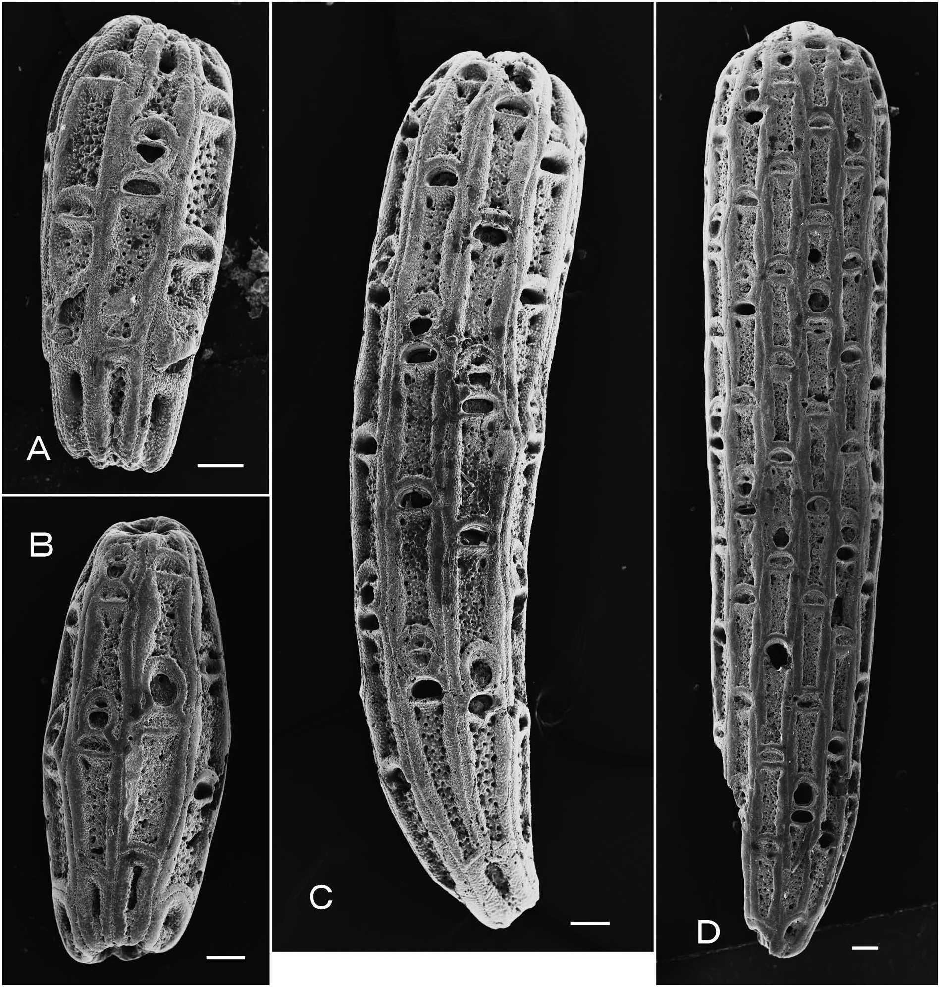
Fig. 6. Microporina japonica Canu and Bassler, 1929, A, NMNS PA 16842A; B, NMNS PA 16842F; C, NMNS PA 18152; D, NMNS PA 16844: A, B, small internodes; C, medium-sized internode; D, large internode. Scale bars: 200µm.
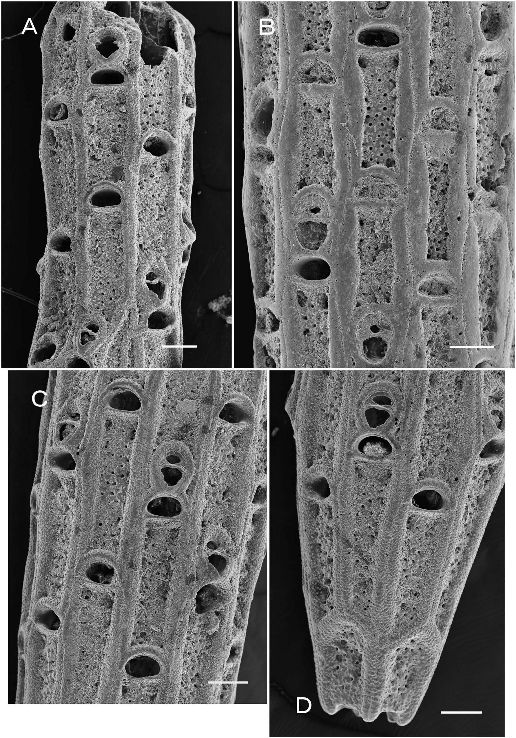
Fig. 7. Microporina japonica Canu and Bassler, 1929, A, NMNS PA 16842 H; B, NMNS PA 16844; C, D, NMNS PA 16843 B: A, zooids with thin mural rim; B, zooids with greatly thickened lateral rim; C, D, zooids with immediately thickened lateral rim. Scale bars: 200µm.
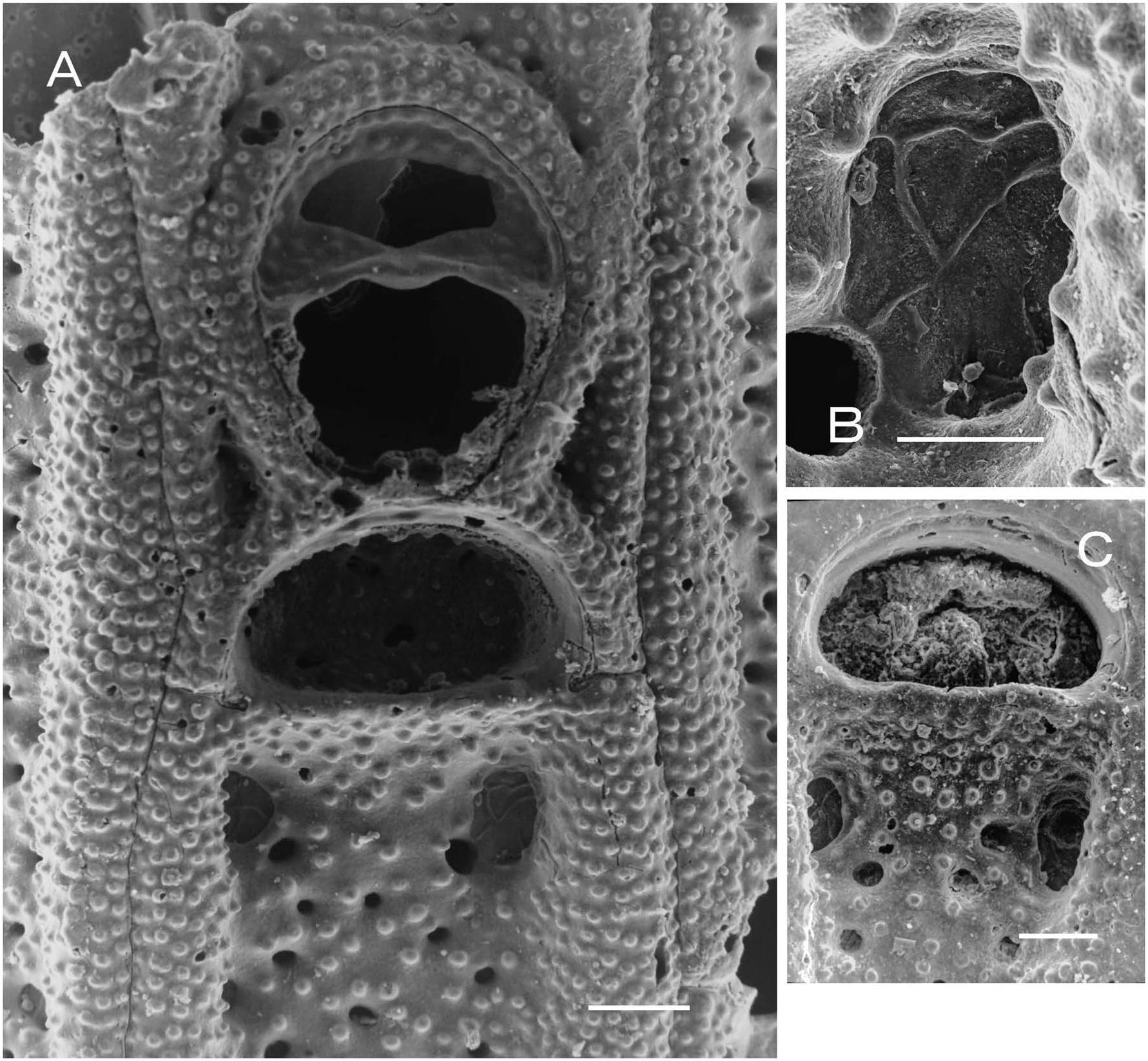
Fig. 8. Microporina japonica Canu and Bassler, 1929, A, B, NMNS PA 18153; C, NMNS PA 18152: A, orifice, opesiules, and avicularium; B, enlargement of opesiule in A; C, orifice and opesiules. Scale bars:50µm (A, C); 20µm (B).
| NMNS |
National Museum of Natural Science |
No known copyright restrictions apply. See Agosti, D., Egloff, W., 2009. Taxonomic information exchange and copyright: the Plazi approach. BMC Research Notes 2009, 2:53 for further explanation.
|
Kingdom |
|
|
Phylum |
|
|
Class |
|
|
Order |
|
|
SubOrder |
Neocheilostomina |
|
InfraOrder |
Flustrina |
|
SuperFamily |
Microporoidea |
|
Family |
|
|
Genus |
Microporina japonica Canu and Bassler, 1929
| Arakawa, Shinji 2016 |
Microporina articulata
| Arakawa, S. 1999: 57 |
| Nishizawa, Y. 1997: 152 |
| Arakawa, S. 1995: 80 |
| Nishizawa, Y. 1987: 182 |
| Arakawa, S. 1984: 74 |
| Sakakura, K. 1936: 259 |
Microporina japonica Canu and Bassler, 1929: 139
| Canu, F. & Bassler, R. S. 1929: 139 |
1 (by felipe, 2021-03-04 21:19:55)
2 (by ExternalLinkService, 2021-03-04 22:00:31)
3 (by felipe, 2021-03-05 11:34:07)
4 (by tatiana, 2021-09-24 16:54:28)
5 (by ExternalLinkService, 2021-09-24 17:04:47)
6 (by ExternalLinkService, 2021-09-24 18:30:04)
7 (by ExternalLinkService, 2021-10-29 01:36:15)
8 (by plazi, 2023-11-01 23:14:45)