
Bayadera serrata Davies and Yang, 1996
|
publication ID |
https://doi.org/10.11646/zootaxa.4894.1.5 |
|
publication LSID |
lsid:zoobank.org:pub:22CBBD7B-2B08-4E1B-81B8-C1AE95D745D7 |
|
DOI |
https://doi.org/10.5281/zenodo.4324005 |
|
persistent identifier |
https://treatment.plazi.org/id/DA06D64B-B023-FF83-0482-F8B9DA6CFF66 |
|
treatment provided by |
Plazi (2020-12-10 22:03:56, last updated 2024-11-28 00:24:04) |
|
scientific name |
Bayadera serrata Davies and Yang, 1996 |
| status |
|
Bayadera serrata Davies and Yang, 1996 View in CoL
Bayadera serrata Davies & Yang (1996) View in CoL , 145–146; Hämäläinen (2013), 47–48; Hämäläinen (2017), 9–10; Phan et al. (2018), 164–166.
Material examined. THAILAND, 5 exuviae (last stadium larvae at collecting, then reared in laboratory): 1 ³, emerged 11/IV/2014; 1 ³, 1 ♀, emerged 12/IV/2014; 1 ³, emerged 19/IV/2014; 1 ³ 20/IV/2014, Yakruae stream at Nam Nao national National Park, (16˚44´27.92̎ N, 101˚34´46.52̎ E; elevation 832 m), Phetchabun province, J. Sripanya leg ; 2 last stadium larvae: 1³, 18/IV/2014; 1♀, 11/V/2014, Yakruae stream at Nam Nao National Park , (16˚44´27.92̎ N, 101˚34´46.52̎ E; elevation 832 m), Phetchabun province, R. Somnak leg.
Description of larva. Larvae stout, almost flat on ventral side and semi-cylindrical shape, coloration mutable, yellowish-brown to brownish-black ( Fig. 2 View FIGURE 2 ).
Head: broad and smooth in dorsal view, roughly squashed pentagon in outline; labrum ( Fig. 3a View FIGURE 3 ) with sparse long SS on distal half and scattered RLS on basal half; frons and vertex smooth, with three prominent ocelli; compound eyes broad and rounded protruding to antero-laterally side; occiput with scattered RLS, anterior margin with two long SS and scattered RLS along margin of compound eyes, posterior margin with row of RLS; postocular lobes well developed, anterior margin with scattered RLS and SPS, posterior margin intermingled with small spine, long SS and RLS; genae ( Fig. 3 View FIGURE 3 b–c) large, with spine-like projection, row of SPS and RLS along ventral margin of compound eyes, anterior margin of left gena ( Fig. 3b View FIGURE 3 ) with row of 2–4 small spines (each spine bearing a SS), 2–3 dominant spines and row of RLS inserted with SPS and SS, respectively, anterior margin of right gena ( Fig. 3c View FIGURE 3 ) with row of 4–7 small spines (each spine bearing a SS), 1–3 long spines and row of RLS inserted with SPS and SS, respectively ( Fig. 3d View FIGURE 3 ); antennae ( Fig. 3e View FIGURE 3 ), filiform, 7–segmented, with A3 the longest, relative length of antennomeres 0.4: 0.68: 1(0.64 mm): 0.48: 0.4: 0.24: 0.12; prementum ( Fig. 4a View FIGURE 4 ) subpentagonal in shape extending posteriorly up to the middle of the fore coxae or mid coxae, with a row of 14–16 (left side) and 13–17 (right side) distinct spines and numerous tiny spines (each spine bearing SS) along lateral margin of each side ( Fig. 4b View FIGURE 4 ), with a pair of long SS on middle of ventro-posterior side ( Fig. 4c View FIGURE 4 ), with 4–6 SPS between basal of ligula and socket of labial palp; ligula ( Fig. 4d View FIGURE 4 ) forming a finely serrate arch, with tiny median cleft and faint traces, with two pairs of long SS and a pair protuberance; labial palp ( Fig. 4e View FIGURE 4 ) as long as 0.32× as long as prementum, outer margin with scattered SS and RLS, serrate on inner margin, distal end with three teeth ( Fig. 4f View FIGURE 4 ), the outer teeth forming uncinate teeth and undulate on outer margin, middle lobe forming uncinate teeth and smooth, inner lobe forming small truncate with tiny teeth, movable hook slender and about 0.63 × as long as labial palp, acuminate, bent inwards; mandible ( Fig. 5 View FIGURE 5 a–d) with mandibular formula: L 1+1’234 0 a(m 1,2,3,4)b / R 1+1’234 y a(m 0)b, unsymmetrical, brawny with well-developed long teeth on each incisor lobe, with molar crest; right mandible with five incisor teeth, molar crest with two teeth (a>b), with an additional tooth; left mandible with five incisor teeth, molar crest with six teeth (a>1=2=3=4=b); maxilla ( Fig. 5 View FIGURE 5 e–f) galeolacinia with seven teeth, four dorsal teeth of approximately the same size, apical teeth largest, three ventral teeth of small size.
Thorax: narrower than the head, with scattered RLS intermingled with SS; prothorax almost saddle-shaped, anterior margin reaching concave of postocular lobe, posterior margin forming rounded, with a row of RLS, anterolateral side with small supracoxal spines; wing pads pale brown to dark brown, with glabrous, parallel, anterior and posterior wing pads reaching half of abdominal S3 to almost abdominal S5; legs broad, robust, covered with dense SS intermingled with RLS, forelegs shortest and hind legs longest; tibial comb ( Fig. 5g View FIGURE 5 ) with numerous SPS intermingled with SS, lateral side with RLS and long SS distal margin end with a row of RLS; tarsi with two rows of SS and scattered long SS, tarsi formula: 3–3–3; two claws simple with pulvilliform empodium.
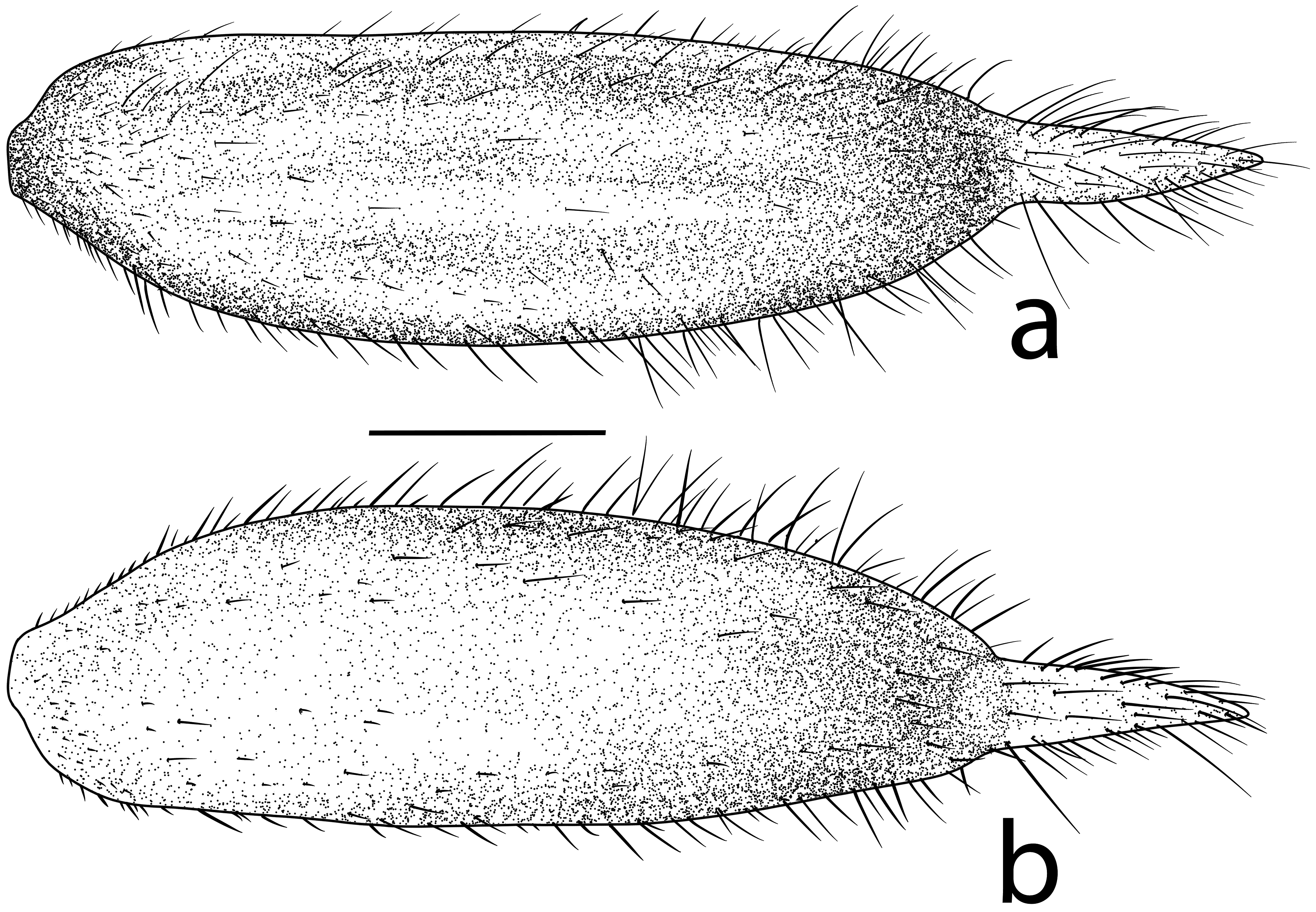
Abdomen: narrowing caudally, scattered RLS and SS; abdominal terga, with a row of RLS and SS on posterior margin, abdominal terga S3–S8 or S9 with a cluster of long SS on the middle of posterior margin; abdominal pleura scattered SS; abdominal sterna smooth, with a pale network of tracheoles, seven pairs of abdominal gills with progressively slenderer from base to end, with dense tiny curving SS on abdominal sterna S2–S8; male gonapophyses ( Fig. 6 View FIGURE 6 a–b) well developed, base stout, blunt on the tip, widely convergent in ventral view, reaching anterior margin of abdominal sternite S10, with the plate-like spine on ventral side; gonopore small, oval-shaped embossed and fissure on the middle to posterior; female gonapophyses ( Fig. 6 View FIGURE 6 c–d) comprising two pairs of long valves, with lateral valves slightly arching from anterior margin of abdominal sternite S8, with the plate-like spines on the ventral side, with rounded process tips, extending to middle of abdominal sternite S8; central valves smooth, slender, apically rounded, and slightly shorter than lateral valves; male cerci slightly concave, convergent with RLS; female cerci concave ventrally with small RLS; caudal gills ( Fig. 7 View FIGURE 7 ) forming brownish-black swollen saccoid shape with yellowish-white terminal filament, covered with SS. Length of median gill about 1.05 × as long as lateral gills.
Measurements. (in mm; n = 7 (5 exuviae and 2 alcoholic specimens)): total length of body without caudal gills = 13.59–18.84; length of caudal gills = 5.16–7.60; width and length of head = 2.97–4.71 and 2.37–3.40; length of antenna = 3.04–3.29; width and length of prementum = 2.18–3.16 and 3.81–4.86; length of movable hook = 1.21–1.59; length of inner and outer wing pads = 2.64–5.69 and 2.54–5.90; length of femora (fore: mid: hind) = 2.73–3.07: 2.92–3.39: 4.18–4.80; length of tibiae (fore: mid: hind) = 3.21–4.04: 3.12–3.96: 3.79–3.96.
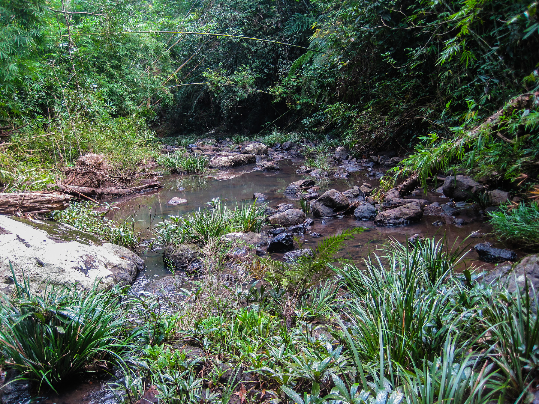
Biological notes. The larvae of B. serrata inhabit around open habitats of headwater streams surrounded by trees, herbs, and shrubs. The composition of the habitat was as follows: cobble (65%), leaf litter (15%), pebble/ gravel/sand/slit (10%), aquatic plants (5%), small boulders (4%), and large boulders (1%). They cling under cobbles and pebbles in a slow riffle of the stream ( Fig. 8 View FIGURE 8 ) and are generally found together with larvae of the genus Euphaea Selys, 1840 . Stream width was 2.81 m, water depth was 16.25 cm, orthophosphate concentration was 0.53 mg /l, nitrate-nitrogen concentration was 0.49 mg /l, dissolved oxygen concentration was 5.18 mg /l, total dissolved solid was 265.59 ppm, electrical conductivity was 531.58 µS/cm, pH was 8.75, and water temperature was 23.32ºC.
Davies, D. A. L. & Yang, B. (1996) New Species of Bayadera Selys and Schmidtiphaea Asahina from China (Odonata, Euphaeidae). Tijdchrift voor Entomologie, 139, 145 - 154.
Hamalainen, M. (2013) Description of Bayadera kinnara sp. nov. from Burma, with taxonomic notes on its congeners (Odonata: Euphaeidae). Tombo-Acta Odonatologica Japonica, 55, 45 - 49.
Hamalainen, M. (2017) The Caloptera damselflies of Thailand-Distribution maps by provinces (Odonata: Calopterygoidea). Faunistic studies in South-East Asian and Pacific Island Odonata, 19, 1 - 28.
Phan, Q. T., Kompier, T., Karube, H. & Hayashi, F. (2018) A synopsis of the Euphaeidae (Odonata: Zygoptera) of Vietnam, with descriptions of two new species of Euphaea. Zootaxa, 4375 (2), 585 - 592. https: // doi. org / 10.11646 / zootaxa. 4375.2.1
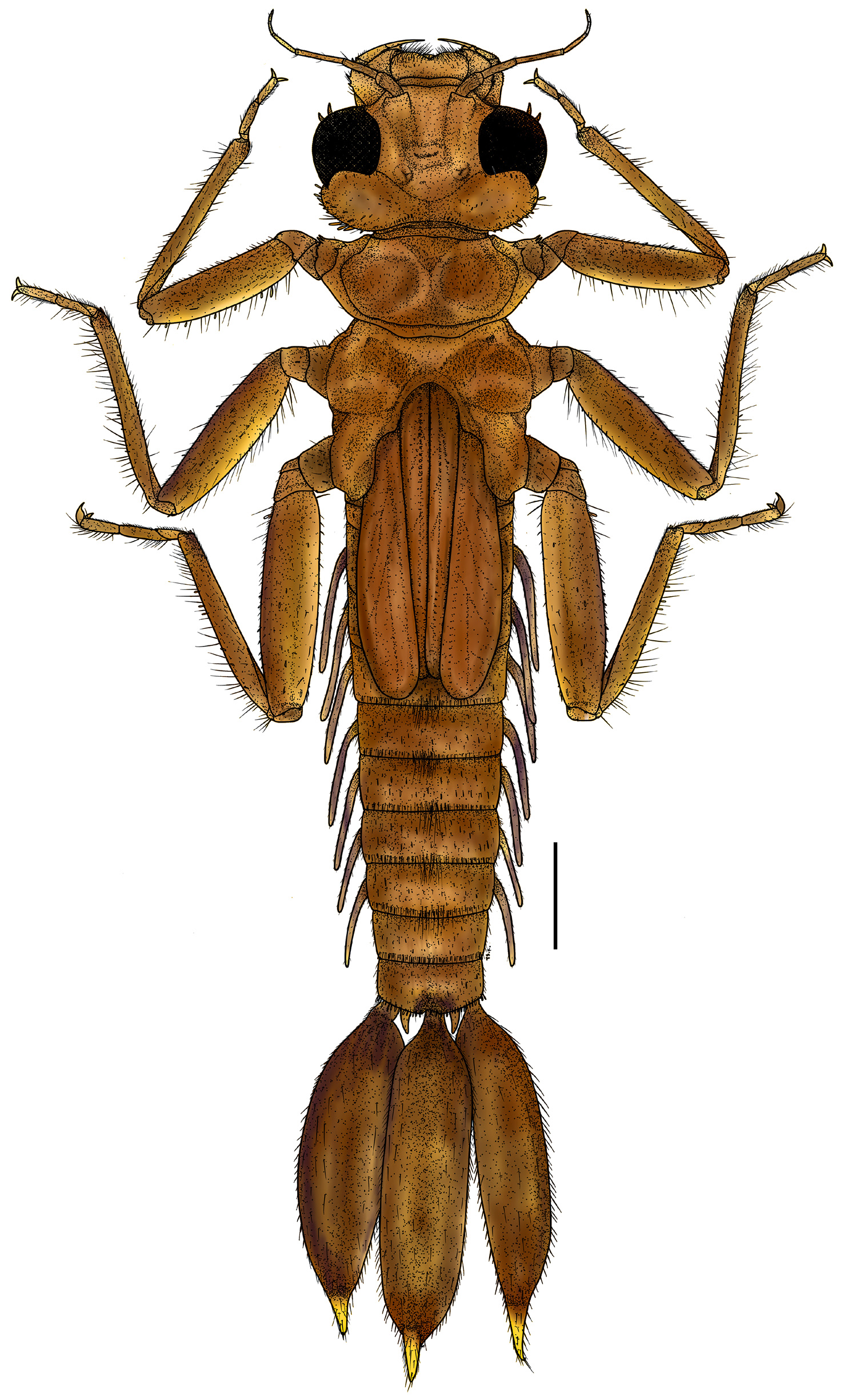

FIGURE 3. Detail morphological of labrum, genae and antenna of Bayadera serrata: (a) labrum, dorsal view; (b) right genae; (c) left genae; (d) manner of spine and setae along margin of genae; (e) antenna; Scale bar = (a, e) 0.2 mm, (d) 0.5 mm, (b, c) 1 mm.
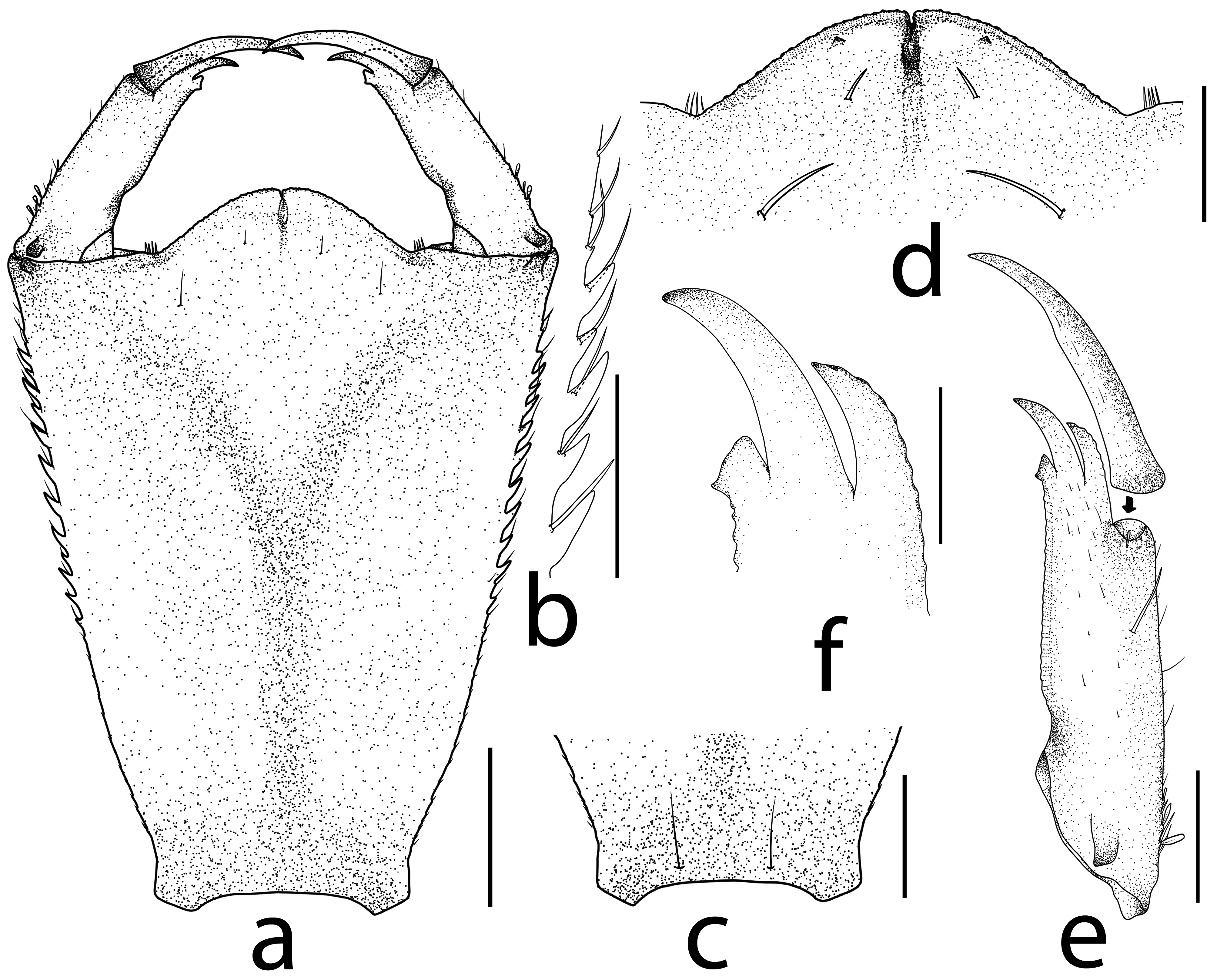
FIGURE 4. Labium of Bayadera serrata: (a) prementum, dorsal view; (b) lateral spines of prementum; (c) setae on posterior margin of prementum, ventral view; (d) ligula, dorsal view; (e) right labial palp, dorsal view; (f) distal end of labial palp, dorsal view. Scale bars = (a–e) 0.2 mm, (f) 0.1 mm.

FIGURE 5. Mouthparts and distal part of leg of Bayadera serrata: (a) right mandible, ventrointernal view; (b) right mandible, ventral view; (c) left mandible, ventral view; (d) left mandible, ventrointernal view; (e) right galeolacinia, ventral view; (f) right galeolacinia, dorsal view; (g) tibial comb and tarsi. Scale bar = 0.5 mm.

FIGURE 6. Abdominal S8-S10 of Bayadera serrata: (a) male gonapophyses, lateral view; (b) male gonapophyses, ventral view (Arrow = gonopore); (c) female gonapophyses, lateral view; (d) female gonapophyses, ventral view. Scale bar = 0.5 mm.
No known copyright restrictions apply. See Agosti, D., Egloff, W., 2009. Taxonomic information exchange and copyright: the Plazi approach. BMC Research Notes 2009, 2:53 for further explanation.
|
Kingdom |
|
|
Phylum |
|
|
Class |
|
|
Order |
|
|
Family |
|
|
Genus |
Bayadera serrata Davies and Yang, 1996
| Keetapithchayakul, Tosaphol Saetung, Sripanya, Jutamas, Phlai-Ngam, Sirikamon & Tungpairojwong, Nisarat 2020 |
Bayadera serrata
| Davies & Yang 1996 |
1 (by plazi, 2020-12-10 22:03:56)
2 (by ExternalLinkService, 2020-12-10 22:14:39)
3 (by valdenar, 2020-12-15 17:36:54)
4 (by ExternalLinkService, 2020-12-15 17:42:14)
5 (by ExternalLinkService, 2020-12-16 00:20:35)
6 (by ExternalLinkService, 2021-10-29 01:36:11)
7 (by ExternalLinkService, 2021-10-29 03:57:26)
8 (by plazi, 2023-11-01 00:12:22)