
Ripartitella degreefii Rizinde, Desjardin, Amalfi, & Decock, 2023
|
publication ID |
https://doi.org/10.11646/phytotaxa.597.3.1 |
|
DOI |
https://doi.org/10.5281/zenodo.7963417 |
|
persistent identifier |
https://treatment.plazi.org/id/D86887AA-FF88-FF87-2F9B-951BFA49F726 |
|
treatment provided by |
Plazi (2023-05-22 21:20:51, last updated 2024-11-28 06:16:31) |
|
scientific name |
Ripartitella degreefii Rizinde, Desjardin, Amalfi, & Decock |
| status |
sp. nov. |
Ripartitella degreefii Rizinde, Desjardin, Amalfi, & Decock , sp. nov. Figs. 2 View FIGURE 2 , 3 View FIGURE 3
[Mycobank: MB842786]
Diagnosis:—The species is similar to R. brasiliensis in the basidioma habit, but differs in the paucity of pleurocystidia, a pileipellis as a cutis, and its habitat in mountain areas of tropical Africa.
Description:— Basidiomata cespitose, small to medium-sized ( Fig. 2A, B View FIGURE 2 ). Pileus 25–70 mm diam., convex to planoconvex, becoming applanate and depressed, with (or without) a small obtuse umbo; surface dull, dry, pure white to off-white, disc with (or without) tiny, brownish orange (ferruginous 7C6–7]) scales when young, wearing off with age. Margin decurved to sometimes uplifted in age, centrally pure white to cream (4A3) with white appendiculate veil remnants. Context 1–2 mm thick, soft, white. Lamellae shallowly adnexed to adnate, crowded to very crowded, unequal, (24 (L+l) / cm), with 2–4 series of lamellulae, narrow (2–3 mm deep), with smooth edge, pale yellowish white (4A2). Stipe central to slightly eccentric, terete, subclavate to bulbous, 15–70 (–100) mm long, 3–10 mm broad, solid, annulate, concolorous with pileus, squarrose, with superficial, scattered white velar remnants toward base, glabrous above the annulus. Annulus evanescent, single, membranous, felted, often incomplete, attached to the upper quarter of the stipe. Odor fungoid, taste not tested. Spore print white Hyphal system monomitic, composed of generative hyphae with clamp connections in all tissues. Pileipellis a cutis, made up of repent, subparallel, radially oriented, cylindric hyphae, 3.5 – 7 µm diam, smooth, thin-walled. Pileus squamules composed of subcatenulate, rarely encrusted hyphae, 5 – 10 µm diam. Pileitrama made up of interwoven hyaline, smooth, thin- to slightly thick-walled, hyphae, 2.5–7 µm diam. Lamellar trama similar to pileitrama. Stipitipellis a layer of ±parallel, longitudinally oriented hyphae, with scattered clusters of subcatenulate elements similar to those of pileipellis ( Fig. 3C View FIGURE 3 ), individual cells 3.5 – 7 µm diam, smooth, thin-walled.
Basidiospores hyaline ( Fig. 3G, H View FIGURE 3 ), broadly ellipsoid to subglobose, thin-walled, verrucose, inamyloid, acyanophilous, (3.5–) 4–5 × 3–3.8 µm (n = 30, mean = 4.3 × 3.4 µm, Q = 1.13–1.5). Basidia with a basal clamp, clavate, with 4 sterigmata ( Fig. 3F View FIGURE 3 ), not siderophilous, 14–24 × 6–8 µm. Pleurocystidia absent or present ( Fig. 3D View FIGURE 3 ), then scarce, difficult to observe, with a basal clamp, lageniform, thin- to thick-walled, apically smooth or slightly, finely or coarsely incrusted ( Fig. 3E View FIGURE 3 ), 32–40 × 5.0–7.5 µm, the apical digitate part 2–2.5 µm diam (mean =36 × 6.5 µm, 2.3 µm in the apical section). Cheilocystidia not observed.
Distribution:—AFRICA. Known from the Eastern Democratic Republic of the Congo and S„o Tomé.
Ecology (substrate, host, habitat):— On fallen trunks of angiosperms, including, in DRC, Xymalos monospora (Harv.) Baill. ( Monimiaceae , locally named “Cinyalubombo”), mountain forests, at 1100 and 2139 m a.s.l.
Etymology:—The species name is a tribute to Jérôme Degreef, scientific director at Meise Botanic Garden for his devotion to African mycology and his efforts for promoting the training of students from tropical Africa.
Material examined:—AFRICA. Democratic Republic of the Congo , South Kivu : Kahuzi Biega mountain range, Kahuzi Biega National Park, 2.331733°S, 28.74848°W, 2139 m a.s.l., on fallen trunk of Xymalos monospora (Harv.) Baill. (Monimiaceae) , November 2018, J.C. Rizinde leg., RHJ 305 (BR #5020189043549, Holotype), culture exholotype MUCL 57374; GoogleMaps SÃO TOMÉ: Macambrara radio antenna area; 0.27595°N, 6.608767°E, 1100 m elev., 25 April 2008, D. E. Desjardin leg. DED 8323; GoogleMaps SÃO TOMÉ; Macambrara radio antenna area; 0.27595°N, 6.608767°E; 1100 m elev., April 2006, D. E. Desjardin leg., DED 7937 (but material lost in transit to the USA) GoogleMaps .
Notes:— Ripartitella degreefii is phylogenetically distant from all other Ripartitella known to date ( Fig. 1 View FIGURE 1 ). Morphologically, R. degreefii is similar in many respects to R. brasiliensis ( Capelari & Asai 2009, Desjardin & Perry 2017), but differs in having a pileipellis as a cutis ( Fig. 3C View FIGURE 3 ) as opposed to a trichoderm ( Capelari & Asai 2009), scarce ( Fig. 3D–E View FIGURE 3 ) in contrast to abundant pleurocystidia ( Capelari & Asai 2009), and a distribution in Afromontane forests in contrast to lowland neotropics. Ripartitella brasiliensis also has dense, brown to reddish brown squamules, covering the whole cap when young ( Capelari & Asai 2009, Battistin et al. 2016). Squamules are variably present in young specimens of R. degreefii ( Fig. 2 View FIGURE 2 ), but soon wear off with age.
Ripartitella degreefii is known so far from two collections originating from two spots of medium elevation forests, in the continental and insular Afromontane range, in the Albertine rift ( DRC) and S„o Tomé. It is the first Ripartitella described from Tropical Africa. Pegler (1977) reported R. brasiliensis from Eastern Africa, in Kenya, Tanzania, and Uganda. He described the eastern African collections as lacking cystidia ( Pegler 1977). Pleurocystidia were observed in R. degreefii , in both specimens available. However, they are scarce and difficult to see. Desjardin and Perry (2017) reported R. degreefii under R. brasiliensis from S„o Tomé, stating that it matched “nicely the description of African material provided by Pegler (1977) ”. A closer examination of the S„o Tomé specimen revealed the presence of scarce pleurocystidia. The identity of the specimens cited by Pegler (1977) remains to be ascertained but they could represent R. degreefii .
Battistin, E., Picciola, P. & Campo, E. (2016) Some considerations on Ripartitella brasiliensis. Rivista Micologica Romana, Bollettion dell'Associazione Micologica Ecologica Romana 32 (2): 45 - 51.
Capelari, M. & Asai, T. (2009) Cystoderma, Cystodermella and Ripartitella in Atlantic Forest, S o Paulo State, Brazil. Hoehnea 36 (2): 339 - 348. https: // doi. org / 10.1590 / S 2236 - 89062009000200011
Desjardin, D. E. & Perry, B. A. (2017) The gymnopoid fungi (Basidiomycota, Agaricales) from the Republic of S o Tome and Principe, West Africa. Mycosphere 8 (9): 1317 - 1391. https: // doi. org / 10.5943 / mycosphere / 8 / 9 / 5
Pegler, D. N. (1977) A preliminary agaric flora of East Africa. Kew Bulletin Additional Series 6: 1 - 615.
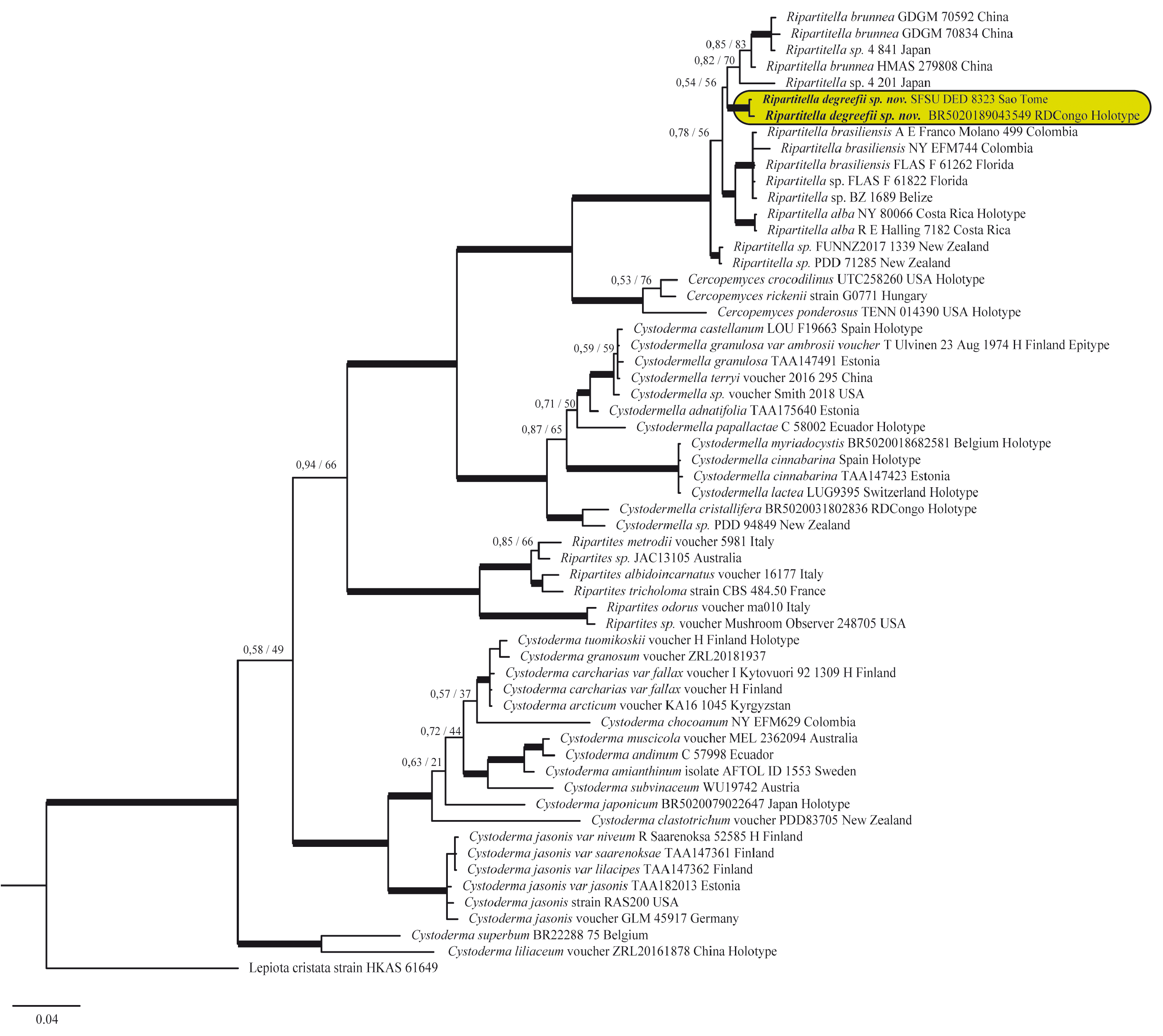
FIGURE 1. The 50 % majority-rule consensus tree from Bayesian Inference of the combined dataset. Thickened branches in bold represent ML BS support greater than 75 % and BPP greater than 0.95; thickened branches in black denote branches supported by either ML BS or BPP; for selected nodes ML BS support value and BPP are respectively indicated to the left and right of slashes; the new taxa are highlighted in the shaded box.
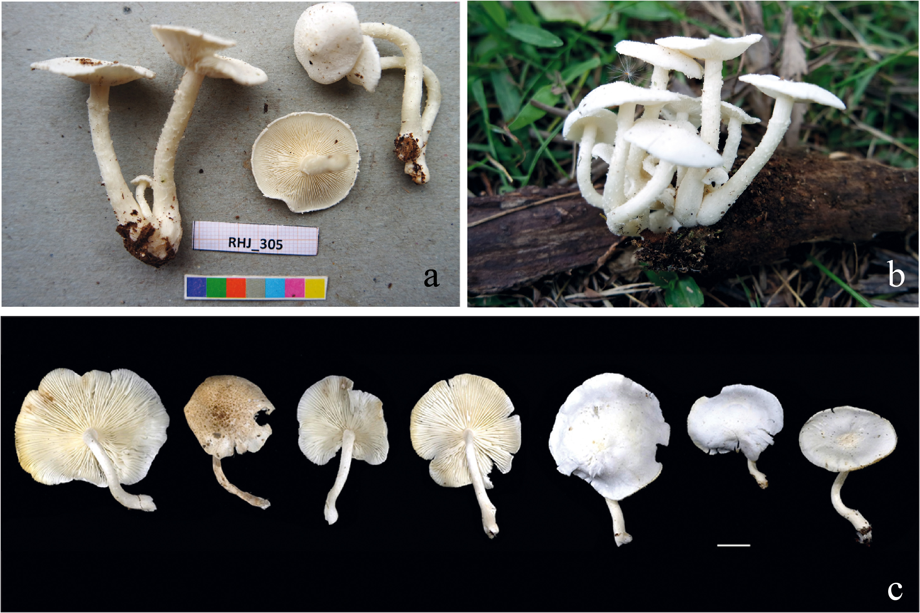
FIGURE 2. Basidiomes of Ripartitella degreefii (A=RHJ 305, holotype; B=DED 8323). Scale bar = 10 mm. Photos by J.C. Rizinde (RHJ 305) and D. Desjardin (DED 8323).
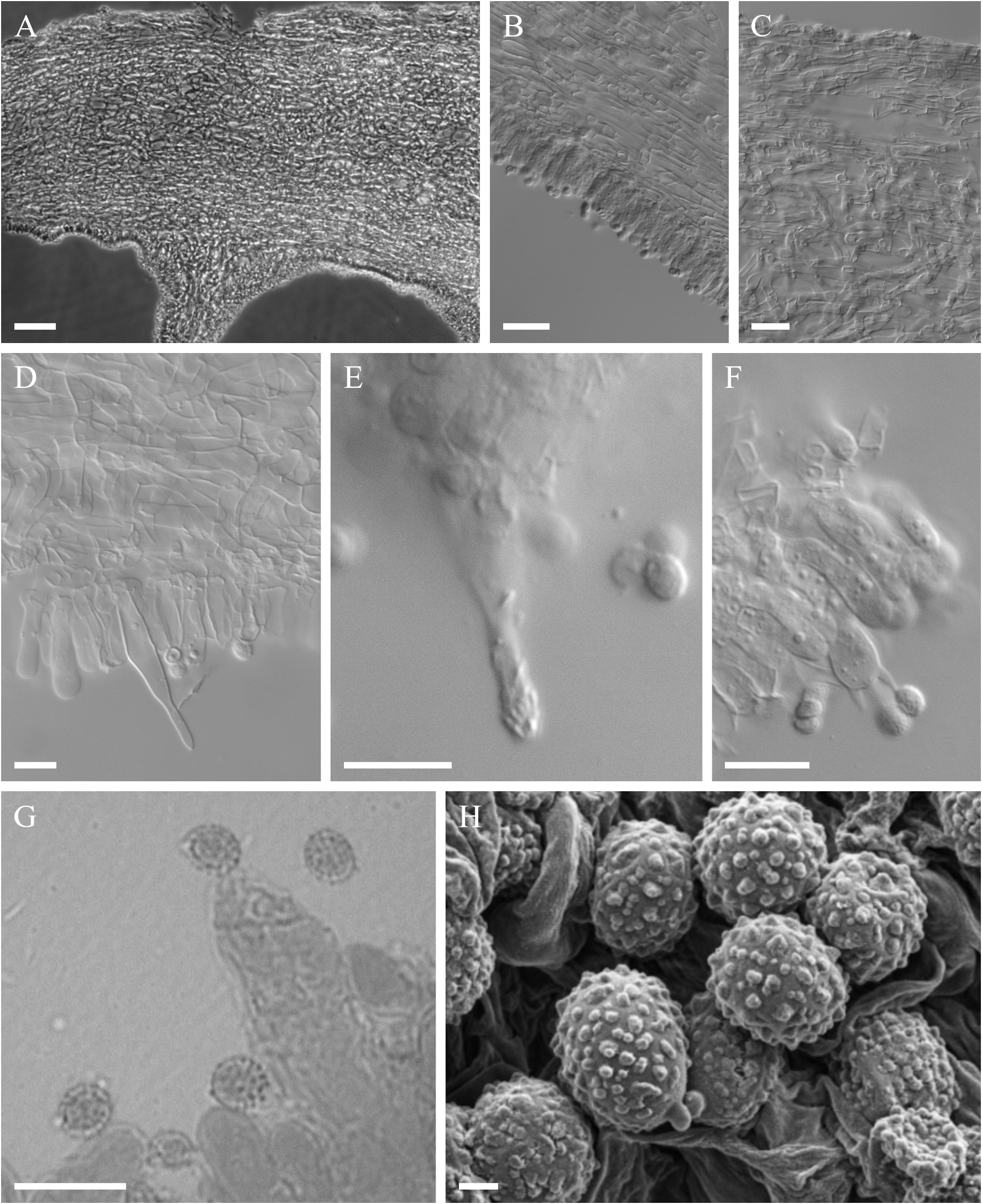
FIGURE 3. A. section of pileus; B. transversal section of lamellae; C. Pileipellis; D, E. Cystidia; F. Basidia; G = basidiospores from RHJ 305, optical microscopy; H = verrucose basidiospores from RHJ 305, SEM. Photos by C. Decock (A–F) and Mario Amalfi (G–H). Scale bars A= 100 µm; B, C = 20 µm; D–G= 10 µm.
No known copyright restrictions apply. See Agosti, D., Egloff, W., 2009. Taxonomic information exchange and copyright: the Plazi approach. BMC Research Notes 2009, 2:53 for further explanation.
|
Kingdom |
|
|
Phylum |
|
|
Class |
|
|
Order |
|
|
Family |
|
|
Genus |