
Macropygium subsulcatum (Amyot & Serville, 1843)
|
publication ID |
https://doi.org/10.11646/zootaxa.4958.1.36 |
|
publication LSID |
lsid:zoobank.org:pub:1616997B-867D-45CD-8470-515C39815F89 |
|
DOI |
https://doi.org/10.5281/zenodo.4692615 |
|
persistent identifier |
https://treatment.plazi.org/id/D40F87E7-6F03-FFCF-FF2C-DB79FBAEB7E7 |
|
treatment provided by |
Plazi (2021-04-15 08:51:38, last updated 2024-11-27 08:28:19) |
|
scientific name |
Macropygium subsulcatum (Amyot & Serville, 1843) |
| status |
|
Macropygium subsulcatum (Amyot & Serville, 1843) , REVISED STATUS
( Figs 6E View FIGURE 6 ; 11 View FIGURE 11 ; 12 View FIGURE 12 ; Table 1 View TABLE 1 )
Oxyrhinus subsulcatus Amyot & Serville, 1843: 12 (removed from the synonymy with M. reticulare ).
Macropygium subsulcatum: Dallas, 1851: 158 .
Lectotype female (here designated) ( Fig. 11 View FIGURE 11 ): FRENCH GUYANE, Cayenne. Labels: Cayenne / Coll. Signoret. // subsulcat. / det. Signoret. ( NHM)
Paralectotype female: FRENCH GUYANE, Cayenne. Labels: Cayenne / Coll. Signoret. // subsulcat. / det. Signoret. ( NHM)
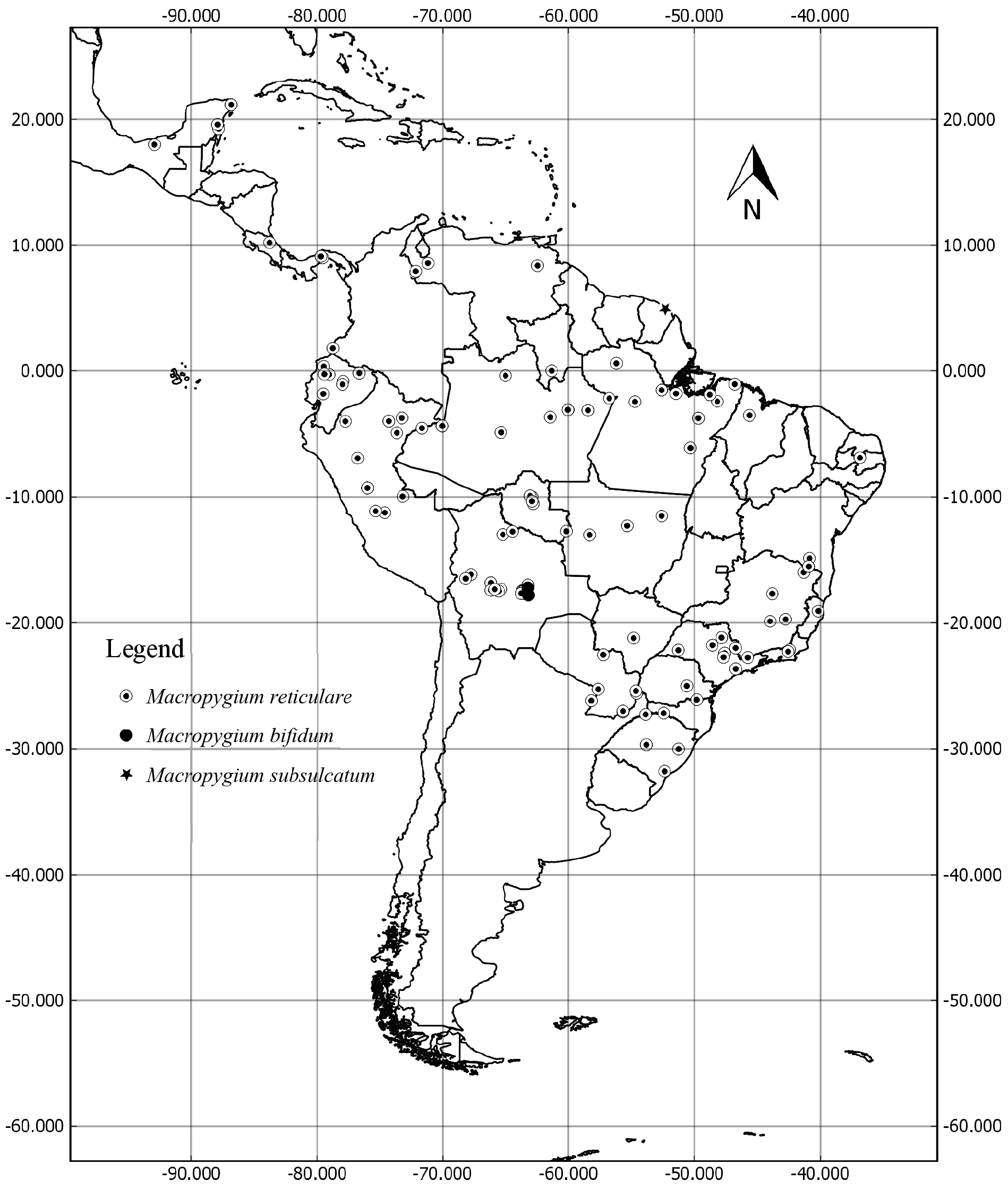
Distribution: French Guyane ( Fig. 12 View FIGURE 12 ).
Diagnosis: Antennae five-segmented, first antennomere slightly longer than the second; sutural angles of valvifers VIII posteriorly projected; lateral angles of valvifers VIII roundly projected, covering the base of laterotergites IX.
Male. Unknown.
Female. Genitalia: valvifers VIII almost as long as wide; lateral angles of valvifers VIII rounded, slightly surpassing posteriorly an imaginary line connecting the spiracles of laterotergites VIII, and partially covering a third of the basal portion of laterotergites IX; sutural angles of valvifers VIII posteriorly projected; posterior margin of valvifers VIII concave near the sutural angles; laterotergites IX with mild lateral depression ( Figs 6E View FIGURE 6 ; 11D View FIGURE 11 ).
Comments. Macropygium subsulcatum is currently known only from the two female types, collected in French Guyane ( Fig. 12 View FIGURE 12 ). Macropygium subsulcatum females are readily distinguished from other Macropygium species by the relatively wide valvifers VIII and with broadly rounded apical angle ( Fig. 6E View FIGURE 6 ).

FIGURE 6. Female genital plates of Macropygium species, ventroposterior view. Macropygium reticulare (Fabricius, 1803) (A); Macropygium spinolae Stål, 1860 (B); Macropygium bifidum (Westwood, 1837) (C); Macropygium graziae Silva & Campos, sp. nov. (D); Macropygium subsulcatum (Amyot & Serville, 1843) (E). Abbreviations: laIX, laterotergite IX; laVIII, laterotergite VIII; vfIX, valvifer IX; vfVIII, valvifer VIII; VII, urosternite VII; X, segment X.

FIGURE 11. Lectotype of Oxyrhinus subsulcatus Amyot & Serville, 1843 [=Macropygium subsulcatum (Amyot & Serville, 1843)]. Dorsal, ventral, and lateral view, respectively (A–C), genital plates in ventroposterior view (D), labels (E).
| NHM |
University of Nottingham |
No known copyright restrictions apply. See Agosti, D., Egloff, W., 2009. Taxonomic information exchange and copyright: the Plazi approach. BMC Research Notes 2009, 2:53 for further explanation.
|
Kingdom |
|
|
Phylum |
|
|
Class |
|
|
Order |
|
|
Family |
|
|
Genus |
Macropygium subsulcatum (Amyot & Serville, 1843)
| Silva, Layse Mitsue Harada Da & Campos, Luiz Alexandre 2021 |
Macropygium subsulcatum:
| Dallas 1851: 158 |
1 (by plazi, 2021-04-15 08:51:38)
2 (by ExternalLinkService, 2021-04-15 08:59:34)
3 (by diego, 2021-04-23 18:25:59)
4 (by ExternalLinkService, 2021-04-23 18:37:49)
5 (by ExternalLinkService, 2021-09-19 03:14:32)
6 (by ExternalLinkService, 2021-10-29 01:37:09)
7 (by plazi, 2023-11-02 06:54:35)