
Limipolycystis sicilicula, Artois, Tom, Willems, Wim, Revis, Nathalie, Martens, Paul & Schockaert, Ernest, 2012
|
publication ID |
https://doi.org/ 10.5281/zenodo.209538 |
|
DOI |
https://doi.org/10.5281/zenodo.6180212 |
|
persistent identifier |
https://treatment.plazi.org/id/D17687D8-FFEE-FFB9-6890-FAA6FEA51B49 |
|
treatment provided by |
Plazi |
|
scientific name |
Limipolycystis sicilicula |
| status |
sp. nov. |
Limipolycystis sicilicula View in CoL n. sp. Artois, Willems & Schockaert
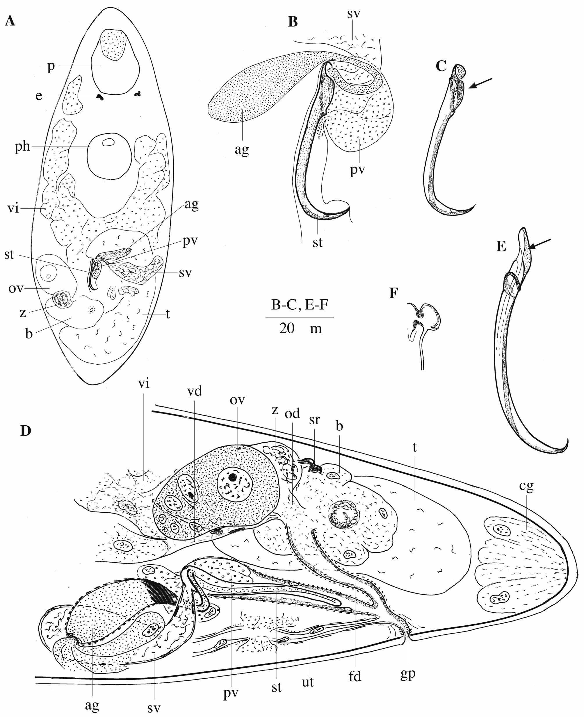
( Figs 1 View FIGURE 1 A–D, 2A–B)
Holotype. A whole mount, France, Bay of Marseille, Cap Canaille, near Cassis, 16 m deep, fine-grained sand, 7 July 1966 ( SMNH Type-8312).
Paratypes. One whole mount and seven serially-sectioned specimens from the type locality (HU 516–523).
Other material. One whole mount (on the same slide as the holotype of Rogneda reticulata Brunet, 1969 : SMNH 3048), France, Bay of Marseille, Cap Canaille, near Cassis, 16 m deep, fine-grained sand, 4 July 1966. Two whole mounts, France, Corsica, bay of Calvi, sand from the main beach, 3 m deep, 5 May 1982 (HU V.2.36–37). Five whole mounts (two on the same slide) and one serially-sectioned specimen, France, Banyuls-sur-Mer, northeast from Ile Grosse, 42°28'57.81"N 3°8'15.42"E, well-sorted, relatively fine sand, 12 m deep, 06 August 2007 (HU V.2.38–42). Live observation, Italy, Sardinia, Budelli Island, Il Cavaliere, 41°17’29.76”N 9°20’52.04” E medium- to fine-grained clean sand, 33–37 m deep, 7 September 2010.
Etymology. Species name refers to the shape of the stylet, which has the shape of a sickle. Sicilicula (Lat.) : small sickle.
Diagnosis. Species of Limipolycystis with two eyes. Prostate vesicle of type III, well developed. With a distally-curved, sickle-shaped stylet, which is an accessory style type II, 46–59 µm long, with strengthened proximal rim and pointed distal end. Proximally from the stylet opening, the stylet forms a gutter-shaped protrusion. Seminal receptacle consisting of a proximal (towards the ovary) tubular and distal (towards the bursa) broader part.
Description. The animals are small, approximately 0.4–0.6 mm long. They are colourless with two eyes ( Fig. 1 View FIGURE 1 A: e). The epidermis is syncytial, with numerous small, globular rhabdites arranged in a row just beneath the surface of the epithelium.
The proboscis ( Fig. 1 View FIGURE 1 A: p) measures 1/5 of the body length, with a construction identical to that of species of Myobulla (for a detailed description, see Artois & Schockaert 2000), except for the fact that in L. sicilicula n. sp. the cone retractors do not run parallel to each other, but are divided into three groups, as is the case in most Polycystididae (see Meixner 1925 for an overview). There are no nuclei in the contact-zone. The internal circular muscles of the proboscis bulb are about 1/20 of the proboscis diameter thick. There are four pairs of proboscis retractors and one pair of ventral integument retractors. The four proximal dilatators of the proboscis sheath are very thick.
The pharynx is situated in the first body half and is slightly inclined forwards ( Fig. 1 View FIGURE 1 A: ph). It is of the normal polycystidid construction (for a detailed description, see Meixner 1925). The distal part of the prepharyngeal cavity is covered with a frayed, degenerating epithelium (pseudociliation). There are 24 internal longitudinal muscles.
The gonads are unpaired. Both the ovary and the testis are situated in the caudal part of the body, the testis at the right, and the ovary at the left ( Fig. 1 View FIGURE 1 A: t, ov). The vitellarium consists of two branches connected to each other behind the pharynx ( Fig. 1 View FIGURE 1 A: vi). The genital pore is situated at ± 75 %. The common genital atrium is narrow and covered with a low, anucleated epithelium ( Fig. 1 View FIGURE 1 D).
The male genital atrium is rather narrow distally, and broadens towards its proximal end. It is covered with a low epithelium, which contains a few nuclei distally. One hard stylet, which is an accessory stylet type II in the terminology of Artois & Schockaert (2003), lies within the male atrium ( Fig 1 View FIGURE 1 D: st). It is a relatively simple, singlewalled tube. It curves abruptly at approximately 2/3 of its length and tapers towards a sharp point, which gives it the overall impression of a sickle ( Figs 1 View FIGURE 1 B–C; 2A–B). The proximal rim is thickened and shows a very broad opening. Proximally from this opening, the stylet has a plate-like protrusion, which forms a gutter (arrow in Figs 1 View FIGURE 1 C, 2A–B). The stylet is 46–59 µm long (x = 52 µm; n = 9; one specimen from Corsica could not be measured). It is 3–7 µm wide, measured at the proximal opening (x = 6 µm; n = 9). The stylet shows a thick ridge running over its entire length on the concave side.
The prostate vesicle (of type III following the terminology of Artois & Schockaert 2003) is relatively large and contains basophilic and eosinophilic secretion (pv in Figs 1 View FIGURE 1 A–B, D; 2A). It enters the male genital atrium proximally, near the proximal rim of the stylet, and bulges deeply into the atrium. The prostate secretion enters the stylet through its large proximal opening, which is at an angle with the longitudinal axis of the stylet. The seminal vesicle is surrounded by a weak muscle layer and narrows towards the male atrium, forming a broad seminal duct. This duct enters the male atrium near the prostate vesicle. The accessory vesicle (of type II; see Artois & Schockaert 2003) contains a coarse-grained basophilic secretion (ag in Figs 1 View FIGURE 1 A–B, D; 2A). Its efferent duct is very narrow and lined with basement membrane only (pseudocuticula). Both the vesicle and the duct are surrounded by thick, moreor-less circular muscles. The efferent duct of the accessory vesicle winds around the seminal duct, and enters the stylet proximally, in which it can be followed for some distance.
The female genital duct (of type I; see Artois & Schockaert 2005) enters the common genital atrium caudally ( Fig. 1 View FIGURE 1 D: fd). It is rather long, lined with a low anucleated epithelium and surrounded by circular muscles. Proximally it ends in the large bursa. The oviduct enters the female duct at the transition from female duct to bursa. It is broad and thin-walled, and receives the vitelloduct ventrally. As far as could be seen on the sections, the seminal receptacle ( Fig. 1 View FIGURE 1 D: sr) consists of a somewhat swollen part, which is connected to the bursa, and a sclerotised tube. As this structure is homologised with the seminal receptacle of the other species of Typhlopolycystidinae by Artois & Schockaert (2000), we will continue to use the term. However, it has apparently lost its function as spermreceiving organ, as sperm were never observed in it. This function seems to be covered by the proximal part of the oviduct (z in Figs 1 View FIGURE 1 A, D), to which the sclerotised tube is connected.
| SMNH |
Saskatchewan Museum of Natural History |
No known copyright restrictions apply. See Agosti, D., Egloff, W., 2009. Taxonomic information exchange and copyright: the Plazi approach. BMC Research Notes 2009, 2:53 for further explanation.