
Melolontha, Fabricius, 1775
|
publication ID |
https://doi.org/10.11646/zootaxa.5263.2.2 |
|
publication LSID |
lsid:zoobank.org:pub:B07051F2-A15B-49EC-9A2F-91B9420FEA4A |
|
DOI |
https://doi.org/10.5281/zenodo.7814556 |
|
persistent identifier |
https://treatment.plazi.org/id/BC1F87FF-137F-FFF9-FF74-FD0EFA6EBF8C |
|
treatment provided by |
Plazi (2023-04-05 06:52:47, last updated 2024-11-26 05:29:02) |
|
scientific name |
Melolontha |
| status |
|
Key to species groups of Melolontha known from India (adopted and modified from Li et al. 2010)
1. Elytral disc glabrous or setiferous; colour of elytra light olive to dark green........................................ 2
– Elytral disc with dense setae; colour of elytra dark brown to yellowish-brown...................................... 3
2. Pronotum shining dorsally, lacking metallic reflection; elytron with one post-humeral and 2 to 3 basal setiferous patches ( Fig. 1 View FIGURES 1–4 ); apex of interval one deeply depressed along elytral disc; parameres with ventroapical process, distinctly elongate and gradually acuminate in lateral view ( Fig. 53 View FIGURES 53–64 ), lateral convexity completely absent ( Fig. 41 View FIGURES 41–46 )............... guttigera group
– Pronotum with dorsal metallic with green reflection; elytra without patches of setae at base ( Fig. 2 View FIGURES 1–4 ); apex of interval one weakly depressed along elytral disc; parameres without a ventroapical process ( Fig. 54 View FIGURES 53–64 ), lateral convexity weakly developed ( Fig. 42 View FIGURES 41–46 ) ........................................................................................ virescens group
3. Basal segment of antenna swollen at apex, with antennomeres 2 and 3 compressed (see Li et al. 2010, fig. 1) ( Figs. 26 View FIGURES 25–32 , 34 View FIGURES 33–40 , 66 View FIGURES 65–71 ); antennal club strongly to moderately curved outwardly ( Figs 25 View FIGURES 25–32 , 33 View FIGURES 33–40 , 66 View FIGURES 65–71 ); length of dense setae more on vertex than those of clypeus ( Figs. 25 View FIGURES 25–32 , 33 View FIGURES 33–40 , 66 View FIGURES 65–71 ); setae on inner surface of meso- and metatibiae stout and long ( Figs. 31 View FIGURES 25–32 , 37 View FIGURES 33–40 )................ … 4
– Basal segment of antennae moderately swollen apically, antennomeres 2 and 3 elongate (see Li et al. 2010, fig. 2); antennal club weakly to moderately curved outwardly (see Li et al. 2010, figs. 4, 5, 6); length of setae on vertex same as those on clypeus; setae on inner surface of meso- and metatibiae stout and short.................................................. 5
4. Antennal club strongly curved outwardly, 3.5 times as length of basal segments (see Li et al. 2010, fig. 3) ( Figs. 25 View FIGURES 25–32 , 33 View FIGURES 33–40 , 66 View FIGURES 65–71 ); outer metatibial spur subequal in length to length of metatarsomeres 1 and 2 combined ( Figs. 31 View FIGURES 25–32 , 37 View FIGURES 33–40 ); apical portion of pygidium not narrowed, more or less rounded ( Figs. 15, 16 View FIGURES 13–18 ); parameres symmetrical, apical process of left paramere not raised ( Figs. 43, 44 View FIGURES 41–46 , 70 View FIGURES 65–71 )....................................................................... phupanensis group
– Antennal club moderately curved outwardly, three times as length of basal segments; outer metatibial spur subequal to length of metatarsomere 1; apical portion of pygidium narrowed to form an elongate process ( Figs 17, 18 View FIGURES 13–18 ); parameres asymmetrical, apical process of left paramere relatively raised ( Figs 45, 46 View FIGURES 41–46 )...................................... aeneicollis group
5. Antennal club relatively shorter, weakly curved (see Li et al. 2010, fig. 6) ( Fig. 7 View FIGURES 5–8 ); pronotum with irregular serrations laterally; parameres with sharply developed pointed process at outer margin in dorsal view ( Fig. 47 View FIGURES 47–52 ).............. .. chinensis group
– Antennal club usually longer and moderately curved (see Li et al. 2010, figs. 4, 5) ( Figs. 8–12 View FIGURES 5–8 View FIGURES 9–12 ); pronotum with serrations developed anteriorly with basal one–half to one–third of margin smooth (see Li et al. 2010, fig. 8); parameres without pointed process ( Figs. 48–52 View FIGURES 47–52 ).................................................................................. 6
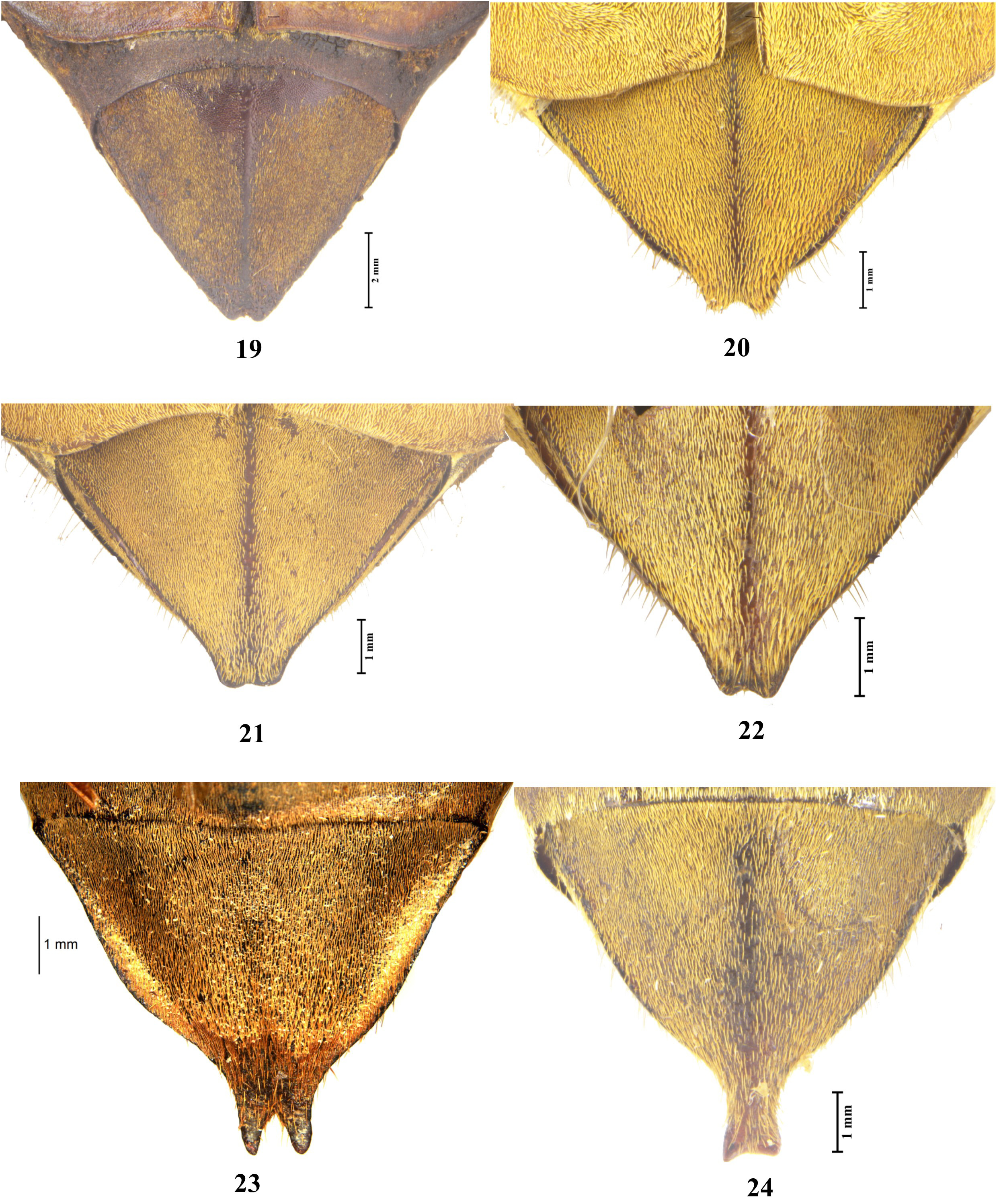
6. Central disc of pronotum evenly and densely punctate; midline of pygidium ridged completely or incompletely ( Figs. 20–22 View FIGURES 19–24 ); apical and dorsal process ( Figs. 48–50 View FIGURES 47–52 ) and lateral convexity of paramere well developed ( Figs. 60–62 View FIGURES 53–64 )..... .. carinata group
– Central disc of pronotum unevenly punctured; midline of pygidium smooth ( Figs 23–24 View FIGURES 19–24 ); apical process of paramere distinctly enlarged posteriorly, fusing with dorsal process ( Figs. 51–52 View FIGURES 47–52 ), lateral convexity broadly swollen laterally ( Figs. 63–64 View FIGURES 53–64 )............................................................................................... incana group
Li, C. - L., Yang, P. - S. & Wang, C. - C. (2010) Revision of the Melolontha guttigera group with a key and an annotated checklist of the East and South-East Asian Melolontha groups. Annals of the Entomological Society of America, 103 (3), 341 - 359. https: // doi. org / 10.1603 / AN 09088
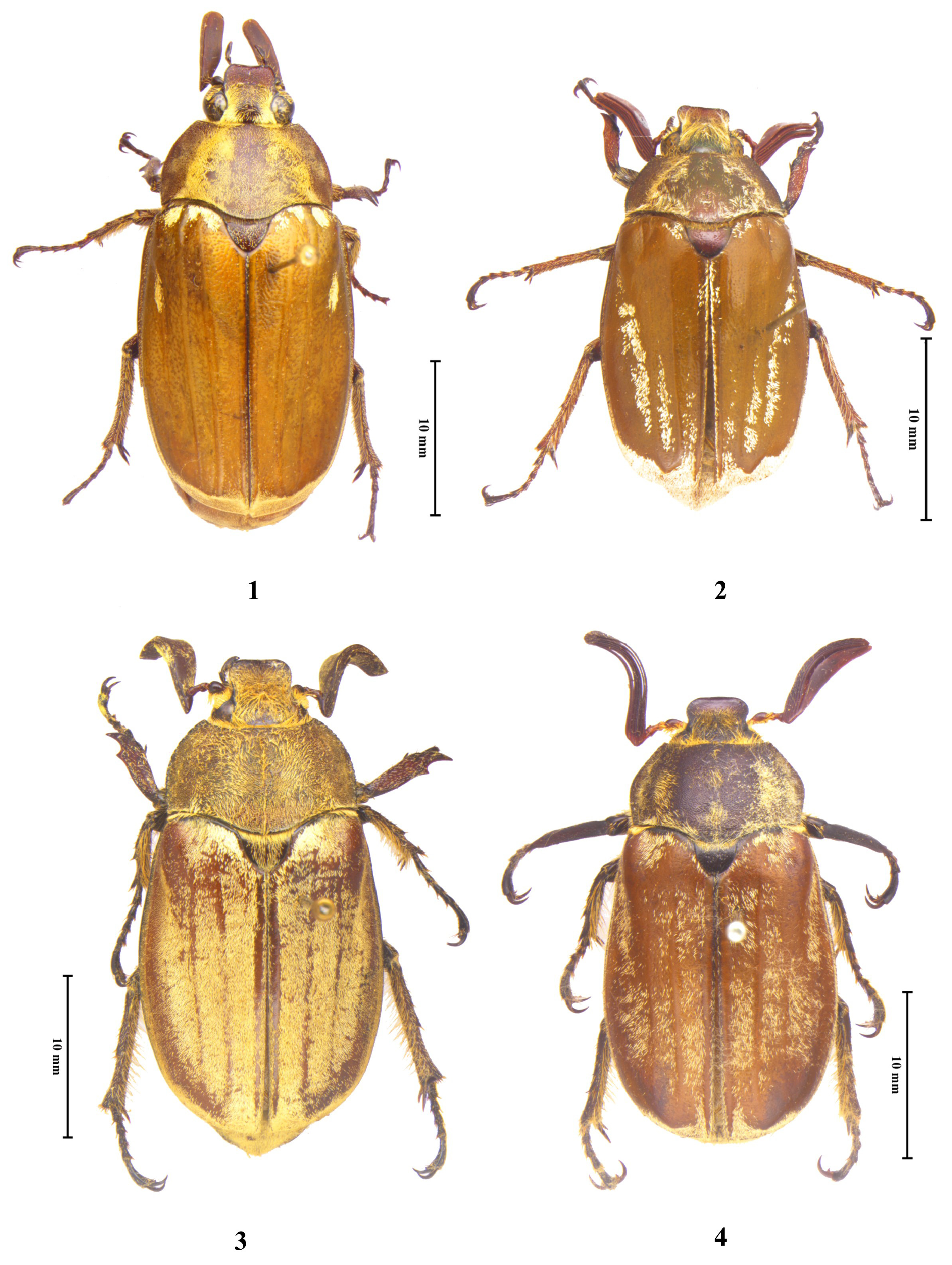
FIGURES 1–4. Habitus of Melolontha species. 1, M. guttigera; 2, M. virescens; 3, M. arunachalensis new species (holotype); 4, M. lachungensis new species (holotype).
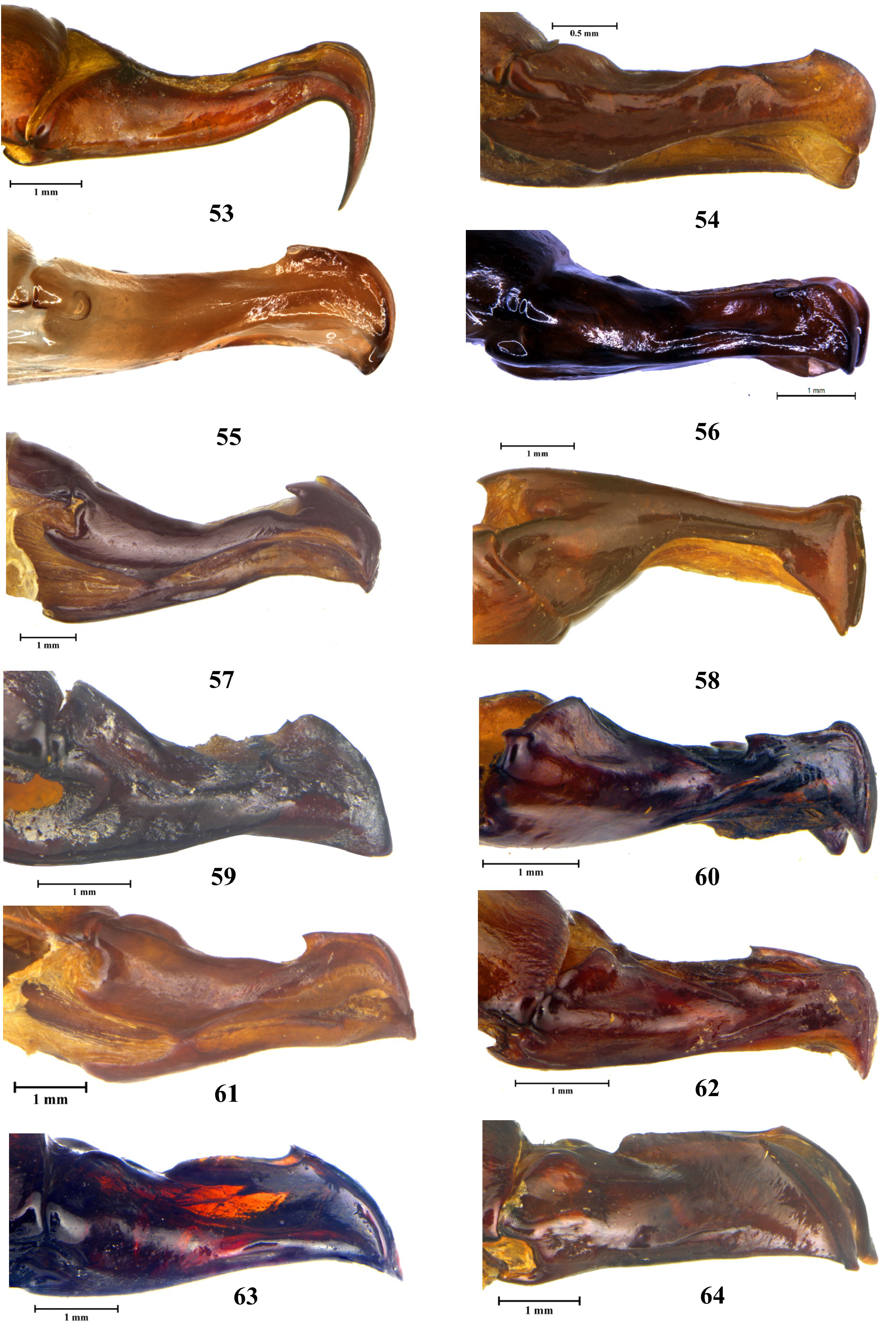
FIGURES 53–64. Parameres of Melolontha species in lateral view. 53, M. guttigera; 54, M. virescens; 55, M. arunachalensis new species (holotype); 56, M. lachungensis new species (holotype); 57, M. aeneicollis; 58, M. cuprescens; 59, M. chinensis; 60, M. nepalensis; 61, M. carinata; 62, M. indica; 63, M. flabellata; 64, M. furcicauda.
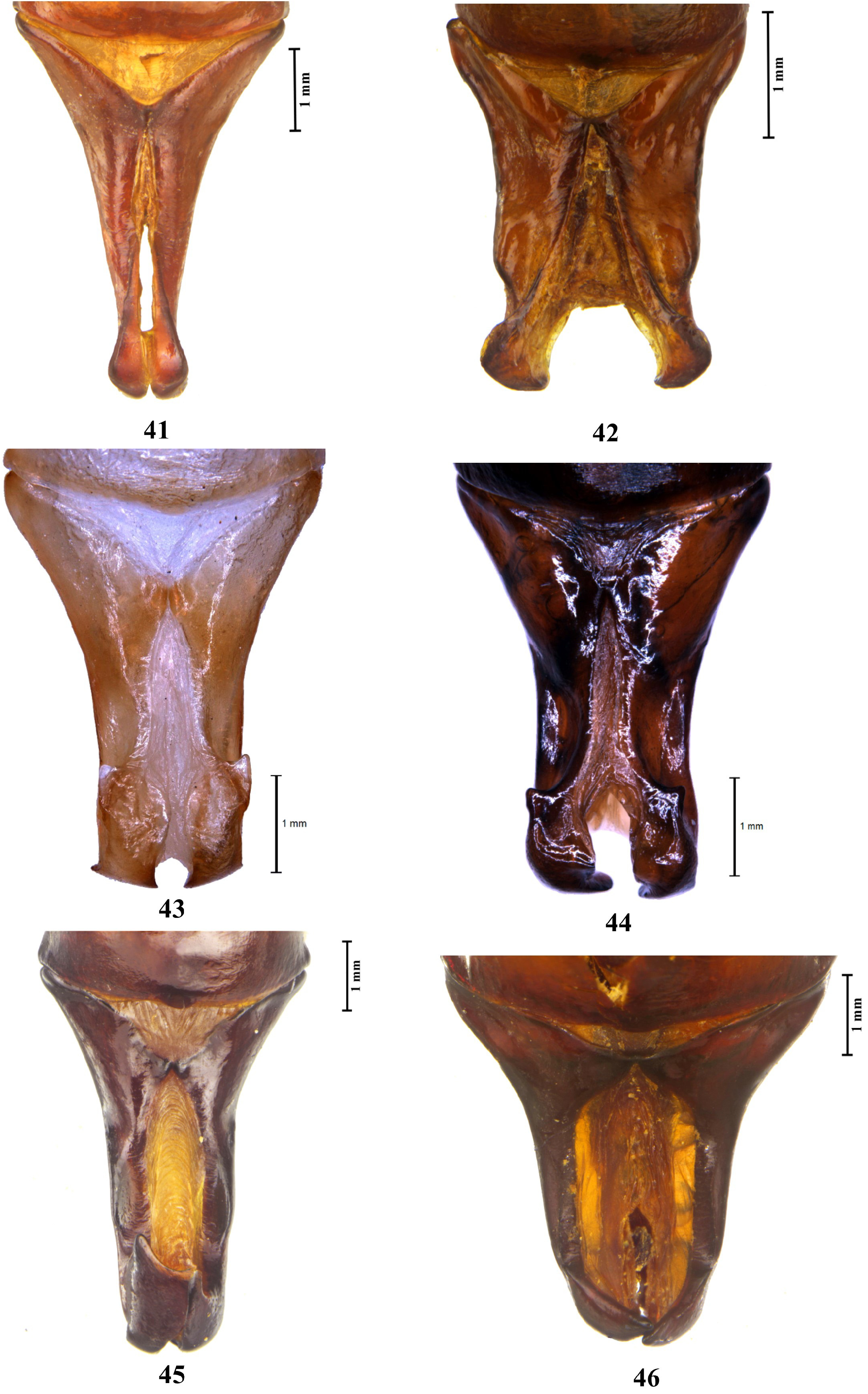
FIGURES 41–46. Parameres of Melolontha species in dorsal view. 41, M. guttigera; 42, M. virescens; 43, M. arunachalensis new species (holotype); 44, M. lachungensis new species (holotype); 45, M. aeneicollis; 46, M. cuprescens.

FIGURES 25–32. Melolontha arunachalensis new species (holotype). 25, head and antennae; 26, basal segments of antennae; 27, pronotum; 28, scutellar plate; 29, protibia; 30, mesometasternal process; 31, metatarsomeres; 32, abdomen.
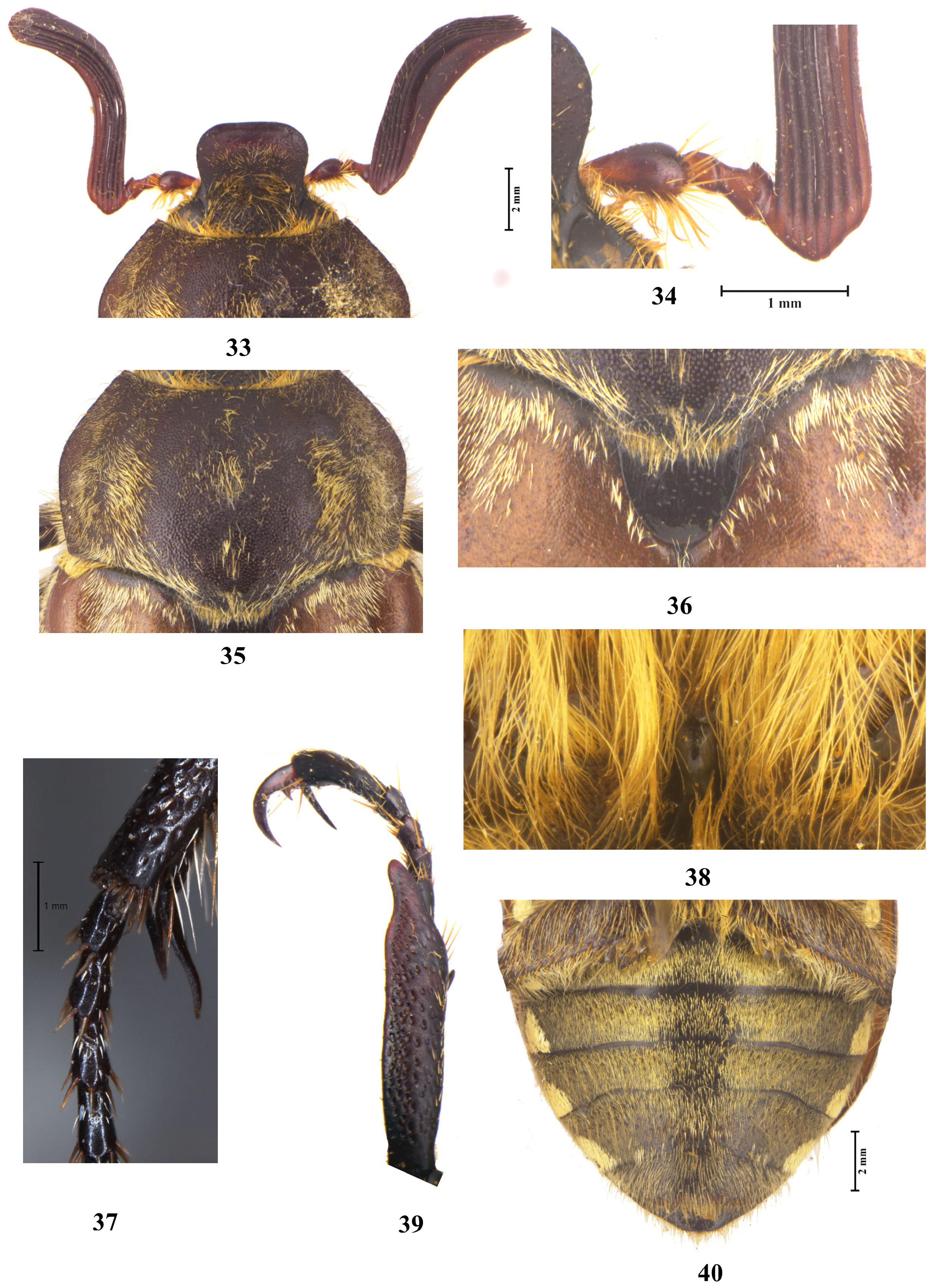
FIGURES 33–40. Melolontha lachungensis new species (holotype). 33, head and antennae; 34, basal segments of antennae; 35, pronotum; 36, scutellar plate; 37, protibia; 38, mesometasternal process; 39, metatarsomeres; 40, abdomen.
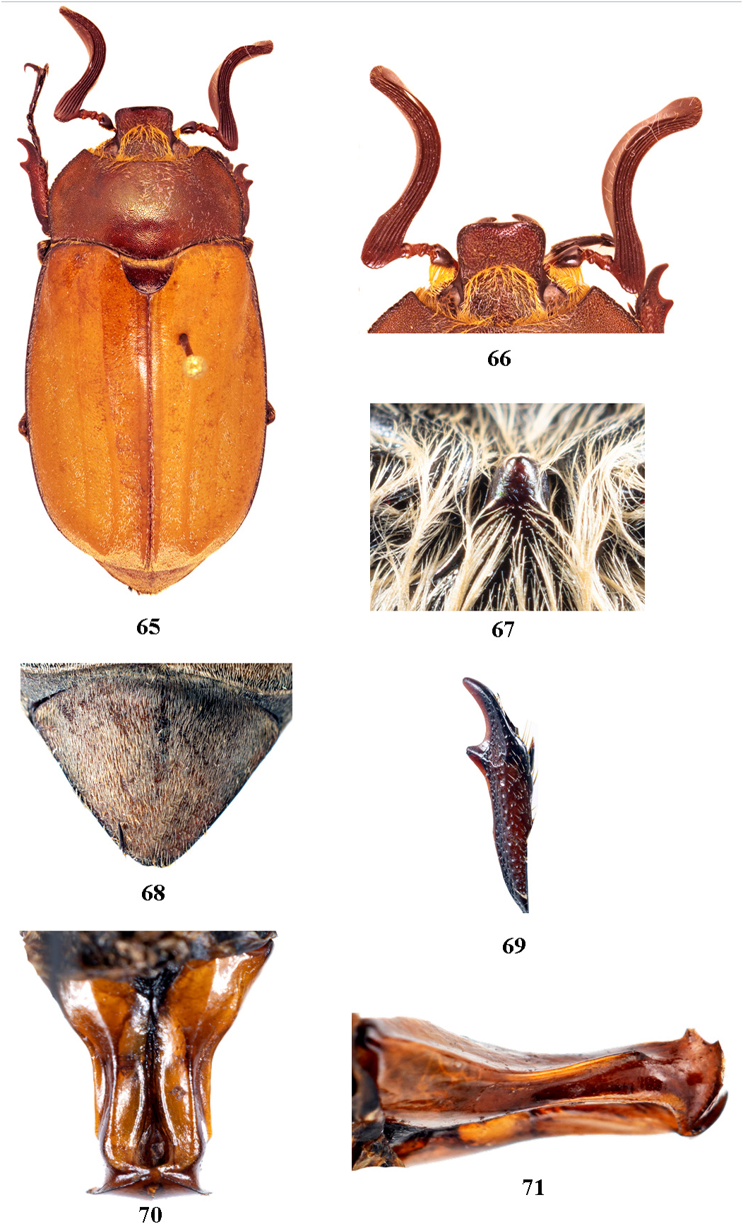
FIGURES 65–71. Melolontha phupanensis (paratype, male). 65, habitus; 66, head and antennae; 67, mesometasternal process; 68, pygidium; 69, protibia; 70, parameres in dorsal view; 72, parameres in lateral view.
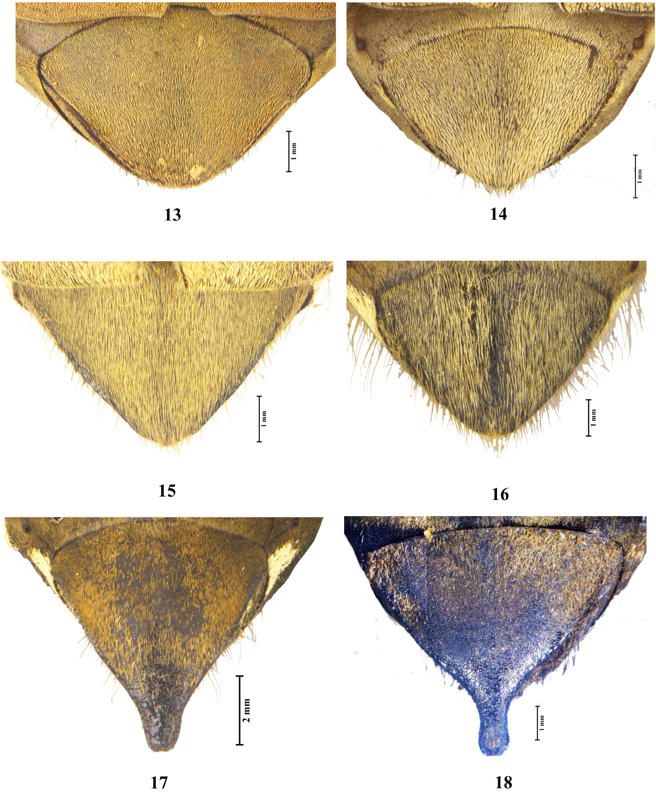
FIGURES 13–18. Pygidium of Melolontha species. 13, M. guttigera; 14, M. virescens; 15, M. arunachalensis new species (holotype); 16, M. lachungensis new species (holotype); 17, M. aeneicollis; 18, M. cuprescens.
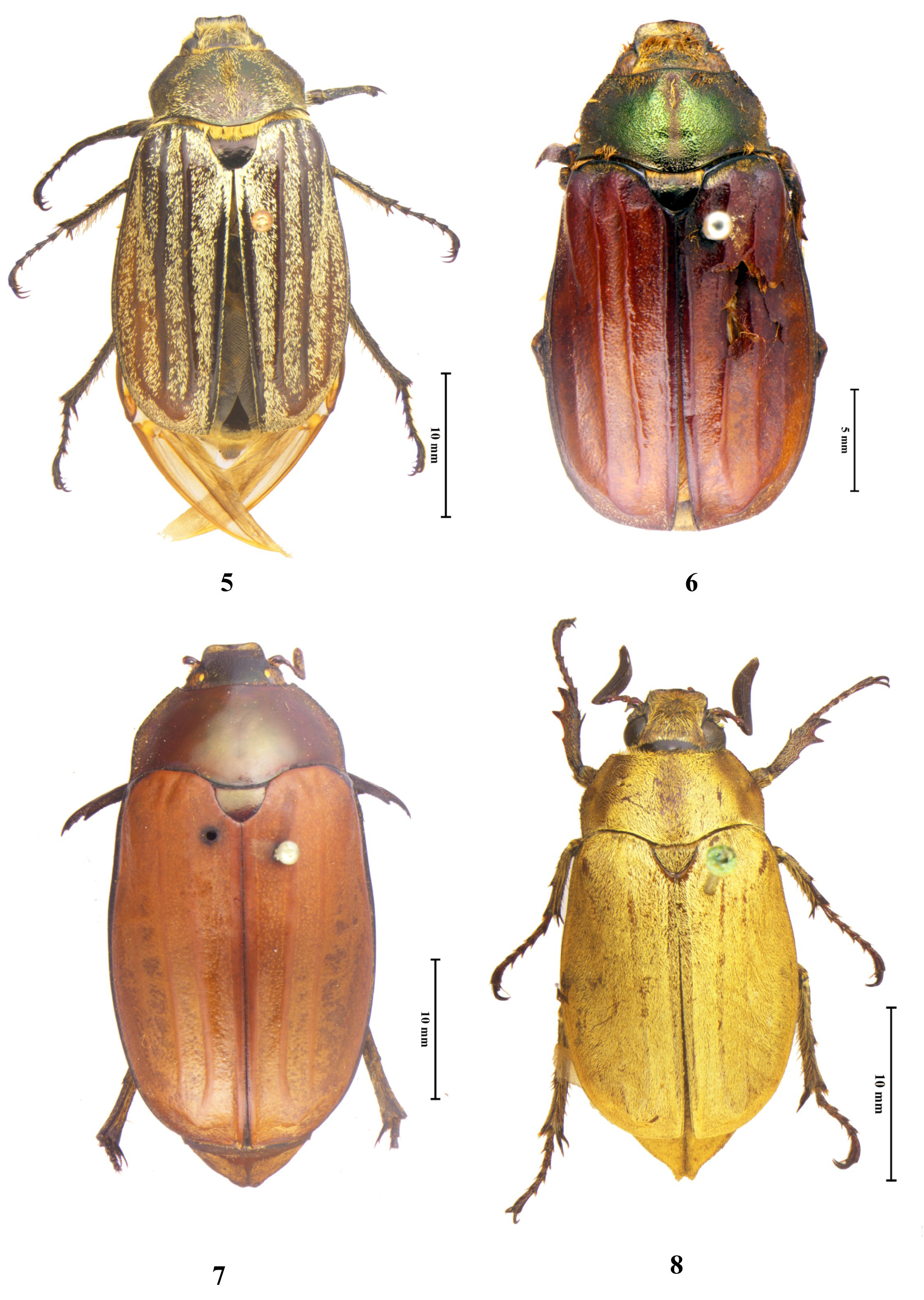
FIGURES 5–8. Habitus of Melolontha species. 5, M. aeneicollis; 6, M. cuprescens; 7, M. chinensis; 8, M. nepalensis.
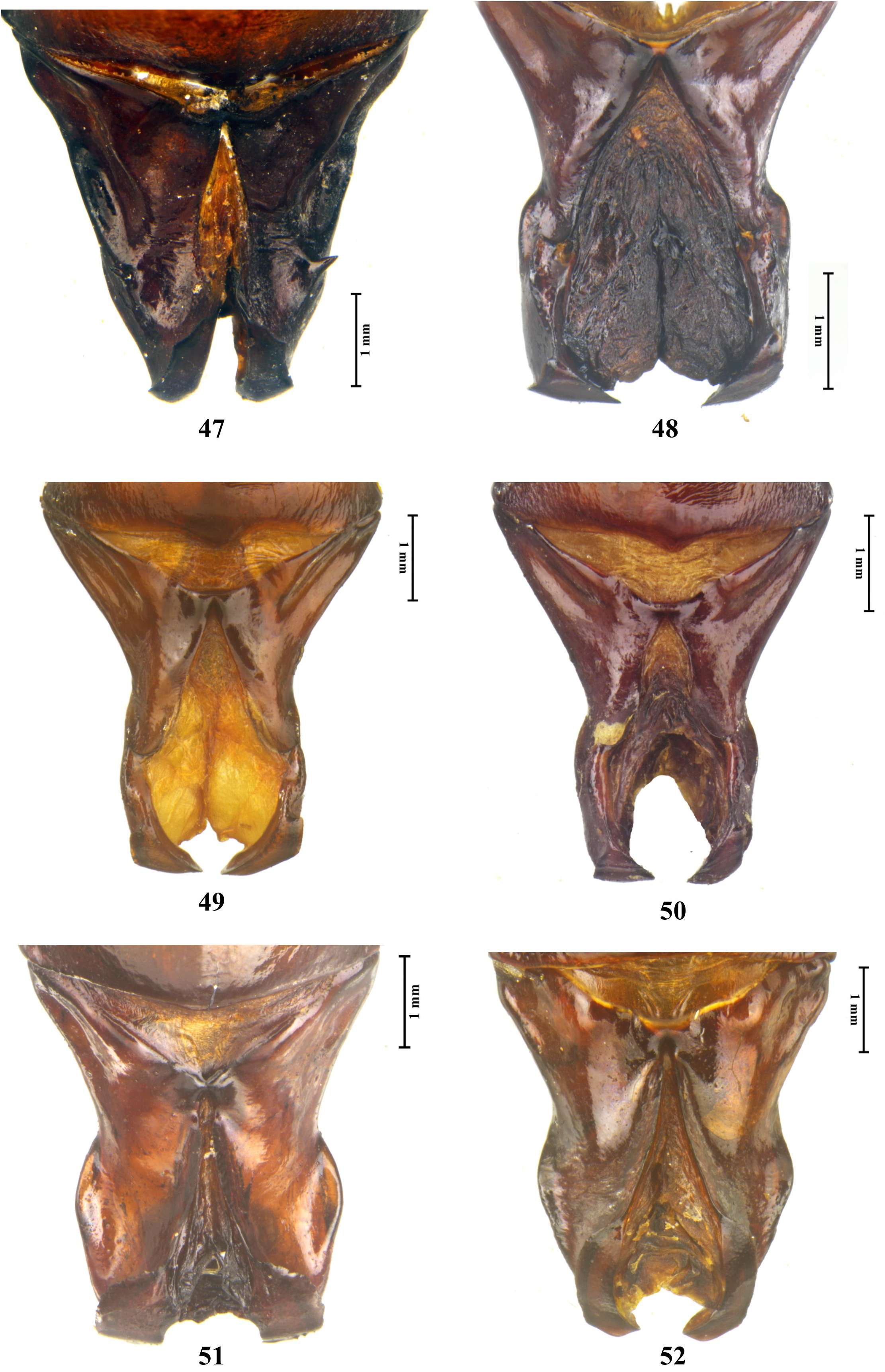
FIGURES 47–52. Parameres of Melolontha species in dorsal view. 47, M. chinensis; 48, M. nepalensis; 49, M. carinata; 50, M. indica; 51, M. flabellata; 52, M. furcicauda.
No known copyright restrictions apply. See Agosti, D., Egloff, W., 2009. Taxonomic information exchange and copyright: the Plazi approach. BMC Research Notes 2009, 2:53 for further explanation.
|
Kingdom |
|
|
Phylum |
|
|
Class |
|
|
Order |
|
|
Family |
|
|
SubFamily |
Melolonthinae |