
Gomphidictinus tongi, Zhang & Guan & Wang, 2017
|
publication ID |
https://doi.org/ 10.11646/zootaxa.4344.2.7 |
|
publication LSID |
lsid:zoobank.org:pub:BACD8FC5-66FB-4514-B4AA-629D5A8F84AC |
|
DOI |
https://doi.org/10.5281/zenodo.6049664 |
|
persistent identifier |
https://treatment.plazi.org/id/BA221469-C04E-A15B-9FDD-FB3B2F59B3CE |
|
treatment provided by |
Plazi |
|
scientific name |
Gomphidictinus tongi |
| status |
sp. nov. |
Gomphidictinus tongi View in CoL sp. nov.
Holotype. ♂ (deposited in the Odonata Collection of Kunming Institute of Zoology, Chinese Academy of Sciences , Yunnan Province, China), China, Lingshui County, Hainan Province, Mt. Diaoluoshan (18°43'23.16"N, 109°52'10.37"E; 930 m a.s.l.), 28 May 2014, Hao-miao Zhang leg. GoogleMaps
Paratypes. 2♂♂, 1♀, same data as holotype GoogleMaps ; 1♂, China, Guangxi Province, Chongzuo , 30 May 2017, Haomiao Zhang leg. All paratypes deposited at the same place as holotype.
Etymology. The species name, a noun in the genitive case, is dedicated to Professor Xiao-li Tong, from South China Agricultural University, the Ph.D. supervisor of the first and second authors. He passed on professional knowledge of entomology to Hao-miao Zhang and gave great help for his study on Odonata .
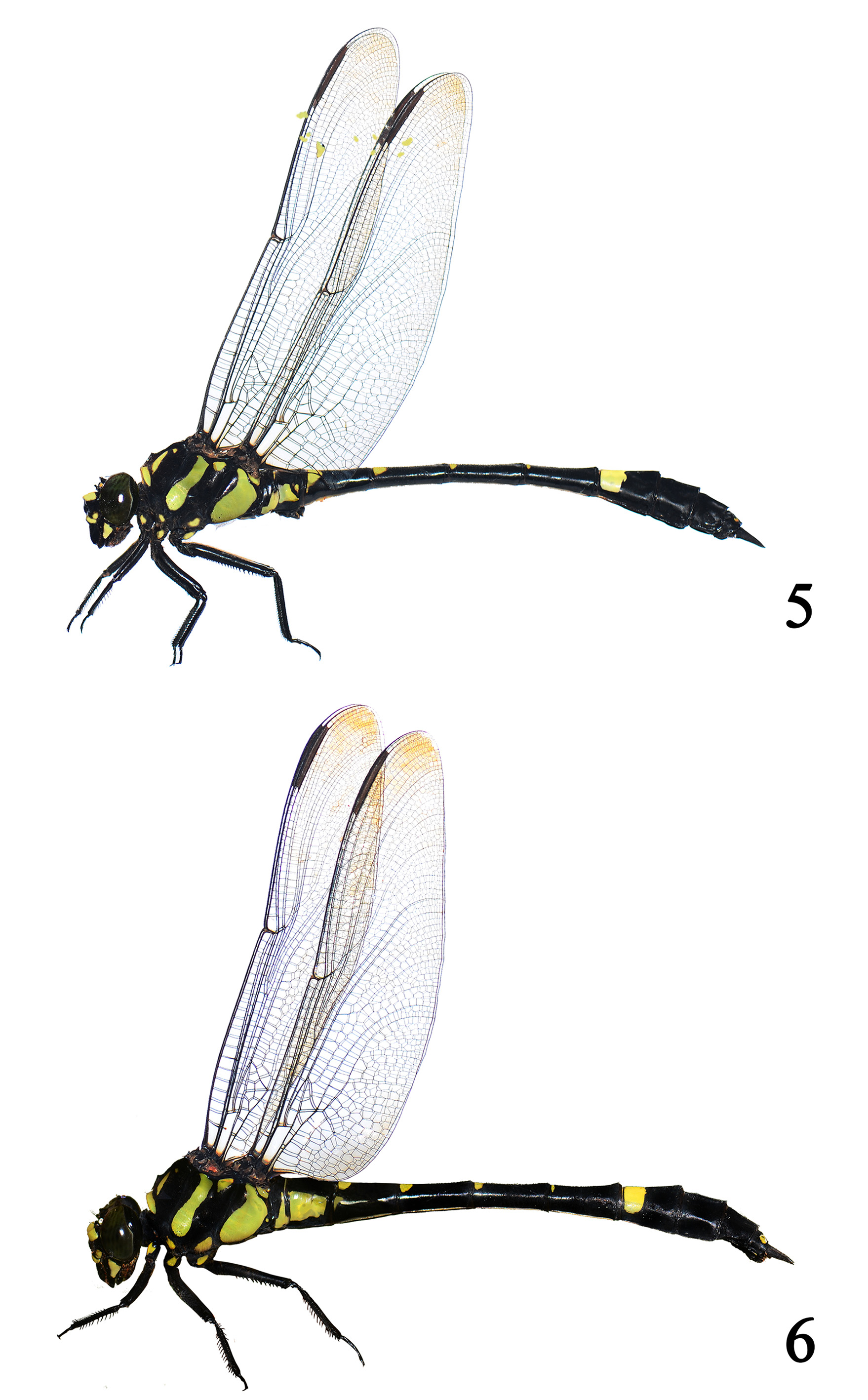
A big and robust dragonfly. Body black with yellow maculation and hyaline wings ( Figs 5–6 View FIGURES 5 – 6 ).
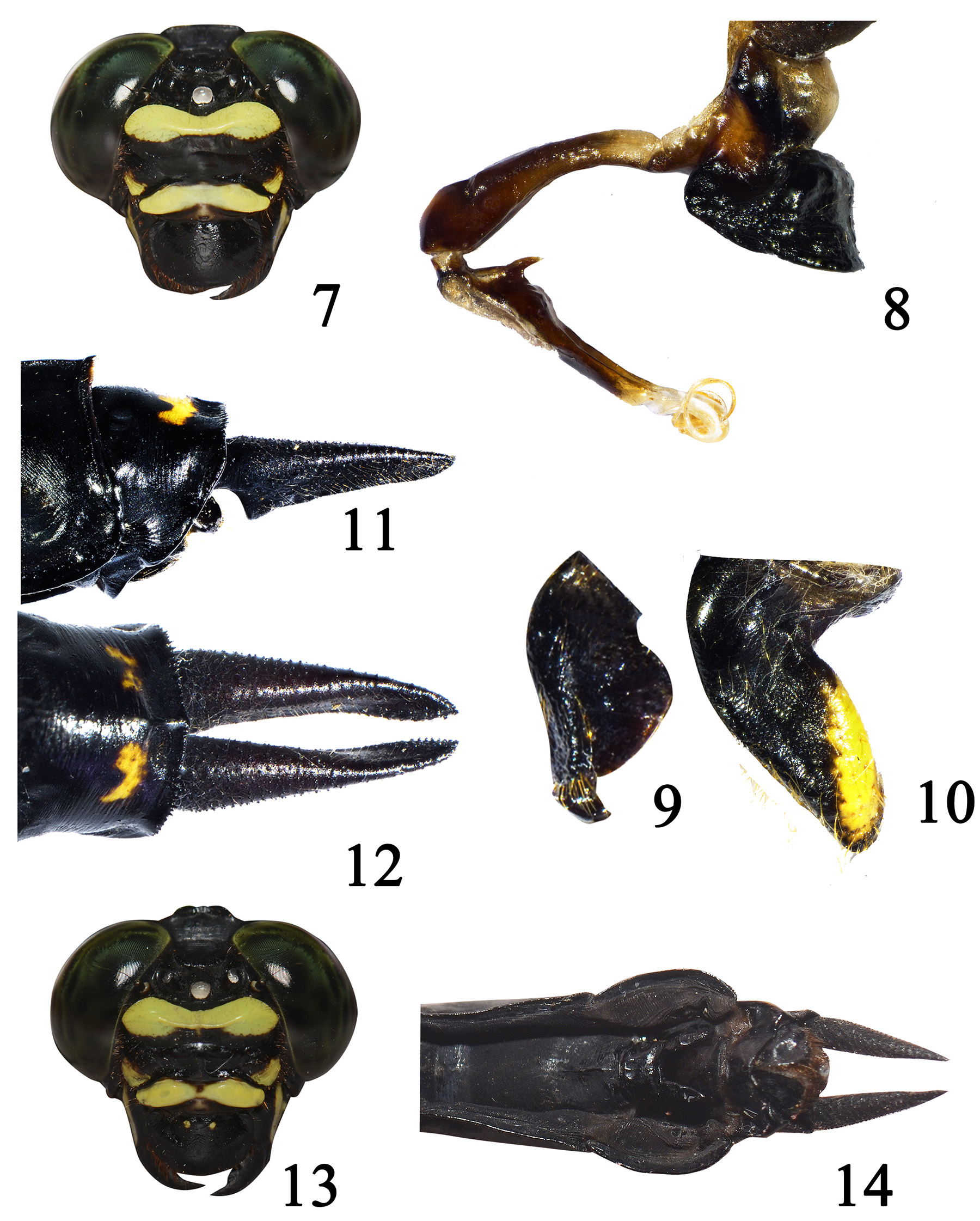
Description of the holotype. Head. Eyes dark green when alive ( Fig. 7 View FIGURES 7 – 14 ). Face black with yellow markings ( Fig. 7 View FIGURES 7 – 14 ). Labium almost black with indistinct yellow spots at the posterior of each lobe; mandible bases yellow; labrum black; anteclypeus entirely yellow; postclypeus largely black with paired yellow spots at the lateral sides; antefrons black below, with the upper half yellow, top of frons with anterior 2/3 yellow; vertex above lateral ocelli triangularly projected, black with paired triangularly projections behind lateral ocelli; occiput black with a very short median tubercle, occipital margin almost straight.
Thorax. Generally black with yellow markings ( Fig. 5 View FIGURES 5 – 6 ). Prothorax black. Synthorax with mesothoracic collar stripes connecting at midpoint, dorsal stripes not connecting with collar stripes; antehumeral spot small, comma shaped; sides of synthorax mostly black with yellow markings: mesepimeron with a broad yellow stripe, metepisternum with a triangular shaped spot near the upper margin, metepimeron largely yellow, mesokatepisternum with a lower spot, metakatepisternum largely yellow. Poststernum with paired large spots. Legs black with yellow markings. Coxae with yellow spots in all legs; fore leg femur with yellow stripes in the inner side, mid and hind legs entirely black.
Wings. Hyaline, slightly tinted with amber brown, venation black. Forewings: 22 antenodals above Sc and 18 antenodals below Sc; 18 (left) and 19 (right) postnodals above R1. Hindwing: 15 (left) and 16 (right) above Sc, 15 antenodals below Sc; 18 (left) and 20 (right) postnodals above R1. Triangle 4-celled in forewings and 3-celled in hindwings, supertriangle 3- or 5-celled in forewings, 3-celled in hindwings, subtriangle 3-celled in forewings and 1-celled in hindwings. Anal triangle 5-celled. Anal loop 5-celled. Pterostigma long, 7mm in length, color dark brown, covering 9 cells below.
Abdomen. Black with pale marking as follows ( Fig. 5 View FIGURES 5 – 6 ): S1 with a large lateral spot close the lower margin, and very fine stripe along the hind margin dorsally; S2 with a large anterior central arrow-shaped spot dorsally, laterally with two spots, anterior one covering auricle; S3 with a large basal spot, triangular shaped; bases of S4–6 with paired spots, connecting at mid-carina; basal half of S7 with a large spot; S8–10 and anal appendages almost black, S10 with paired tiny spots dorsally.
Accessory genitalia and anal appendages. Vesicle with a huge ventral projection. Median segment of penis organ with a sharp basal spine, distal segment with twisted flagellae ( Fig. 8 View FIGURES 7 – 14 ). Anterior hamuli stout, gently expanded apically, hooked backwards in apex ( Fig. 9 View FIGURES 7 – 14 ); posterior hamuli broad and strongly curved posteriorly ( Fig. 10 View FIGURES 7 – 14 ). Anal appendages black. Cerci about 1.65 times as long as S10, conical and tapering distally in lateral view, with a ventral spine basally ( Fig. 11 View FIGURES 7 – 14 ); in dorsal view, depressed at apical 2/5, with a row of 7–9 denticles ( Fig. 12 View FIGURES 7 – 14 ). Epiproct very short, thus forming a big hollow from the ventral spine of cerci, the dorsal surface with two very small spines apically.
Paratype female: Head. Maculation very similar to holotype male. Occiput with a pair of lobed projections posteriorly ( Fig. 13 View FIGURES 7 – 14 ).
Thorax. Almost identical to holotype male, but yellow spots on head and coxae slightly larger than holotype male, wings brown color darkened.
Abdomen. Black with pale marking as follows ( Fig. 6 View FIGURES 5 – 6 ): S1 with a large lateral spot posteriorly and a very fine stripe along the hind margin dorsally; S2 with a triangular central spot dorsally, lateral side with a big rectangular spot; S3 with anterior semicircle spot dorsally and anterior lateral small stripes; S4–6 with anterior spot dorsally; S7 with a large basal spot; S8–10 and cerci almost black except for the small paired spots on S10 dorsally. Cerci long, about 1.5 times as long as S10. Vulvar lamina very long, reach almost the end of S9, deeply depressed medially ( Fig. 14 View FIGURES 7 – 14 ).
Variation in paratype males. Two paratype males have collar stripes interrupted at midpoint. Paired yellow spots larger. Fore leg femur with less yellow markings. Anal triangle 5- or 6-celled.
Measurements (mm). Holotype male: total length 85.0, abdomen (including anal appendages) 63.0, hind wing 51.5; Paratype males: total length 78.0–79.0, abdomen (including anal appendages) 58.0–60.0, hind wing 48.0–49.0; Paratype female: total length 81.0, abdomen (including anal appendages) 61.0, hind wing 54.0;
Distribution. China (Hainan and Guangxi).
Diagnosis. Male of G. tongi sp. nov. is similar to G. kompieri , but can be distinguished from it by the following characters: 1) the epiproct shorter, with a bigger hollow between cerci and epiproct in lateral view; 2) the spine on the median segment of the penis is situated more basally; 3) the subtriangle of the hindwings is not reticulated. Male of G. tongi sp. nov. is quite different from G. perakensis by the absence of the whale-tail shaped tubercle on ventral surface of S1 and the shape of the male anal appendages. Female of G. tongi sp. nov. can be separated from the other congeners by longer vulvar lamina which is reaching the end of S9.
More detailed characters between the three species are listed in Table 1.
Notes on biology. Only a few adults were collected from Mt. Diaoluoshan Mountain in Hainan Island. The population is quite small and all the males were hidden in the very dense forested streams. Males held the territory quite near water, usually perch on the short trees or top of sticks, which is also seen in Gomphidia males. They always perch but seldom fly, and only appear in the sunny days when the sunlight can pass through the dense tropical rain forest. Species co-occurring include the endemic Chlorogomphus gracilis Wilson & Reels, 2001 , the famous Pseudolestes mirabilis Kirby, 1900 and various gomphids including Gomphidia kruegeri . In Guangxi the stream is less shady and the paratype male and two males of G. kruegeri were collected in the same tree.
No known copyright restrictions apply. See Agosti, D., Egloff, W., 2009. Taxonomic information exchange and copyright: the Plazi approach. BMC Research Notes 2009, 2:53 for further explanation.
|
Kingdom |
|
|
Phylum |
|
|
Class |
|
|
Order |
|
|
Family |
|
|
Genus |