
Bitabulata paoma, Goldschmidt, 2008
|
publication ID |
https://doi.org/ 10.11646/zootaxa.1954.1.1 |
|
persistent identifier |
https://treatment.plazi.org/id/B03B8797-6916-FFC0-FF21-FF41595CFEE8 |
|
treatment provided by |
Felipe |
|
scientific name |
Bitabulata paoma |
| status |
sp. nov. |
Bitabulata paoma sp. nov.
( Figs 184–196 View FIGURES 184–190 View FIGURES 191–196 , Table 10)
Type: Holotype male, MD 11 d, Anjozorobe (Antananarivo), Ravoandrina, Rivière Ampanakamonty near campsite, 1280 m asl, 12.8 °C, 78 µS/cm, 21.07.2001, mounted.
Additional material examined: MD 63, Andohahela (Tulear), Isaka, stream exposition west 1 km north from the village, 250 m asl, 19.7 °C, 136 µS/cm, 07.09.2001, 0/1/0 mounted; MD 71a, Andohahela (Tulear), Isaka, spring area south pass RIP 118 (km 36), 700 m asl, 16.0–18.4 °C, 55–60 µS/cm, 10.09.2001, 0/1/0 mounted, 0/1/0 unmounted.
Habitat: Streams and spring at 250–1280 m asl.
Distribution: Madagascar (Central Highland and Southern mountain regions).
Derivatio nominis: Paoma (Malagasy) — apple; referring to the broad, rounded shape of the anterior coxae of the species.
Diagnosis: Anterior and posterior dorsal plate with antero-lateral ridge; most platelets of glandularia in dorsal furrow elongated; Cx-I and -II apically rounded; ventral shield with distinct “shoulder corners”; caudal margin of Cx-IV nearly transverse, genital field only slightly surpasses genital bay caudally (especially in male); Vgl-3 far posteriorly on ventral shield; Vgl-2 and -4 free, posterior ventral shield in indentations of caudal margin; legs as characterised for the genus; capitulum short, compact; palp mid-sized.
Description, male (n = 1): Idiosoma rectangular-oval ( Fig. 184 View FIGURES 184–190 ), purple-brownish; broad dorsal shield (L/ W 677/441), anterior plate trapezoid, laterally undulated, caudally concave (L/W 221/394), bearing post-ocular setae; posterior plate broad rectangular, anteriorly straight to convex, caudally undulated, semi-circular (L/ W 452/441), Dgl-4 fused with antero-lateral corners, Dgl-5 fused postero-laterally; central area of plates slightly raised against antero-lateral margins ( Fig. 185 View FIGURES 184–190 ); Dgl-2 on irregularly elongated platelet, Dgl- 3 in lateral indentation of anterior plate, Lgl-4 and Dgl- 6 in indentations at caudal margin of posterior plate; l1 posteriorly attached to Dgl-2, l2 posterior Dgl-3, l3 and l4 directly posterior Lgl-2 and -3, l5 posterior caudal margin of ventral shield, between Vgl-2 and -4; soft integument in dorsal furrow irregularly undulated lined; setae of Dgl-3 to -5 large, others mid-sized ( Fig. 185 View FIGURES 184–190 ); lateral eyes oval, far separated from each other, free under platelet of Dgl-2 ( Fig. 185 View FIGURES 184–190 ); venter nearly completely covered by ventral shield (L/W 693/536); Cx-I to -III antero-laterally rounded, anteriorly surpassing ventral shield; dorso-lateral to Cx-II/III ventral shield extended, forming clear “shoulder corners” ( Fig. 184 View FIGURES 184–190 ); medial margins of Cx-I overlapping, separate, Cx-I laterally overlapping Cx-II, latero-caudal and medio-caudal margin of Cx-I/II overlapping Cx-III; Cxgl-2 and -4 close together at anterior margin of Cx-III; medial margins of Cx-III approximate, not fused; Cx-IV nearly rectangular, medial margin concave, forming very tight genital bay, medio-caudal corner rounded, caudal margin of Cx-IV slightly oblique to anterior, insertion of leg-IV exactly lateral to genital field, lateral margin of Cx-IV not visible, ridge on Cx-IV from slightly medial of insertion of leg-IV reaching to dorso-laterally of Cx-III ( Fig. 184 View FIGURES 184–190 ); genital field elongated-rectangular, sharp antero-lateral corners, posteriorly more rounded, laterally nearly straight, posteriorly only slightly extended beyond caudal margin of Cx-IV; acetabula elongated-oval, touching each other (Ac1 and Ac3 remote from anterior and caudal end of genital flaps), setae on genital flaps very tiny; very fine area of articular membrane lateral to genital field ( Fig. 184 View FIGURES 184–190 ); genital skeleton relatively slender (not clearly visible in preparation) ( Fig. 186 View FIGURES 184–190 ); setae of Vgl-1 directly posterior to caudal margin of Cx-IV, Vgl-3 far distant from coxal field, near latero-caudal margin of ventral shield; Vgl-2 and Vgl-4 on mid-sized platelets directly posterior of ventral shield in indentations of caudal margin ( Fig. 184 View FIGURES 184–190 ); legs strong, especially leg-IV bearing many heavy setae, segments rather compact, claws on leg-I to -III slen- der, with fine dorsal and strong ventral clawlet ( Fig. 187 View FIGURES 184–190 ); leg-IV-6 with mid-sized, pinnate sub-terminal seta ( Fig. 188 View FIGURES 184–190 ); capitulum short, compact ( Fig. 189 View FIGURES 184–190 ); palp relatively short, P1 with one dorsal seta; P2 with one pointed ventro-lateral seta and six dorsal setae; P3 with three dorsal and one lateral setae; P4 slightly curved ventral setae on very flat humps; P5 tapering ( Figs 189, 190 View FIGURES 184–190 ).
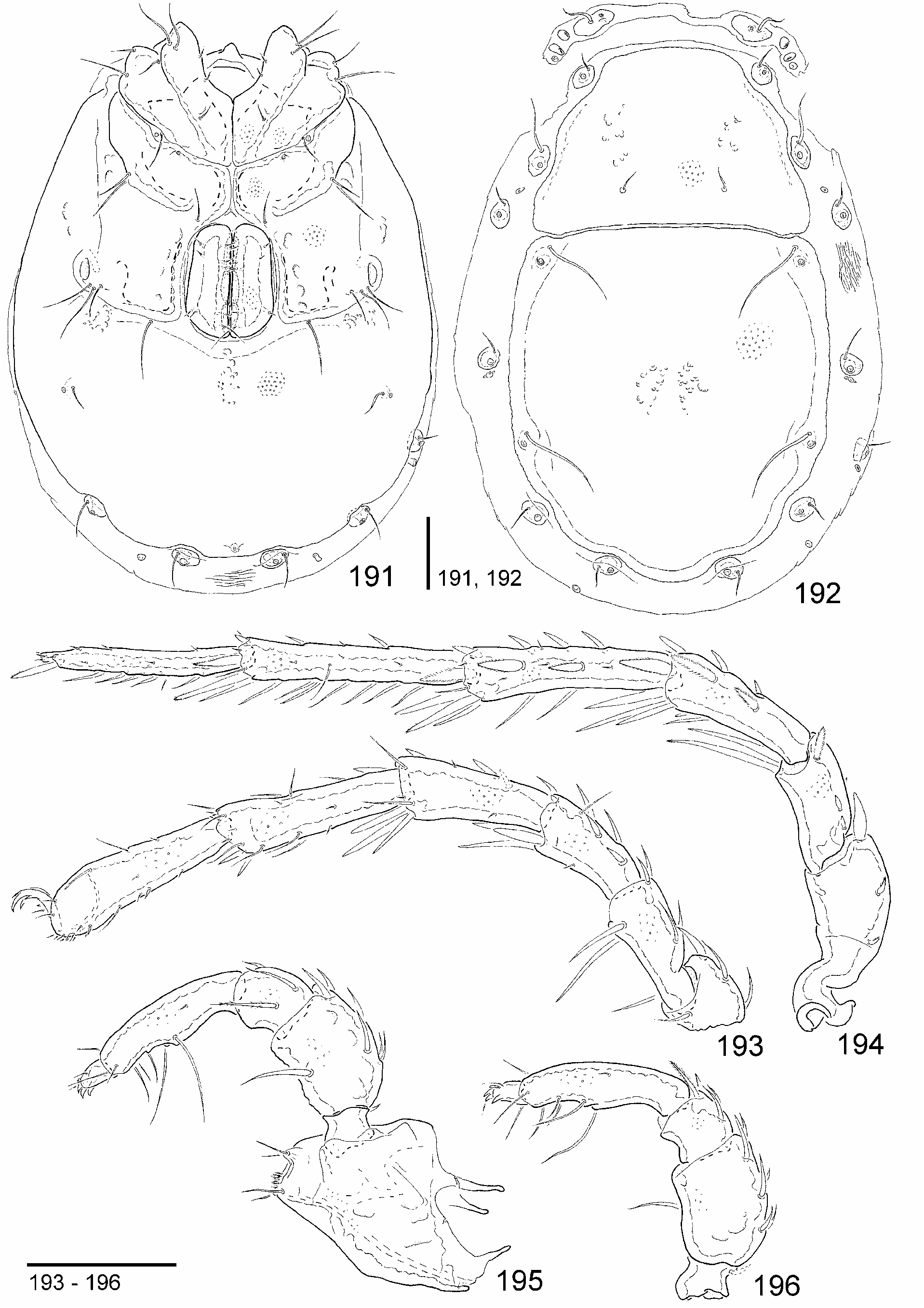
Female (n = 2): Slightly larger and slenderer than male ( Fig. 191 View FIGURES 191–196 ); pale purple-brownish; dorsal plates slenderer ( Fig. 192 View FIGURES 191–196 , Table 10); platelet of Dgl-2 less elongated; ventral shield similar to male, “shoulder corners” less distinct ( Fig. 191 View FIGURES 191–196 ); genital field more compact diverging posteriorly; Vgl-3 (at least in one specimen) closer to Cx-IV ( Fig. 191 View FIGURES 191–196 ); legs and gnathosoma similar to male ( Figs 193–196 View FIGURES 191–196 ).
Remarks: See below under B. sagai .

| MD |
Museum Donaueschingen |
No known copyright restrictions apply. See Agosti, D., Egloff, W., 2009. Taxonomic information exchange and copyright: the Plazi approach. BMC Research Notes 2009, 2:53 for further explanation.