
Coeliccia curua, Kompier & Dow & Steinhoff, 2020
|
publication ID |
https://doi.org/ 10.11646/zootaxa.4766.4.1 |
|
publication LSID |
urn:lsid:zoobank.org:pub:2F8FEDC1-6891-46D1-B372-858CDCBE4051 |
|
DOI |
https://doi.org/10.5281/zenodo.3803422 |
|
persistent identifier |
https://treatment.plazi.org/id/AF6A87D5-4368-6176-FF6A-DDAE4B6B3BD8 |
|
treatment provided by |
Carolina |
|
scientific name |
Coeliccia curua |
| status |
sp. nov. |
2. Coeliccia curua View in CoL sp. nov.
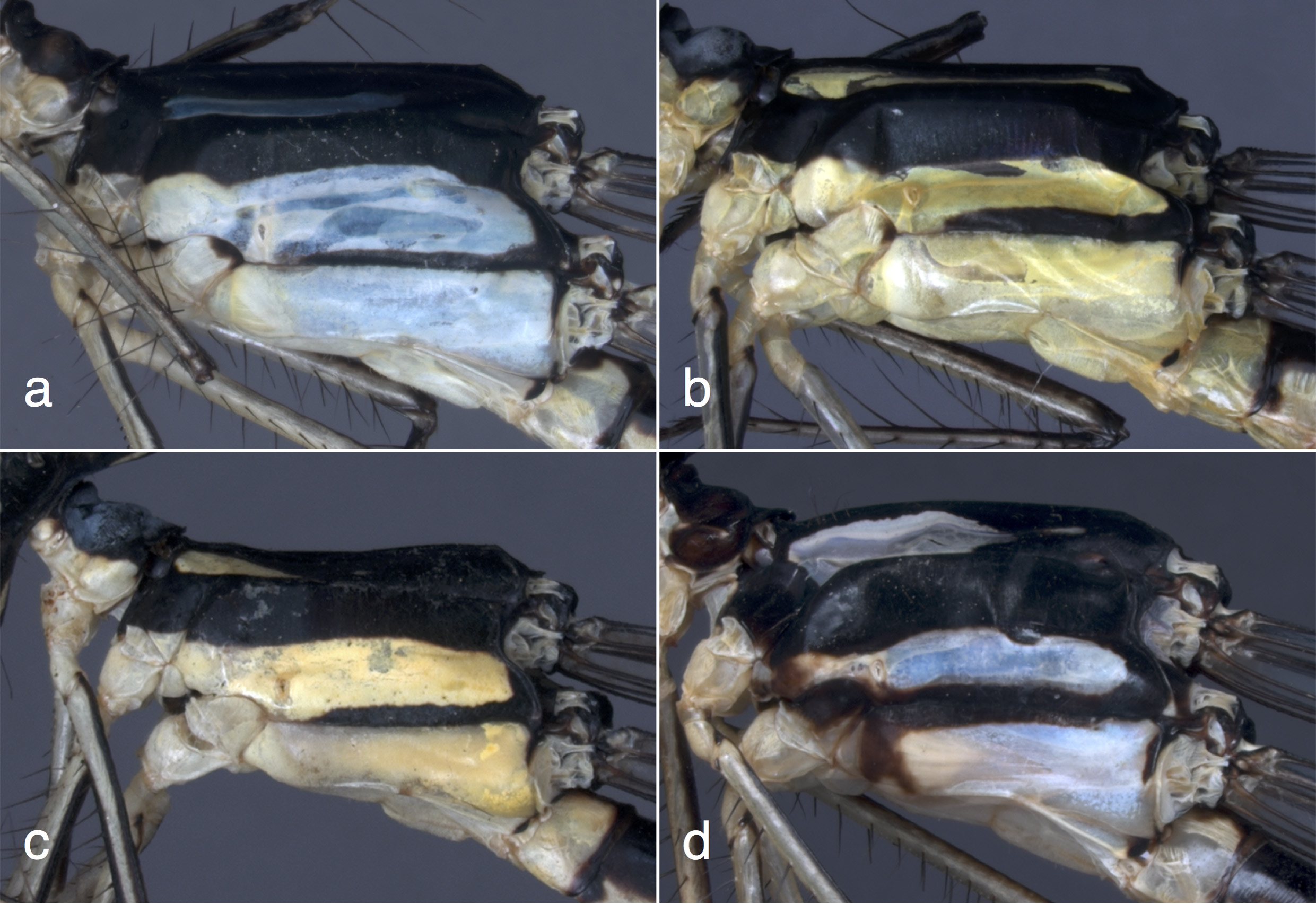
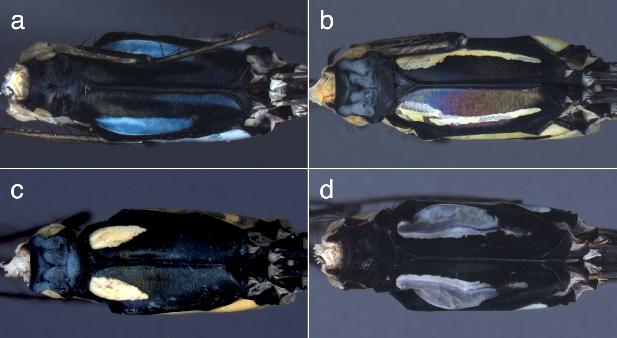
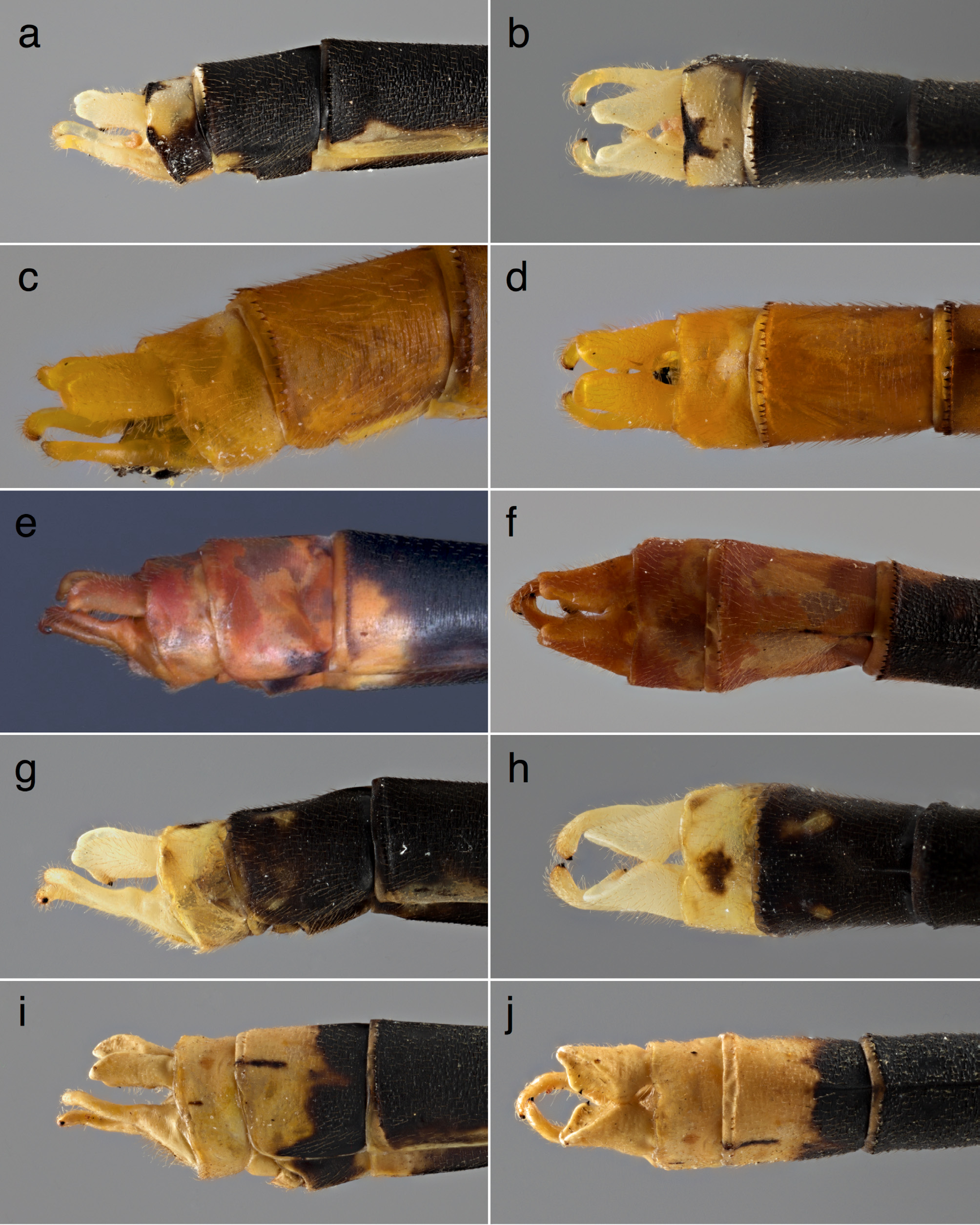
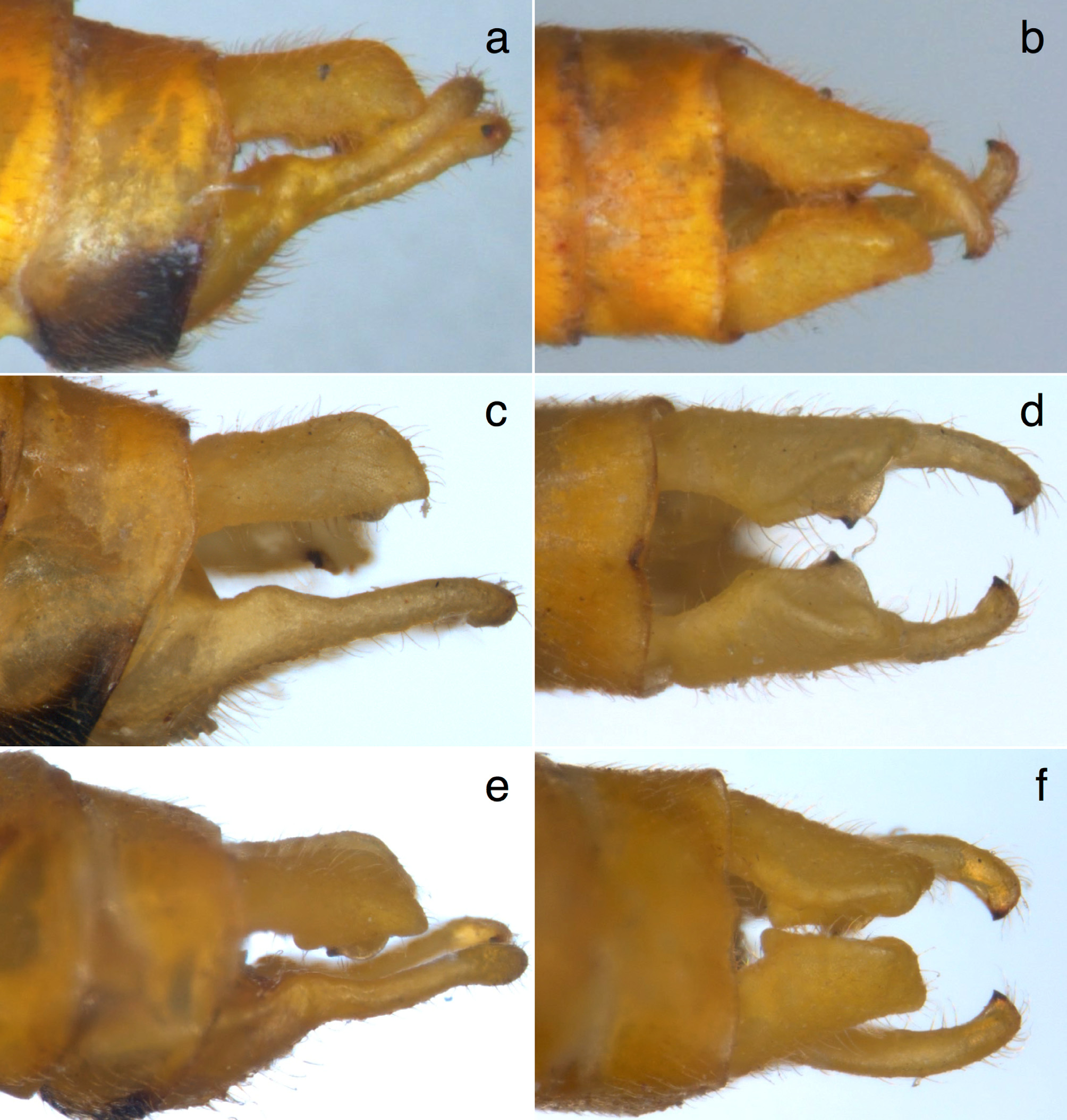
( Figs 1d View FIGURE 1 , 2d View FIGURE 2 , 3d View FIGURE 3 , 4d View FIGURE 4 , 5d View FIGURE 5 , 7 View FIGURE 7 e–f, 9, 10a–b, 12, 25a–d, 29c–d)
Holotype. ♂, Ba Be NP, Bac Kan Prov. (appr. 22.413N, 105.610E; alt. 250 m asl.), 22 vi 2014, TK leg. GoogleMaps
Paratypes. 1 ♂, same location and collector, 25 vii 2013; 1 ♂, Ba Be NP, Bac Kan Prov. (appr. 22.373N, 105.650E), 2 vii 2017, TK leg.; 2 ♂♂, Xuan Son NP, Phu Tho Prov. (appr. 21.141N, 104.932E), 23 vii 2017, TK leg.; 2 ♂♂, same location and collector, 9 vi 2018.
Etymology. The term “ Cụ Rùa ” (great grandfather turtle) was the name used by Hanoi locals to refer to the last Giant Softshell Turtle Rafetus swinhoei (Gray, 1873) alive in the city. The last representative of the species in Hoan Kiem Lake was found dead on January 19, 2016. Its demise serves as a stark reminder of the many threats to the Vietnamese fauna. A noun in apposition.
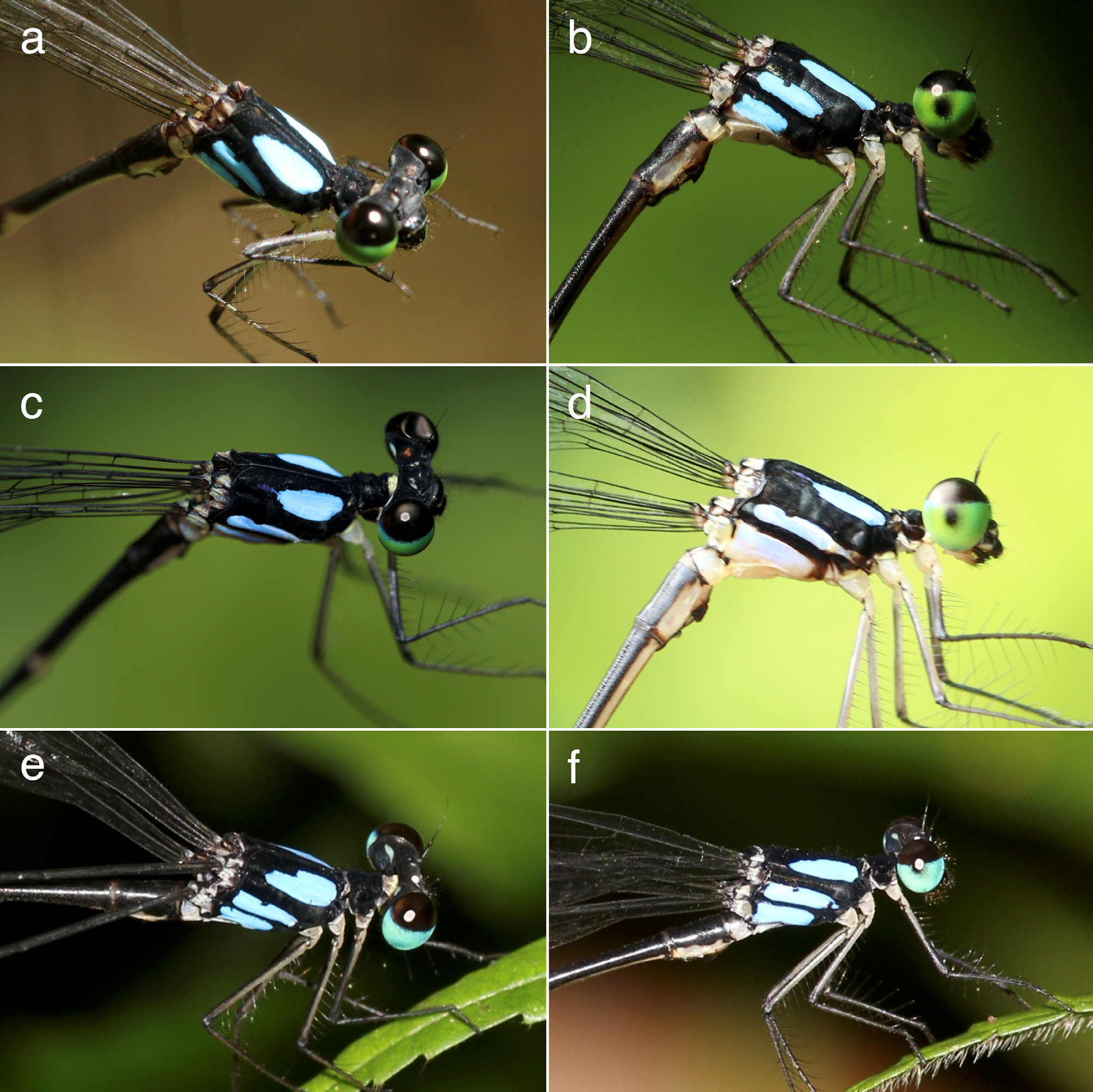
Description of holotype ( Figs 1d View FIGURE 1 , 2d View FIGURE 2 , 3d View FIGURE 3 , 4d View FIGURE 4 , 5d View FIGURE 5 , 7 View FIGURE 7 e–f, 10a–b, 12e–f, 25c–d). Head ( Fig. 1d View FIGURE 1 ). Labrum blackish brown, chestnut brown at free margin and basally. Anteclypeus white, with two darker areas left and right on lower half. Postclypeus shiny black. Base of mandibles black, anteroventral corner white. Genae white, the white narrowly extending over the antefrons above the postclypeus to about one-fourth in, remainder of frons matte black. Dorsal surface of head matte black, apart from two small bluish white spots between base of antennae and lateral ocelli and two oblong bluish white postocular spots. Antennae black, top of first segment white. Compound eyes three-colored in life, black or very dark chestnut above, green below, but posterior border between green lower half and blackish upper half is blue.
Thorax. Prothorax ( Figs 2d View FIGURE 2 , 3d View FIGURE 3 ): matte black, propleuron dark brown with pale edges. Synthorax ( Figs 4d View FIGURE 4 , 5d View FIGURE 5 ): mesepisternum black, with a large oval pale blue antehumeral shield, the inner edge of which is somewhat irregular and the outer edge of which follows the mesopleural suture. Extent of this blue shield is about two-thirds of the width, and three-fifths of length of mesepisternum. Mesepimeron and mesinfraepisternum black. Metepisternum black with large rectangular pale blue area, which forward reaches proximity of mesocoxa. This blue area with a small rounded excision along its upper margin near the wing base, and with black margin along wing base connecting the black of mesepimeron to the well-defined black line over metapleural suture. Metepimeron bluish white with brown, irregular smudge near coxae, metinfraepisternum black. Legs: coxae and trochanters white. Femur grayish with black stripes. Spines and claws dark.
Wings. Hyaline, 25 Px in FW, 23 in HW. Pt dark brown, covering two cells.
Abdomen. S1 narrowly black dorsally, remainder bluish white; S2 upper half black and lower half white; S3–6 black above and white below, and with prominent subapical white annulus; S7 virtually black, but with large subapical white spot; S8 black with posteroventral corner yellow; S9–10 all yellow.
Anal appendages ( Fig. 10 View FIGURE 10 a–b). Yellow (Color in Fig. 10 View FIGURE 10 a–b result of post-mortem changes). In lateral view the cerci twice as thick and a little shorter compared to paraprocts, apex curving downward, forming a small but distinct drooping point, and with subapical ventral expansion, tipped with black tooth. In dorsal view a small tooth at onethird of length of cerci, from where a dorsal ridge and the subapical ventral expansion start. The paraprocts of usual type, apically curved inwards and with black tooth at apex.
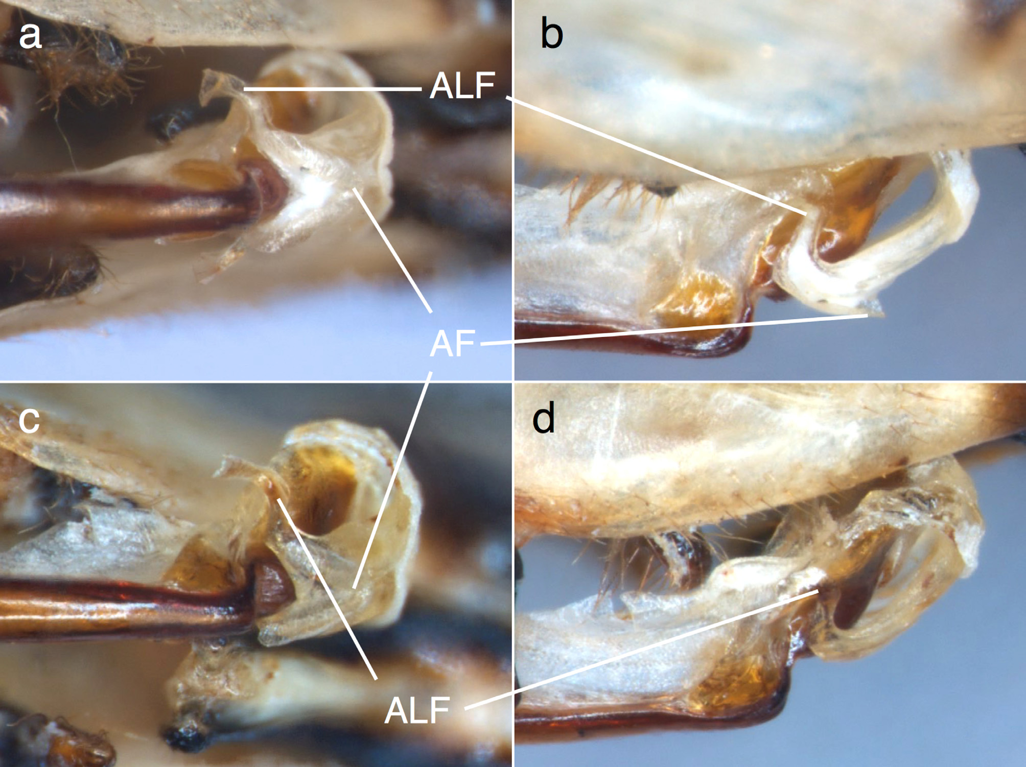
Genital ligula ( Fig. 12 View FIGURE 12 e–f). The apical segment with apical flap and two robust flagella that curve back to base of apical segment and from there extend a little sideways, appearing to taper to a point (but actually ribbon-like and curled up under microscope).
Variation in paratypes ( Figs 9 View FIGURE 9 , 12 View FIGURE 12 a–c, 25a–b). Propleuron with pale central area and less obvious pale edges or completely black. In Xuan Son specimens the antehumeral shields are somewhat larger ( Fig. 25 View FIGURE 25 a–b). Blue stripe on metepisternum without black excision along upper margin or continuing over spiracle almost to reach mesinfraepisternum. Metepimeron without brown smudge near coxae. S3–4 with more white on proximal part of venter, S9 with antero-ventral corner dark brown in one paratype or S10 almost completely yellow. Anal appendages deformed in one mature specimen. In Xuan Son specimens the paraprocts appear more slender and a little longer relative to the cerci ( Fig. 9 View FIGURE 9 ). Apico-lateral flagella of Xuan Son specimens shorter, not extending as obviously laterally in apical third ( Fig. 12 View FIGURE 12 a–c). 24 Px in left FW. Pt covering 1.5 cells (Xuan Son). 19–21 Px in FW, 16–20 in HW (Xuan Son).
Measurements (in mm). Ba Be: HW 33–35; abdomen incl. appendages 51–52; Xuan Son: HW 26–27; abdomen incl. appendages 37–39.
Female. Unknown.
Differential diagnosis. Coeliccia curua has a typical genital ligula of the pyriformis -group. Within this group it shares the combination of large blue antehumeral shields, bold black lines over mesopleural and metapleural sutures and yellow abdominal tip with C. uenoi ( Fig. 25 View FIGURE 25 e–f, 29a–b) and C. phamiha . In C. phamiha the flagella of the genital ligula are directed laterally and posteriorly, before curving inward apically ( Phan & Tran 2018), whereas in C. uenoi they are robust and directed first laterally and then anteriorly ( Fig. 13 View FIGURE 13 ). In C. curua the flagella are curved posteriorly, hidden in ventral view under terminal segment, but apical third directed laterally and visible in ventral view (although limited so or not at all in Xuan Son specimens) ( Fig. 12 View FIGURE 12 ). The antehumeral shields of C. uenoi have a characteristic step on the inner margin and are not oval ( Figs 25 View FIGURE 25 e–f, 29a–b), and the black lines over the metapleural and mesopleural sutures are narrower. Coeliccia phamiha has even larger antehumeral shields that obviously and suddenly narrow at their center, and has black S9–10, besides structurally different and whitish cerci ( Phan & Tran 2018). The caudal appendages of C. curua and C. uenoi are similar in structure ( Figs 9 View FIGURE 9 , 10 View FIGURE 10 ).
Habitat and ecology. In Ba Be NP Coeliccia curua sp. nov. was found at two different locations in karst mountains with seeps over rocks and forest floor, where it occurred with C. pyriformis and Protosticta nigra Kompier, 2016 . In Xuan Son NP it was found at two small and very shallow streams at appr. 900m asl. in degraded primary forest, similarly on karst mountains. Xuan Son NP boasts at least 7 other Coeliccia species, including the similar C. uenoi , but only C. pyriformis was found together with C. curua .


No known copyright restrictions apply. See Agosti, D., Egloff, W., 2009. Taxonomic information exchange and copyright: the Plazi approach. BMC Research Notes 2009, 2:53 for further explanation.
|
Kingdom |
|
|
Phylum |
|
|
Class |
|
|
Order |
|
|
Family |
|
|
Genus |