
Acasta crucibasis, Yu & Chan & Achituv & Kolbasov, 2017
|
publication ID |
https://doi.org/10.5281/zenodo.5358376 |
|
publication LSID |
lsid:zoobank.org:pub:6D4EE4A2-C237-4338-AE8F-77C4F2292B2D |
|
persistent identifier |
https://treatment.plazi.org/id/F2B6EEDE-66D5-4BB8-BCC8-4D3660F8421B |
|
taxon LSID |
lsid:zoobank.org:act:F2B6EEDE-66D5-4BB8-BCC8-4D3660F8421B |
|
treatment provided by |
Valdenar (2021-08-29 08:05:38, last updated by Plazi 2023-11-05 13:42:03) |
|
scientific name |
Acasta crucibasis |
| status |
sp. nov. |
Acasta crucibasis , new species
( Figs. 8–14 View Fig View Fig View Fig View Fig View Fig View Fig View Fig )
Material examined. Seven specimens (holotype and paratypes) in the sponge Topsentia sp. Berg, 1899 , Dabaisha , Green Island, Taitung, Taiwan , 22°38′12″N, 121°29′30″E, SCUBA diving, depth 9 m. Two specimens (paratypes) in the sponge Xestospongia vansoesti Bakus & Nishiyama, 2000 , SER.03/080905/010, Indonesia GoogleMaps , Java Sea, Kepulauan Seribu ( Thousand Islands ), off Jakarta, Damar Kecil Island , NW-side, 5°59′00″S, 106°50′43″E, SCUBA diving and snorkeling. The holotype (dry shell compartments, mouth parts and cirri mounted in glycerol on glass slide) and paratype (shell in EtOH) are deposited in the Zoological Museum of Moscow State University under registration numbers Mg –1226 (holotype) and Mg –1227 (paratype). The other paratypes ( ASIZCR000373 to ASIZCR000375 ) are deposited in the Biodiversity Research Museum, Biodiversity Research Center, Academia Sinica, Taiwan GoogleMaps and ( RMNH. CRUS.C.10243) in Naturalis Biodiversity Center , Leiden, The Netherlands .
Diagnosis. Shell white, semitransparent, globular, with 4 large windows between compartments; wall plates and basis thin and fragile; shorter carinolatus 2 eliminated with large window, does not reach basis; rostrum bifurcated at base; basis cup-shaped, with five processes; scutum latticed; cirrus IV with developed curved teeth on anterior ramus, basidorsal point of penis rudimentary. Embedded in sponges Topsentia sp. Berg, 1899 and Xestospongia vansoesti Bakus & Nishiyama, 2000 .
Description. (Based on holotype). Shell of holotype white, semitransparent, globular, ~ 5.6 mm maximal height and diameter, 4.8 mm rostro-carinal basal diameter, orifice toothed, 2.6 mm maximum length, with four large windows 2.2–2.6 mm high and 1.3–2.2 mm wide between wall plates and corresponding processes of basis ( Fig. 8A, B View Fig ). Basis ( Fig. 8A, B, F View Fig ) deep moderately, cup-shaped, cruciform, with external growth lines concentric, margin denticulated, 1.7 mm high, with five long processes corresponding to wall plates (one for carina, two for carinolatera 1, two for bifurcated base of rostrum). In paratypes basis flat ( Fig. 10A, H View Fig ) or saucer-shaped ( Fig. 10B, C, I View Fig ), with reduced arms of rostral processes and hole at center ( Fig. 10H View Fig ). Parietes ( Figs. 8A–E View Fig , 10A, B View Fig ) consist of axial part connected with processes of basis, two adjacent lateral semitransparent parts eliminated with windows, with conspicuous sharp, dense external projections and fine growth lines, sheath ~1/2 total length, with horizontal striations, inner lamina beneath sheath smooth, sometimes with few small indistinct basal longitudinal ribs. Radii and alae with summits oblique, ~1/2 length of parietes, radii with fine, oblique, external striations. Rostrum of holotype with basal part bifurcated ( Fig. 8D View Fig ). Basal part of carinolatus 2 eliminated with big window, twice shorter than carinolatus 1, paries developed, ~1/2–1/3 width of carinolatus 1 ( Figs. 8A, B, E View Fig , 10A, B View Fig ).
Scutum ( Figs. 9A–D View Fig , 10D–G View Fig ) white (after treatment with bleach), semitransparent, height slightly longer than width, latticed, distinct external growth ridges crossed by prominent radial ribs, occludent margin strongly dentate. Articular ridge ~2/3 length of tergal margin, slightly prominent, lower end smooth, not truncated; adductor ridge feeble, pits for adductor and depressor muscles not developed. Scutum of biggest specimen from Indonesia has internal sculpture different from that in Taiwanese specimens ( Fig. 10D, E View Fig ): articular ridge distinctly prominent; pits for adductor and depressor muscles conspicuous.
Tergum ( Fig. 9E–H View Fig ) semitransparent, white, growth lines distinct; apex beak-shaped, scutal margin concave; spur short, truncated, ~half width of basal margin, distinctly separated from basiscutal angle; spur furrow wide, distinct. Articular ridge feeble, ~1/3 length of scutal margin, depressor muscle crests rudimentary.
Labrum ( Figs. 11A, B View Fig , 13A View Fig ) with deep medial notch, 3 tiny, delicate teeth on each crest hidden by dense, thin, small setae. Crests of labrum with rows of ctenoid scales ( Fig. 13A View Fig ). Mandibular palps ( Figs. 11A View Fig , 13A, B View Fig ) club-shaped, with long, small, thin, dense, setulate and simple setae on distal ends, small, simple, dense setae and rows of ctenoid scales on lateral surfaces. Mandible ( Figs. 11C–F View Fig , 13C–E View Fig ) cutting edge with 5 teeth decreasing in size from upper to lower, teeth 1–3 well separated from each other, teeth 2–4 bifid, inferior angle sharp, with group of sharp denticles and setae, inner margin with long, simple setae, outer margin and upper part of blade with long, thin, omniserrate setae, lateral surface of blade with dense, small, biserrate setae ( Fig. 13D, E View Fig ). Maxillules ( Figs. 11G, H View Fig , 13F, G View Fig ) with 8–10 cuspidate setae of different lengths along straight cutting edge; notch absent; upper and lower pairs of cuspidate setae largest; tuft of small, sharp cuspidate setae on inferior angle; long, simple, serrate setae along outer and inner margins; dense biserrate setae on lateral surfaces. Maxillae ( Fig. 13H–J View Fig ), bilobed; dense, long simple and serrate setae distally and along inner margins of upper and lower lobes; ctenoid scales on lateral surfaces of upper lobes; field of stub setae in upper-lateral part of lower lobe; dense carpet of sharp, biserrate setae and fine small setae in fused basal parts.
Cirrus I with rami unequal; anterior ramus (22 annuli) almost 3 times longer than posterior ramus (7 annuli); both rami covered with dense setae. Cirrus II ( Fig. 14A View Fig ) with rami unequal; anterior ramus (12 annuli) longer than posterior ramus (9 annuli); annuli of rami with long, dense, simple and serrate setae on anterior edge; tuft of serrate setae in posterio-distal corner. Cirrus III ( Figs. 12A View Fig , 14A, D, E View Fig ) with anterior ramus (15 annuli) slightly longer than posterior (11 annuli); proximal and middle annuli of anterior ramus with single, sharp upward denticle beneath 2 pairs of long setae and 2 pairs of small, serrate setae; tuft of serrate setae in posterio-distal corner; lateral surfaces of annuli with row of ctenoid scales in upper part ( Fig. 14E View Fig ). Cirrus IV ( Figs. 12B View Fig , 14A, D, F View Fig ) with rami subequal; anterior ramus with 21 annuli, posterior ramus with 22; proximal annuli of anterior ramus with developed curved tooth on anterior edge, group of sharp denticles at tooth base; basis without teeth; ramal annuli with 3 pairs of long, middle setae and short, serrulate setae on upper part of anterior edge, tuft of thin simple setae on posterio-diatal corner; distal annuli of rami with tuft of 6 stout, serrate setae in upper part. Cirrus V ( Figs. 12C View Fig , 14A View Fig ) with broken rami (19, 27 annuli); annuli of anterior ramus without teeth and denticles; ramal annuli with 3 pairs of long, middle setae and short, serrulate setae on anterior edge, tuft of setae on posterio-distal corner. Cirrus VI with broken rami (24, 21 annuli); intermediate annuli of both rami with 3 pairs of long, middle setae and short, serrulate setae; some annuli with few long, thin setae on posterio-distal corner.
Penis ( Fig. 14A–C View Fig ) annulated; basidorsal point rudimentary; gradually tapering distally; tip with few thin, simple setae.
Remarks. The new species Acasta crucibasis is similar to the species of genus Acasta , A. armata Gravier, 1921 (= A. sinica Ren, 1984 ), A. fenestrata Darwin, 1854 , A. foraminifera Broch, 1931 , and A. pertusa Kolbasov, 1990 in having large windows between the wall plates. But only A. forminifera has basal part of carinolatus 2 eliminated with a large window as in A. crucibasis . A. crucibasis differs from A. foraminifera by the cruciform basis, latticed scutum, truncated tergal spur and number of cuspidate setae on cutting edge of maxillules (8–10 instead 5 in A. foraminifera ).
Etymology. The specific name is derived from the Latin ‘crux’ or ‘crucis’ – cross, and ‘basis’, referring to the crossshaped basis of this species.
Bakus GJ & Nishiyama GK (2000) Three species of toxic sponges from Cebu, Philippines (Porifera: Demospongiae). Proceedings of the Biological Society of Washington, 113 (4): 1162 - 1172.
Berg C (1899) Substitucion de nombres genericos. III. Communicaciones del Museo Nacional de Buenos Aires, 1 (3): 77 - 80.
Broch H (1931) Paper from the Dr. Mortensen's Pacific Expedition 1914 - 16. LVI. Indomalayan Cirripedia. Videnskabelige Meddelelser Dansk Naturhistorisk Forening, 91: 1 - 146.
Darwin C (1854) A Monograph on the Sub-Class Cirripedia, With Figures of All the Species. The Balanidae. Ray Society, London, 770 pp.
Gravier C (1921) Sur deux especes de Cirripedes du genre Acasta Leach vivant a la cote francaise des Somalis. Bulletin du Museum d'Histoire naturelle, Paris, 1921: 353 - 357.
Kolbasov GA (1990) Acasta pertusa sp. n. (Cirripedia, Thoracica) from the Red Sea. Zoologichesky Zhurnal, 69 (9): 142 - 145.
Ren X (1984) Studies on Chinese Cirripedia (Crustacea) V. Genus Acasta. Studia Marina Sinica, 23: 183 - 214.
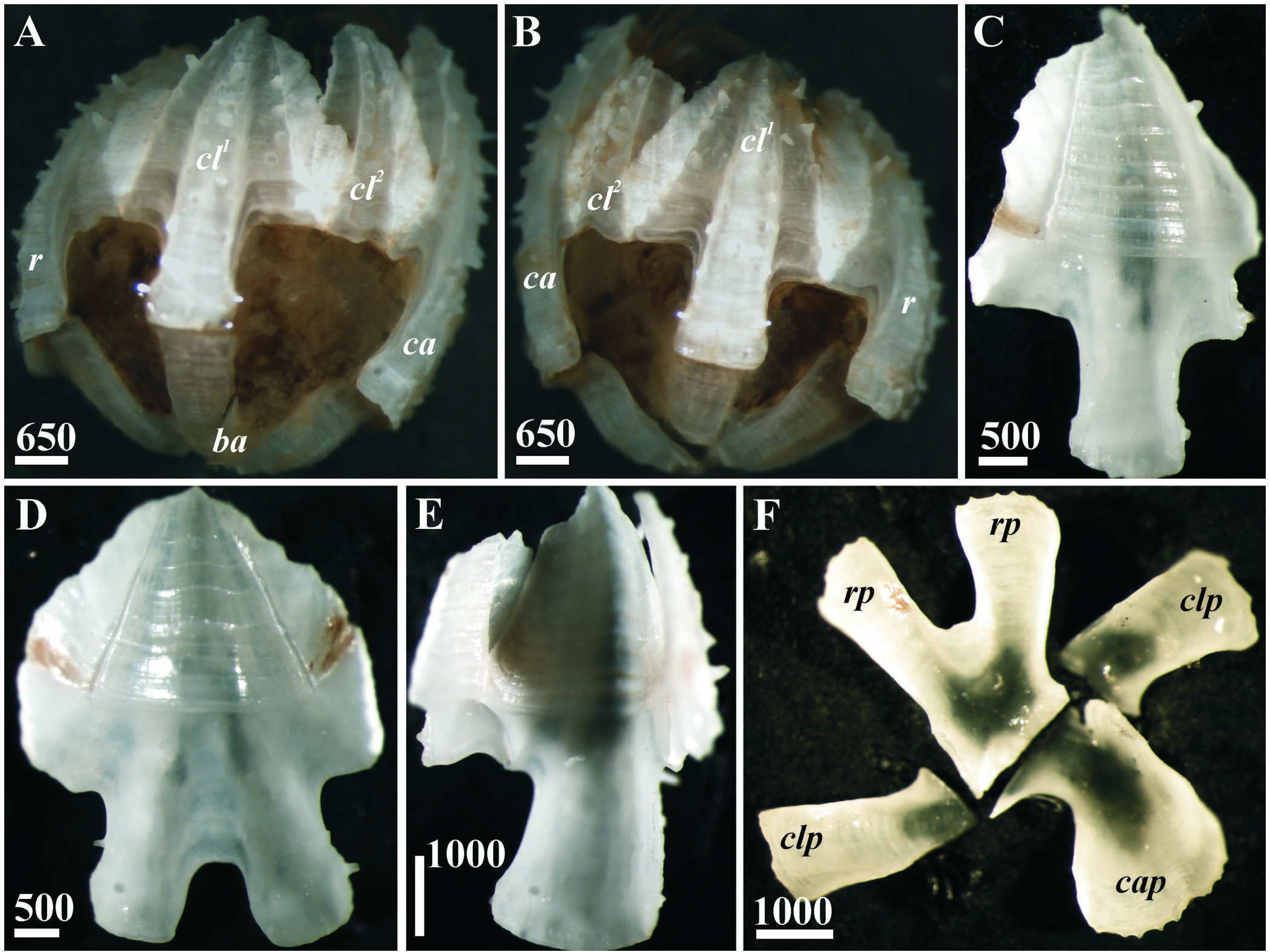
Fig. 8. Acasta crucibasis sp. nov., shell, general view and compartments (holotype). A, B, Complete shell, right and left side; C, Carinolatus 1, internal surface; D, Rostrum, internal surface; E, Carina with rudimental carinolatera 2, internal surface; F, Basis, internal surface. Abbreviations: ba = basis, ca = carina, cap = carinal process of basis, cl1 = carinolatus 1, cl2 = carinolatus 2, clp = carinolateral process of basis, r = rostrum, rp = rostral process of basis. Scale bars in µm.
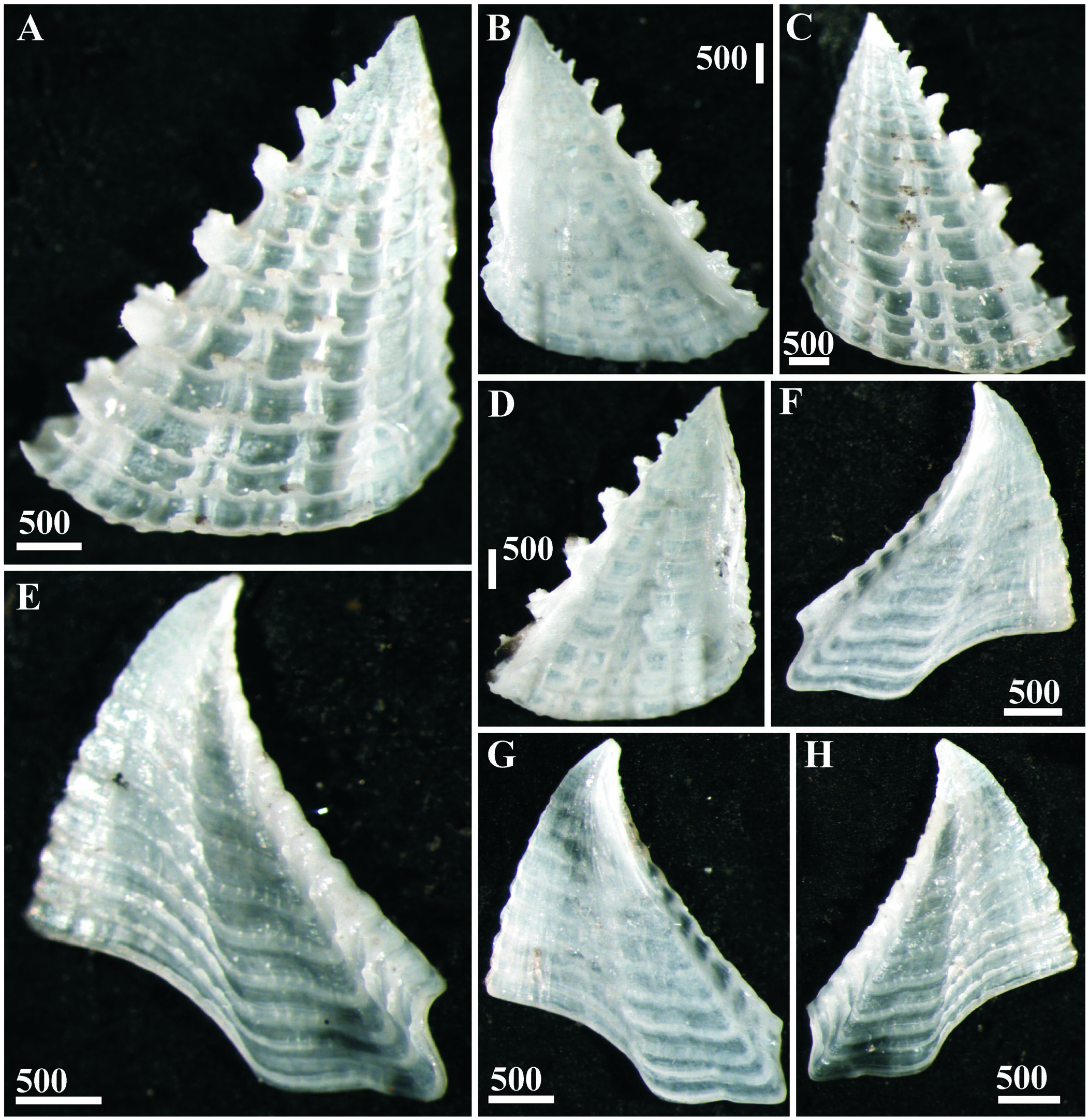
Fig. 9. Acasta crucibasis sp. nov., opercular plates (holotype). A, B, Right scutum, external and internal side; C, D, Left scutum, external and internal side; E, F, Left tergum, external and internal side; G, H, Right tergum, internal and external side. Scale bars in µm.

Fig. 10. Acasta crucibasis sp. nov., shells, general view and compartments (paratypes: A, D, E, H from Java Sea; B, C, F, G, I from Taiwan). A, B, Complete shells, left and right side; C, General view from basis; D, F, Scutum, external surface; E, G, Scutum, internal surface; H, I, Basis, internal surface. Abbreviations: ba = basis, ca = carina, cap = carinal process of basis, cl1 = carinolatus 1, cl2 = carinolatus 2, clp = carinolateral process of basis, r = rostrum, rp = rostral process of basis. Scale bars in µm.
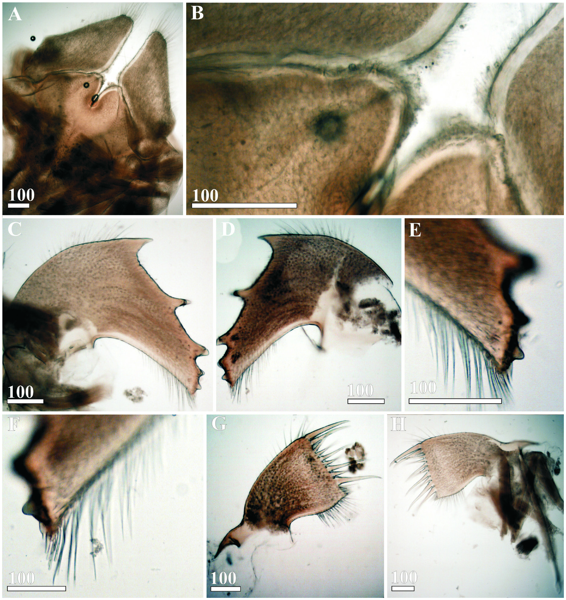
Fig. 11. Acasta crucibasis sp. nov., mouth parts (holotype). A, Labrum and mandibular palps; B, Enlarged crests of labrum; C, D, Mandibles; E, F, Lower angle of mandibles; G, H, Maxillules. Scale bars in µm.
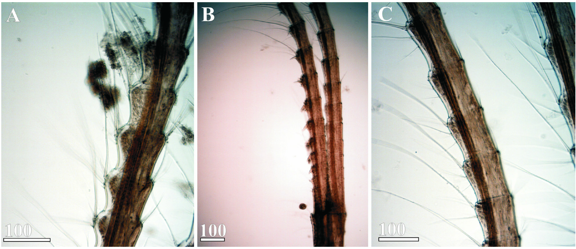
Fig. 12. Acasta crucibasis sp. nov., cirri (holotype). A, Cirrus III, annuli of middle part of anterior ramus; B, Cirrus IV, proximal part; C, Cirrus V, annuli of middle part of anterior ramus. Scale bars in µm.
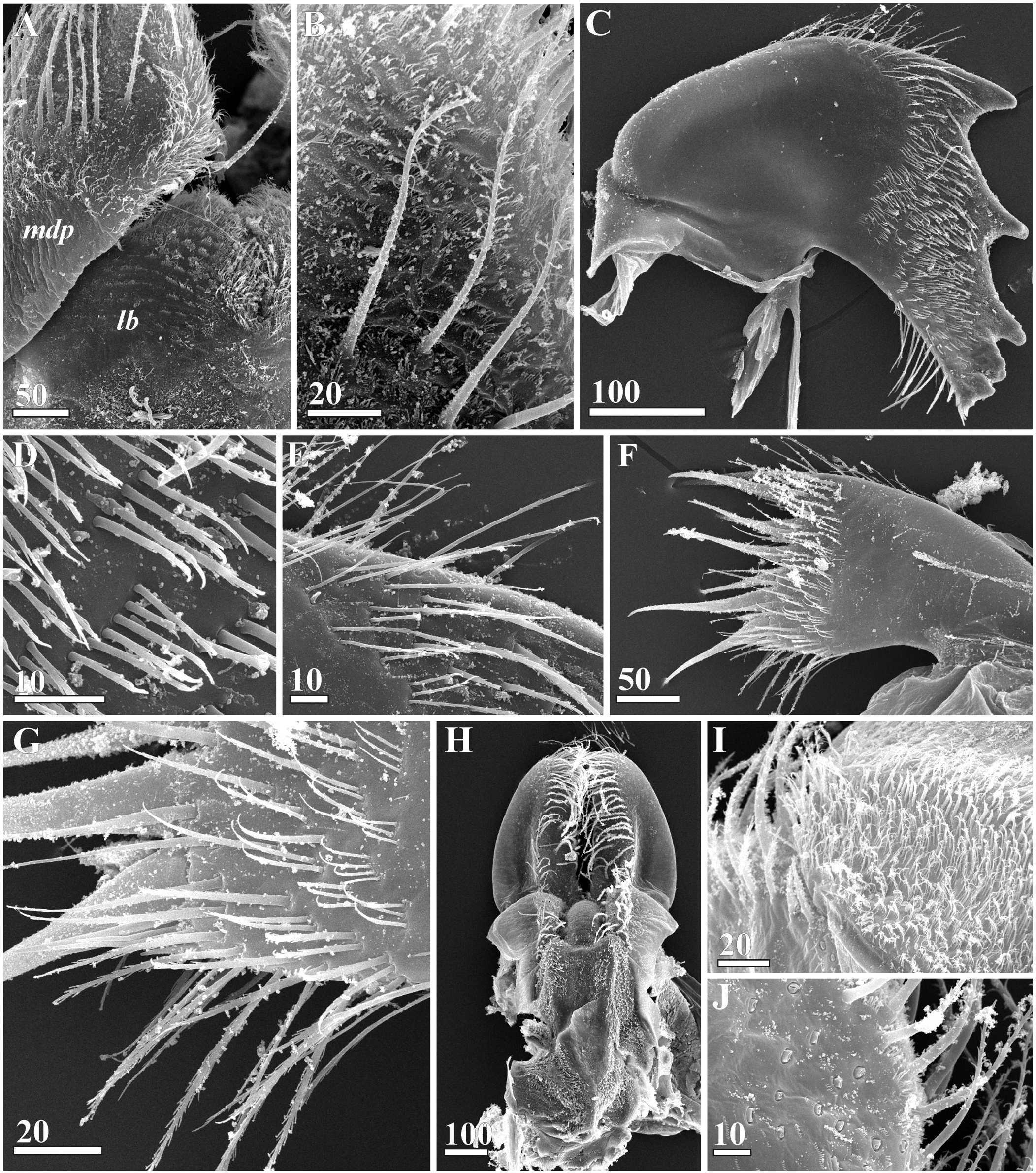
Fig. 13. Acasta crucibasis sp. nov., mouth parts (SEM, paratype, from Taiwan). A, Crest of labrum with mandibular palp; B, Surface of distal part of mandibular palp; C, Mandible; D, Biserrate setae on lateral surface of mandible; E, Setae on upper margin of mandible; F, Maxillule; G, Lower part of maxillule; H, Maxillae, inner surface; I, Small setae on basal part of maxilla; J, Stub setae on proximal lobe of maxilla. Abbreviations: lb = labrum, mdp = mandibular palp. Scale bars in µm.
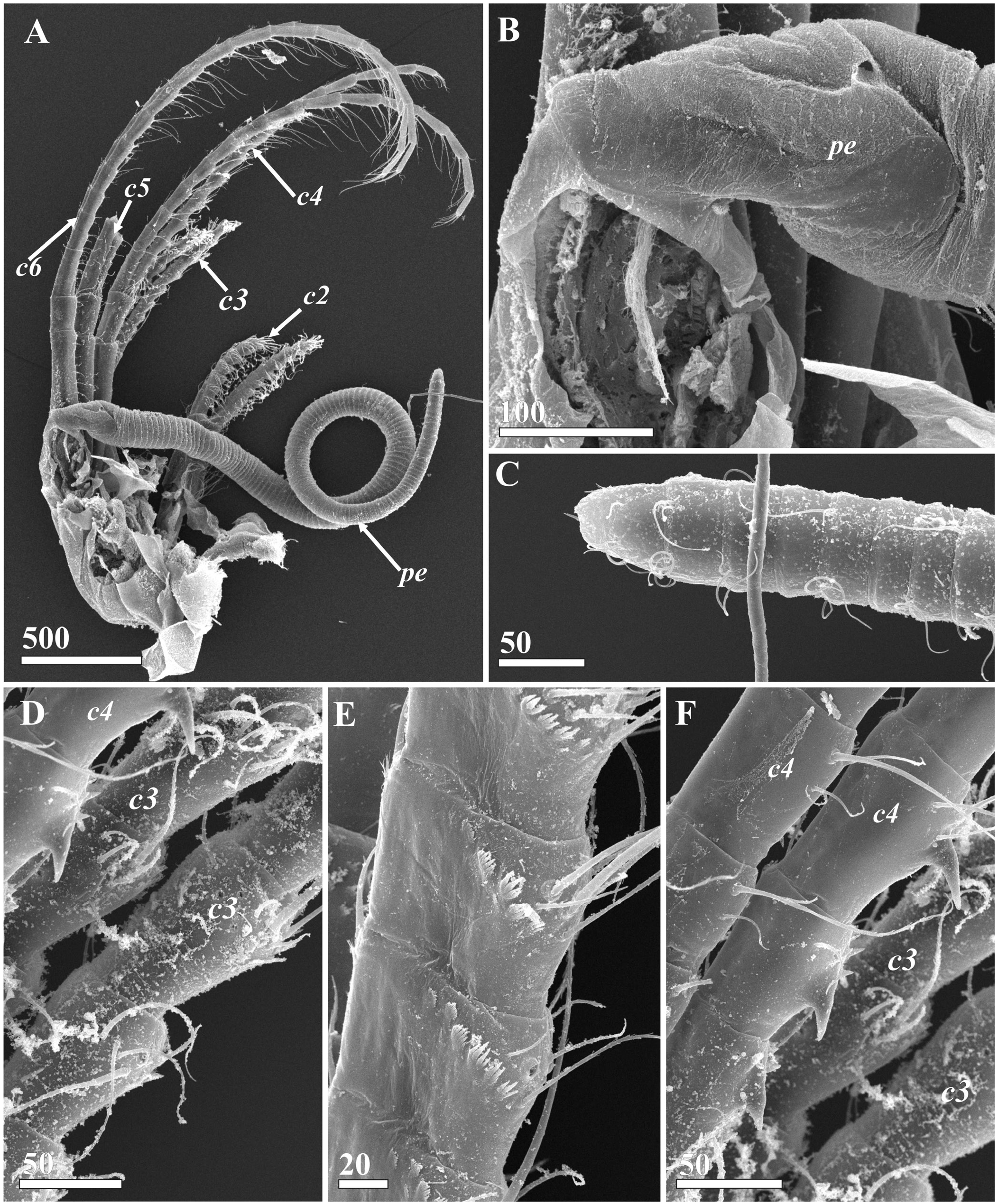
Fig. 14. Acasta crucibasis sp. nov., cirri and penis (SEM, paratype, from Taiwan). A, Right cirri and penis; B, Base of penis; C, Terminal part of cirri; D, Cirrus III, middle parts of rami; E, Cirrus III, annuli in middle part of anterior ramus. F, Cirrus IV, annuli in middle part of rami. Abbreviations: c2 to c6 = cirri II to cirri VI, pe = penis. Scale bars in µm.
| RMNH |
National Museum of Natural History, Naturalis |
No known copyright restrictions apply. See Agosti, D., Egloff, W., 2009. Taxonomic information exchange and copyright: the Plazi approach. BMC Research Notes 2009, 2:53 for further explanation.
|
Kingdom |
|
|
Phylum |
|
|
Class |
|
|
Order |
|
|
Family |
|
|
Genus |