
Adenopygus Bolfarini
|
publication ID |
https://doi.org/10.5281/zenodo.210622 |
|
DOI |
https://doi.org/10.5281/zenodo.6178985 |
|
persistent identifier |
https://treatment.plazi.org/id/A11087C7-FFE9-FF9F-FF58-FD0BFE22F976 |
|
treatment provided by |
Plazi (2016-04-12 16:42:12, last updated 2024-11-27 02:03:18) |
|
scientific name |
Adenopygus Bolfarini |
| status |
|
Adenopygus Bolfarini & de Mello, n. gen.
( Figs. 1–4 View FIGURE 1 View FIGURE 2 View FIGURE 3 View FIGURE 4 )
Type species: A. heikoi , n. sp.
Etymology. The specific epithet alludes to the glandular condition of the supra-anal plate of the male.
Diagnosis. (Based mainly on comparisons with specimens of Ottedana ). Maxillary palpi short and robust, the last joint with a large apical truncation; male supra-anal plate with an unpigmented, nearly glabrous, membranous, area in central portion; male paraprocts without a cavity on ventral surface; male cerci without clasping structure; dorsum and laterals of the phallic complex covered by scutum-shaped membranous hood; pseudepiphallic parameres 1 and 2 without inflatable membranous structure (cf. de Mello & Andrade, 2003 for this character). Ovipositor straight.
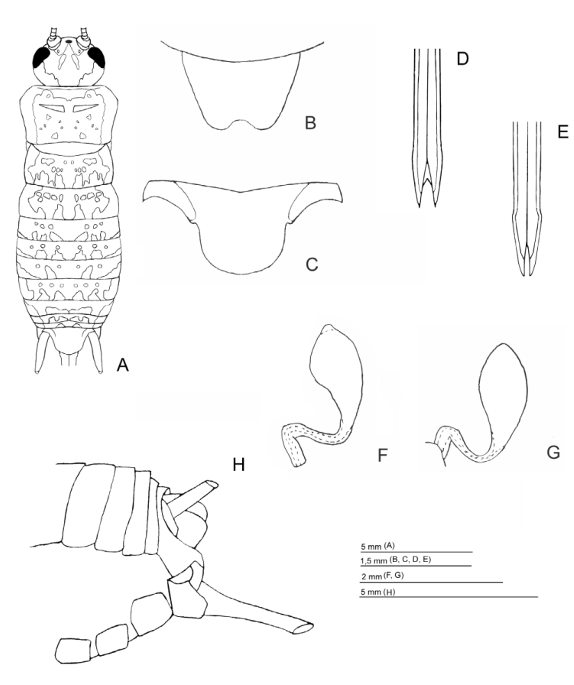
Description. Male: body sub-cylindrical, pubescent; back of head and fastigium with bristles and micro pilosity, its greatest width at eye level; inter-ocular space slightly flattened, oblique in lateral view; eye without unpigmented area ( Fig. 1 View FIGURE 1 E); three ocelli present; maxillary palpi short, last joint with robust apical truncation occupying about half of its length ( Fig. 1 View FIGURE 1 D). Pronotum disk wider than long, covered with bristles, its anterior margin slightly concave, posterior margin straight; lateral lobes with straight ventral margin, antero-ventral angle of nearly 90 degrees, postero-ventral angle rounded; fore wings short, not reaching posterior margin of metanotum, non-glandular, covered with bristles on posterior half and bearing trace of single longitudinal vein ( Fig. 1 View FIGURE 1 F, G), internal margins juxtaposed, lateral margins gradually merging into posterior margin; dorsum of metanotum with granular area; tibia I armed with 2 ventral apical spurs, one internal and one external; tibia II with 4 apical spurs; tibia III with 4 dorsal spurs on both sides, 3 apical spurs on each side, of which median spur is the longest on the internal face and upper spur is the longest on the external. Supra-anal plate broad, glandular, densely haired ( Fig. 1 View FIGURE 1 L and 2K-M), paraprocts horizontally oriented, devoid of cavity on ventral face to accommodate pseudoepiphallic lateral spines (condition present in Ottedana ); cerci short, without claspers. Phallic complex ( Fig. 3 View FIGURE 3 ) with membranous dorso-lateral cap covering much of pseudepiphallus; point of contact of pseudepiphallic parameres 1 and 2, in posterior view, without spherical membranous structure; ectophallic apodemes short with broad, well developed arch, ectophallic fold short; endophallic sclerite small, bearing crest-shaped dorsal apodeme only. Female: similar to male but larger and less cylindrical; apterous; dorsum of head more rounded in lateral view; supra-anal plate not glandular; paraprocts not horizontal; posterior margin of subgenital plate with wide shallow concavity; ovipositor straight, apical valves small.
De Mello, F. A. G., De Andrade, F. (2003) Ottedana cercalis: a new genus and species of phalangopsidae cricket from the Mantiqueira Range of Southeastern Brazil. (Orthoptera: Grylloidea). Journal of Orthoptera Research, 12 (2), 141 - 148.
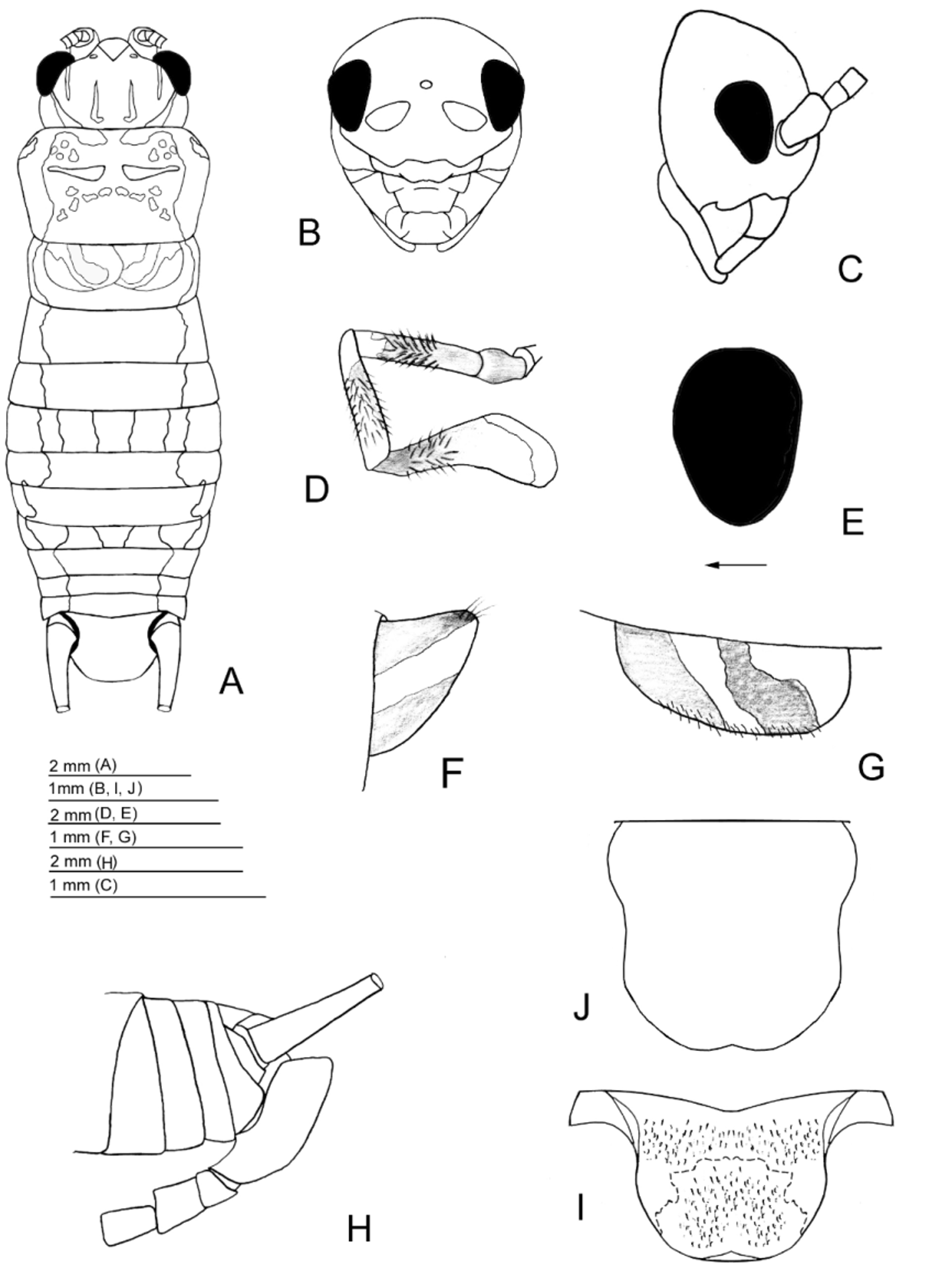
FIGURE 1. Adenopygus heikoi Bolfarini & de Mello sp. n. — male. A—dorsal view of body; B - frontal view of head; C—lateral view of head; D—maxillary palpi; E—perpendicular view of left eye; F, G—lateral and dorsal view of fore wings; H—lateral view of terminalia; I—supra-anal plate; J— subgenital plate.
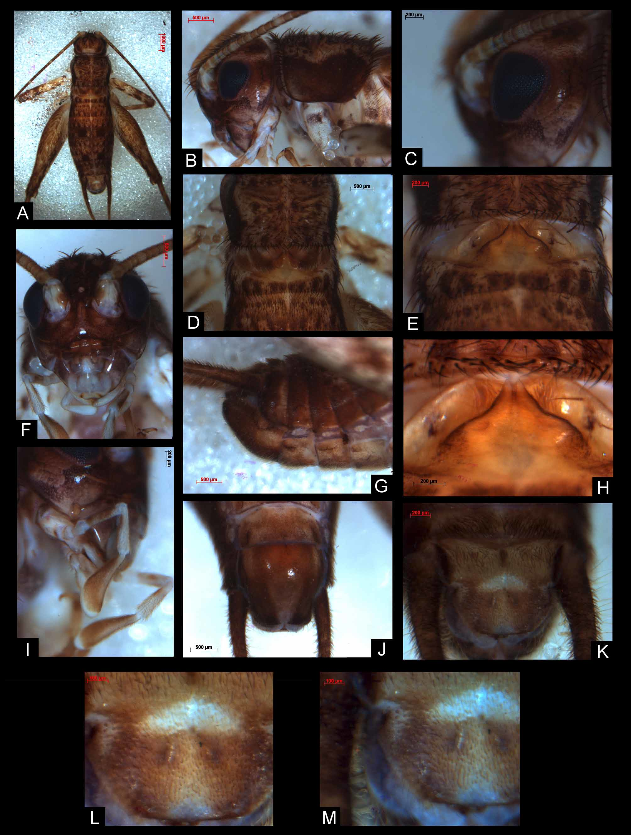
FIGURE 2. Adenopygus heikoi Bolfarini & de Mello sp. n. — male. A—dorsal view of body; B—lateral view of head and pronotum; C—perpendicular view of eye; D—dorsal view of fore wings; E, H—metanotum structure; F—frontal view of head; G—lateral view of terminalia; I—maxillary palpi; J—subgenital plate; K, L, and M—supra-anal plate.
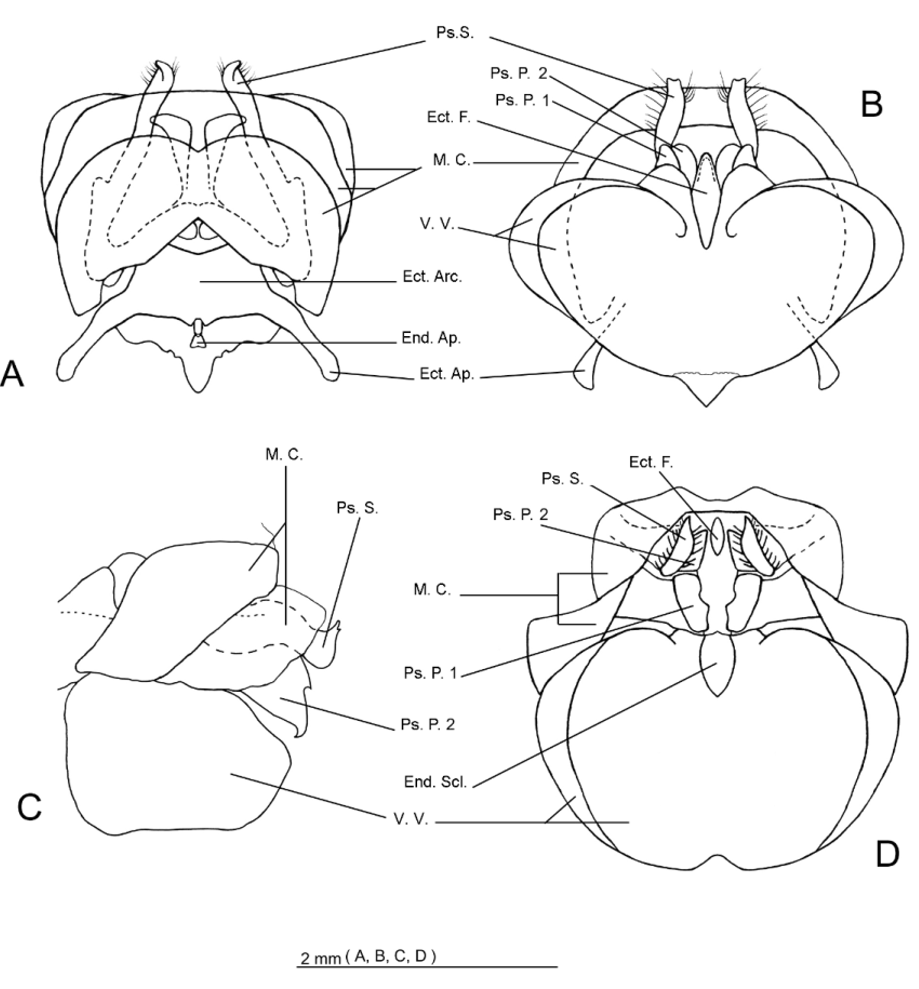
FIGURE 3. Adenopygus heikoi Bolfarini & de Mello sp. n. A—ventral view of phallic complex; B—idem, dorsal view; Cidem, lateral view; D—idem, posterior view. Conventions: Ps. S. — pseudepiphallic spine; Ps. P. — pseudepiphallic paramere; M. C. — membranous cap; Ect. F. — ectophallic fold; Ect. Arc. — ectophallic arch; End. Ap. — endophallic apodeme; Ect. Ap. ectophallic apodeme; End. Scl. — endophallic sclerite; V. V. — ventral valves.
No known copyright restrictions apply. See Agosti, D., Egloff, W., 2009. Taxonomic information exchange and copyright: the Plazi approach. BMC Research Notes 2009, 2:53 for further explanation.
|
Kingdom |
|
|
Phylum |
|
|
Class |
|
|
Order |
|
|
SuperFamily |
Grylloidea |
|
Family |
|
|
SubFamily |
Luzarinae |
1 (by plazi, 2016-04-12 16:42:12)
2 (by ImsDioSync, 2016-12-19 22:33:54)
3 (by ImsDioSync, 2016-12-19 22:34:42)
4 (by ImsDioSync, 2017-06-23 19:55:14)
5 (by ImsDioSync, 2017-06-23 20:51:25)
6 (by ExternalLinkService, 2019-09-26 19:36:39)
7 (by ExternalLinkService, 2022-01-30 12:49:05)
8 (by ExternalLinkService, 2022-02-20 05:37:03)
9 (by plazi, 2023-10-26 08:01:18)