
Pseudogarypus pangaea Henderickx
|
publication ID |
https://doi.org/ 10.5281/zenodo.173709 |
|
DOI |
https://doi.org/10.5281/zenodo.6257012 |
|
persistent identifier |
https://treatment.plazi.org/id/9E2A87D0-5641-FF8A-FE9E-42945668FB2F |
|
treatment provided by |
Plazi |
|
scientific name |
Pseudogarypus pangaea Henderickx |
| status |
sp. nov. |
Pseudogarypus pangaea Henderickx n. sp.
( Figs 1–12 View FIGURES 1 – 2 View FIGURE 3 View FIGURES 4 – 5 View FIGURES 6 – 7 View FIGURES 8 – 12 )
Type material. Holotype in Baltic amber from Jantarny, Kaliningrad, Russia, deposited in the Royal Museum for Central Africa, Tervuren, Belgium ( MRAC 219415).
Etymology. The specific epithet refers to the ancient continent Pangaea and is to be treated as noun in apposition. Based on modern distributions, ancestral Pseudogarypidae were likely to have been distributed across Pangaea , before this continent broke up (mid Jurassic).
Diagnosis. A small Pseudogarypus with broad carapace bearing two posterolateral protuberances, situated anterior to the alae. Anterior and posterior eyes nearly contiguous, femur length less than 1.0 mm, pedipalpal fingers with contiguous, regular teeth.
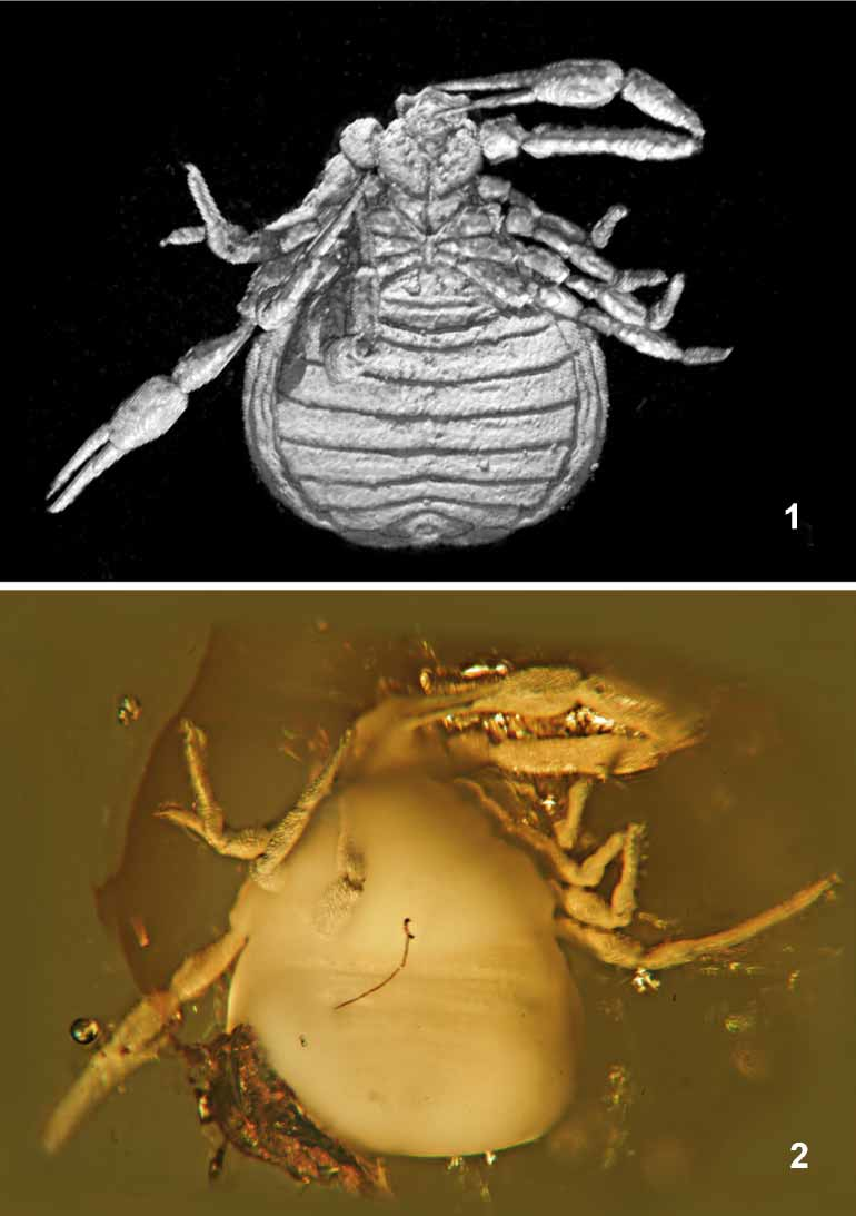
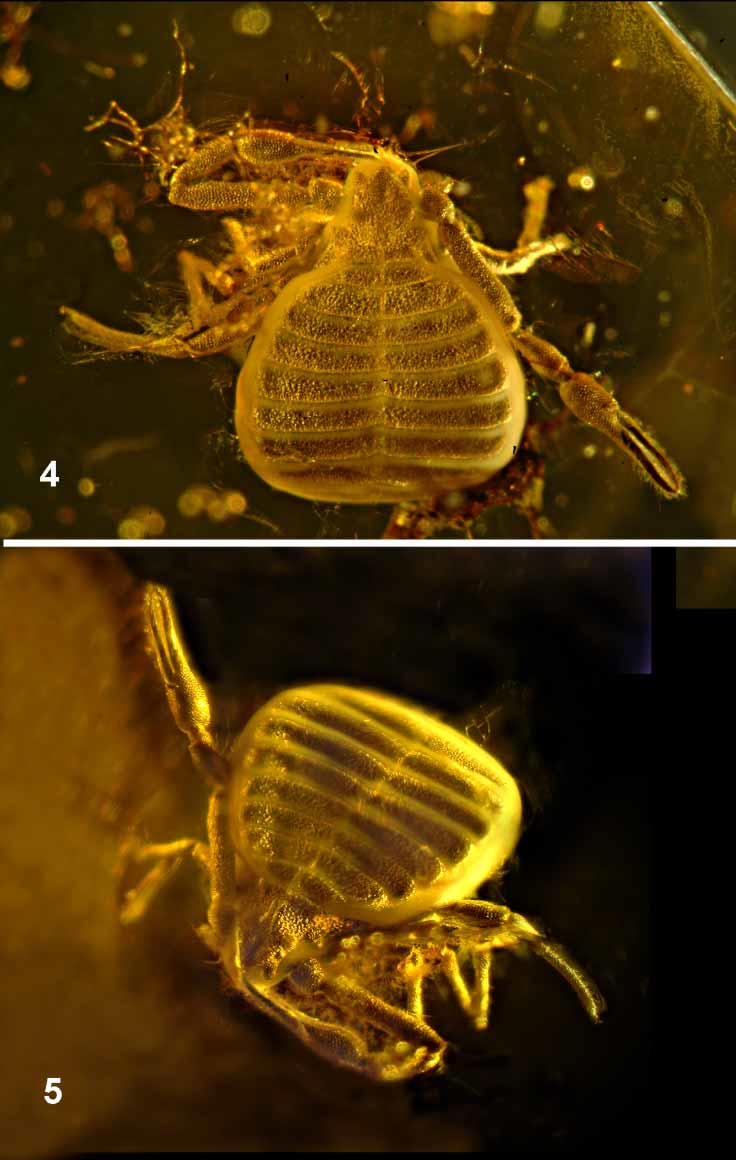
Description. Habitus as in Figs. 3 View FIGURE 3 and 4 View FIGURES 4 – 5 . Body clear, ventral and lateral sides obscured ( Figs. 2 View FIGURES 1 – 2 , 5 View FIGURES 4 – 5 ). No dorsal colour pattern could be observed (as is usual in Baltic amber, the original coloration will have been lost in the fossilization process).
Carapace wider than long (length 0.78× width measured at level of posterior margin or 0.95x when width is measured across posterolateral tips behind the eyes). Carapace very irregular in outline; anterior margin with a relatively deep notch between anterolateral and median protuberances. Posterior margin elevated into a ridge. Anterior and posterior eyes well developed, nearly contiguous, posterior eyes partially covered and facing posteriorly. Cucullar furrow a broad, shallow central depression which becomes obsolete anteriorly, extending forward from elevated median disk. Carapace, sclerites, palp and leg cuticle reticulated with an irregular netlike structure. Chaetotaxy of carapace and opisthosoma not visible, chelicerae not observable.
Opisthosoma wider than long, broad ovate (L/W=0.92). Pleural membranes raised into three folds ( Fig. 6 View FIGURES 6 – 7 arrow b, Fig. 8 View FIGURES 8 – 12 ), no pleural plates or tergal chaetotaxy observable. Figs. 1 View FIGURES 1 – 2 and 8 View FIGURES 8 – 12 show the shape of the sternites, unobservable in visible light, but reconstructed with Xray microCT. No coxal spines observed.
Pedipalp ( Fig. 10 View FIGURES 8 – 12 ) cuticle reticulated, except in the area around the trichobothria. The palpal articles have a remarkable ventral edging, visible with light microscopy along the full length of the femur, patella and chela of both pedipalps. Fig. 7 View FIGURES 6 – 7 shows the Xray CT scan of the pedipalpal hand. The cross section of the cuticle is dropshaped on the outside, whereas the inside is circular. Coxa 1.40×, trochanter 0.95×, femur 4.58×, patella 2.68×, chela (with pedicel) 3.91× and hand (with pedicel) 1.56× longer than broad. Femur 1.36× as long as carapace. Due to local opaqueness of the amber, the position of only 4 trichobothria could be observed on the fixed chelal finger; on the movable finger 4 trichobothria and their position are more clearly visible ( Fig. 9 View FIGURES 8 – 12 ). Fixed finger with 32, movable finger with 26 regular, not curved and not significantly spaced teeth (contiguous to very slightly spaced). Movable finger 1.5× as long as hand.
All legs with similar edging to that found on palp, visible with light microscopy on patella, tibia and tarsus; femur only observable as a contour in transmitted light, coxa and femur reconstructed with Xray microCT. Leg I ( Fig. 12 View FIGURES 8 – 12 ) with trochanter 1.4×, femur 1.4×, patella 2.4×, tibia 1.5× and tarsus 6.2× longer than broad. Leg IV ( Fig. 11 View FIGURES 8 – 12 ) with trochanter 1.9×, femur 1.9×, patella 2.06×, tibia 4.0× and tarsus 8.8× longer than broad. Arolia shorter than claws.
Measurements (in mm). Body length 2.09. Carapace L=0.58, W=0.74 at level of posterior margin, W=0.61 at level of laterally extending tips behind posterior eyes; cucullus L=0.16; anterior and posterior ocular breadth 0.03.
Pedipalp: trochanter 0.19/0.20; femur 0.79/0.17; patella 0.43/0.16; chela (with pedicel) 0.90/0.23; hand (with pedicel) 0.36/0.23; movable finger L=0.54.
Leg I: trochanter 0.13/0.09; femur 0.14/0.10; patella 0.24/0.10; tibia 0.14/0.09; tarsus 0.31/0.05.
Leg IV: trochanter 0.19/0.10; femur 0.19/0.10; patella 0.31/0.15; tibia, 0.36/0.09; tarsus 0.53/0.06.
Distribution. Found in Baltic amber, a fossil Pinus resin from the Eocene Baltic amber forest.
Remarks. Setae are below the resolution of the CT scan in this sample. Fig. 1 View FIGURES 1 – 2 shows a single image from the 3dimensional rotating reconstruction, which is in fact a dynamic observation tool. In a single reconstructed image artefacts can occur, depending on the parameters entered. Therefore a drawing of the ventral parts was made ( Fig. 8 View FIGURES 8 – 12 ), based on the dynamic model, giving more accurate dimensions. The circular inside of the pedipalpal articles is an indication that there was no shape deformation caused by pressure (squeezing) during the formation of the fossil and, though the ‘edging’ of the outside cuticula has not been described for any pseudoscorpion before, it is unlikely to be an artefact of fossilization.


| MRAC |
Musée Royal de l’Afrique Centrale |
No known copyright restrictions apply. See Agosti, D., Egloff, W., 2009. Taxonomic information exchange and copyright: the Plazi approach. BMC Research Notes 2009, 2:53 for further explanation.