
Cycloporus pinkipus, Dittmann & Grosbusch & Bertemes & Egger, 2023
|
publication ID |
https://doi.org/10.11646/zootaxa.5319.2.5 |
|
publication LSID |
lsid:zoobank.org:pub:47B8DCDC-63C9-4DA1-8997-17DDD83B2BD3 |
|
persistent identifier |
https://treatment.plazi.org/id/9D46DD54-FFD5-D14E-FF79-B4BDAE8FFBBD |
|
treatment provided by |
Plazi (2023-07-25 09:46:59, last updated 2023-07-25 09:56:24) |
|
scientific name |
Cycloporus pinkipus |
| status |
|
History of Cycloporus papillosus View in CoL View at ENA /Differential diagnosis
So far, only one Cycloporus species, C. papillosus , was described from the Mediterranean, featuring the eponymous papillae ( Lang 1884). A variety without papillae was named C. papillosus var. levigatus Lang (1884) . Lang (1884) argued that based on the external appearance, C. papillosus could be classified into five to six different species, but decided against it due to the very similar internal morphology. In the meanwhile, the external morphology turned out to be an important character for the species determination of Cycloporus ( Newman & Cannon 2002) .
Lang described C. papillosus in 1884 and synonymised it with Proceros tuberculatus Schmidtlein, 1880 . As Schmidtlein did not provide a description of P. tuberculatus , the material of which he had received from Lang ( Schmidtlein 1880), P. tuberculatus is a nomen nudum and unavailable according to Article 12.1 of the International Code of Zoological Nomenclature ( ICZN 1999). The formal species description under the name C. papillosus was published in Lang (1884). Some years later, Francotte (1897) found that Lang’s description of C. papillosus also fits the description of Planaria schlosseri Giard, 1873 , and also observed variants without dorsal papillae. Bock (1913) synonymised Thysanozoon papillosus Sars, 1878 with C. papillosus of Lang (1884) as C. papillosus ( Sars, 1878) and noted colour variants and specimens without dorsal papillae. He provided a photograph of a sagittal section through the genital region of C. papillosus but remained sceptical that Giard’s Planaria schlosseri was the same species as C. papillosus ( Bock 1913) . We therefore regard P. schlosseri as a nomen dubium. The latest study dealing with C. papillosus was conducted by Noreña et al. (2014), giving a detailed sagittal reconstruction of the genital region, and noting several colour variations, all with dorsal papillae ( Noreña et al. 2014). All studied specimens of C. papillosus are from Atlantic coasts, with the exception of Lang (1884), who worked with material from the Mediterranean. Lang (1884) also introduced a variation without dorsal papillae under the name C. papillosus var. levigatus .
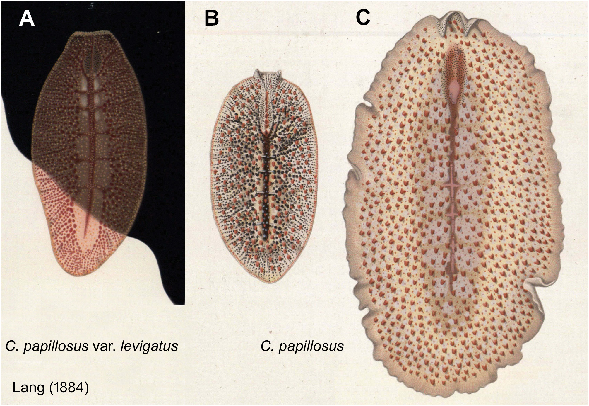
The size and colouration of C. pinkipus sp. n. differ from C. papillosus or C. papillosus var. levigatus . The latter have a length between 10 and 16 mm (except 5 mm in Bock 1913), while C. pinkipus sp. n. measures only between 2 and 4 mm. Lang (1884) describes the colouration of C. papillosus var. levigatus as transparent and slightly reddish, with yellow spots at the positions of the missing papillae, and yellow marginal pores. The carmine red intestinal branches dominate the colour pattern ( Lang 1884, plate 8, fig. 5, Fig. 8 View FIGURE 8 ). The observed colour patterns in C. papillosus are according to Lang (1884) white to yellow/orange, with either white or carmine red dorsal papillae; the marginal pores are conspicuously red-brown. The dorsal colour pattern in C. pinkipus sp. n. is similar to the written descriptions of C. papillosus , but the given pictures are dissimilar ( Fig. 8 View FIGURE 8 ), and there are no dorsal papillae in C. pinkipus sp. n. In addition, the marginal pores are colourless and inconspicuous in C. pinkipus sp. n., in contrast to C. papillosus . The colour patterns given for C. papillosus var. levigatus and C. pinkipus sp. n. are substantially different ( Figs. 1–2 View FIGURE 1 View FIGURE 2 ; 8 View FIGURE 8 ). Our molecular data also confirm the separation of the two species ( Fig. 7 View FIGURE 7 ).
Bock, S. (1913) Studien u ¨ ber Polycladen. Zoologiska Bidrag fra r n Uppsala, 2, 31 - 344.
Francotte, C. P. (1897) Recherches sur la maturation, la fecondation et la segmentation chez les polyclades. In: De Lacaze- Duthiers, H. & Pruvot, G. (Eds.), Archives de Zoologie experimentale et generale: histoire naturelle-histologie-evolution des animaux. Troisieme serie. Librairie C. Reinwald Schleicher freres, editeurs, Paris, pp. 189 - 298.
Giard, A. (1873) Contributions a l'histoire naturelle des synascidies. Archives de Zoologie experimentale et generale, 2, 481 - 514.
ICZN (1999) International Code of Zoological Nomenclature. 4 th Edition. The International Trust for Zoological Nomenclature, London, 306 pp.
Lang, A. (1884) Die Polycladen (Seeplanarien) des Golfes von Neapel und der angrenzenden Meeresabschnitte. Eine Monographie. Fauna und Flora des Golfes von Neapel. W. Engelmann, Leipzig, 688 pp. https: // doi. org / 10.5962 / bhl. title. 10545
Newman, L. J. & Cannon, L. R. G. (2002) The genus Cycloporus (Platyhelminthes: Polycladida) from Australasian waters. Raffles Bulletin of Zoology, 50 (2), 287 - 299.
Norena, C., Marquina, D., Perez, J. & Almon, B. (2014) First records of Cotylea (Polycladida, Platyhelminthes) for the Atlantic coast of the Iberian Peninsula. ZooKeys, 404, 1 - 22. https: // doi. org / 10.3897 / zookeys. 404.7122
Sars, G. O. (1878) s. n. In: Jensen, O. S. Turbellaria ad litora Norvegiae occidentalia. Turbellarier ved Norges vestkyst. J. W. Eided Bogtrykkeri, Bergen, pp. 1 - 97.
Schmidtlein, R. (1880) Vergleichende Ubersicht uber das Erscheinen grosserer pelagischer Thiere und Bemerkungen. uber Fortpflanzungsverhaltnisse einiger Seethiere im Aquarium. Mittheilungen aus der zoologischen Station zu Neapel, 2, 162 - 175.
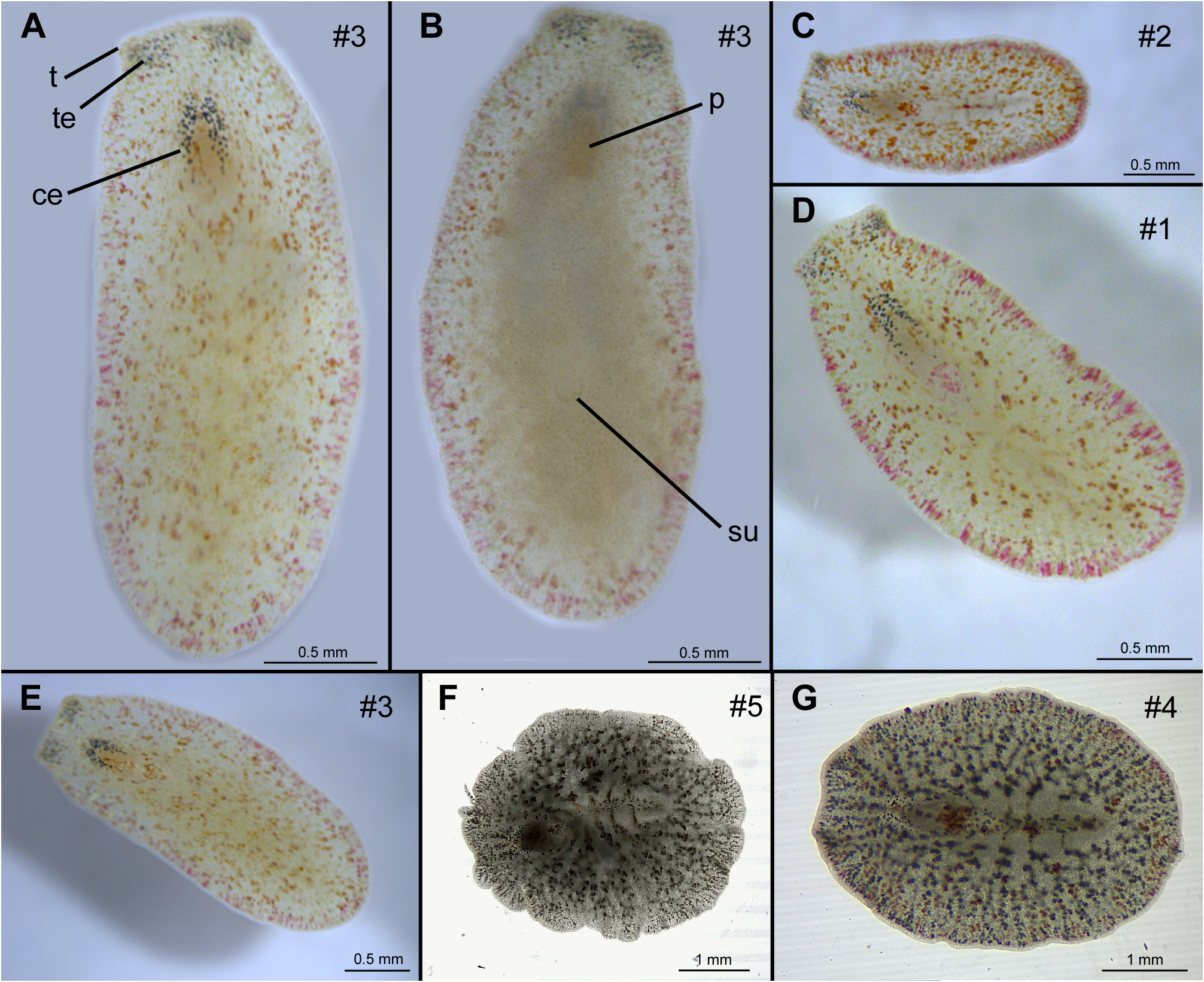
FIGURE 1. Live images of Cycloporus pinkipus sp. n. A–E. Incident light, F–G. Transmitted light. A., E. Dorsal view of the holotype (specimen #3). B. Ventral view of the holotype (specimen #3). C. Dorsal view of specimen #2. D. Dorsal view of specimen #1. F. Dorsal view of paratype 1 (specimen #5). G. Dorsal view of specimen #4. ce = cerebral eyes, p = pharynx, su = sucker, t = tentacle, te = tentacle eyes. Orientation: A–B anterior directed upwards, C–G anterior directed to the left.
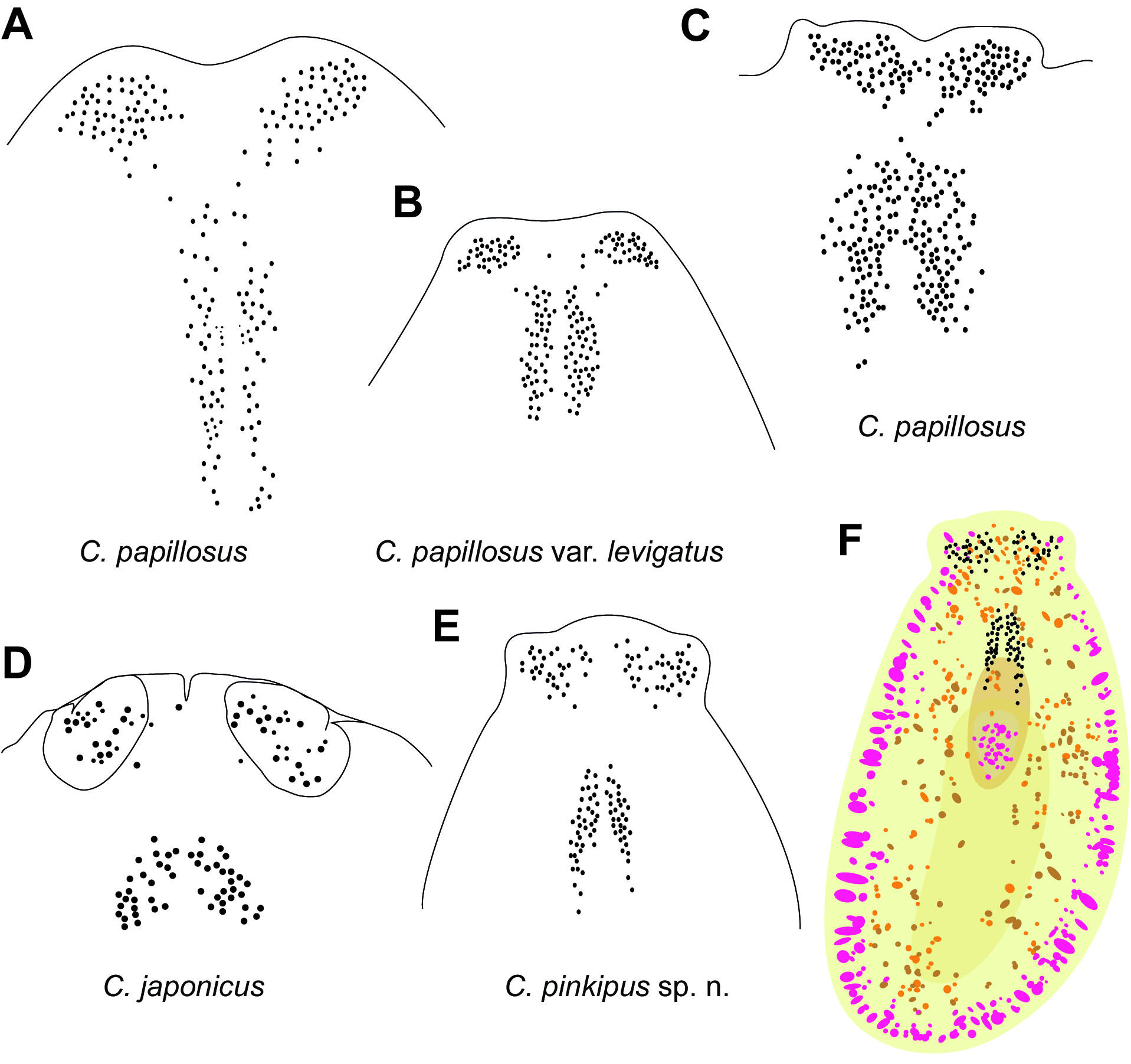
FIGURE 2. Schematic drawings of eye spot arrangement in similar Cycloporus species. A–E. Schematic drawings of eye spot clusters of different species and specimens after Lang (1884) (A–B.), after Marquina et al. (2015) (C.), and Kato (1937) (D.). F. Schematic drawing of the dorsal colouration of Cycloporus pinkipus sp. n. Anterior directed upwards. Not drawn to scale.
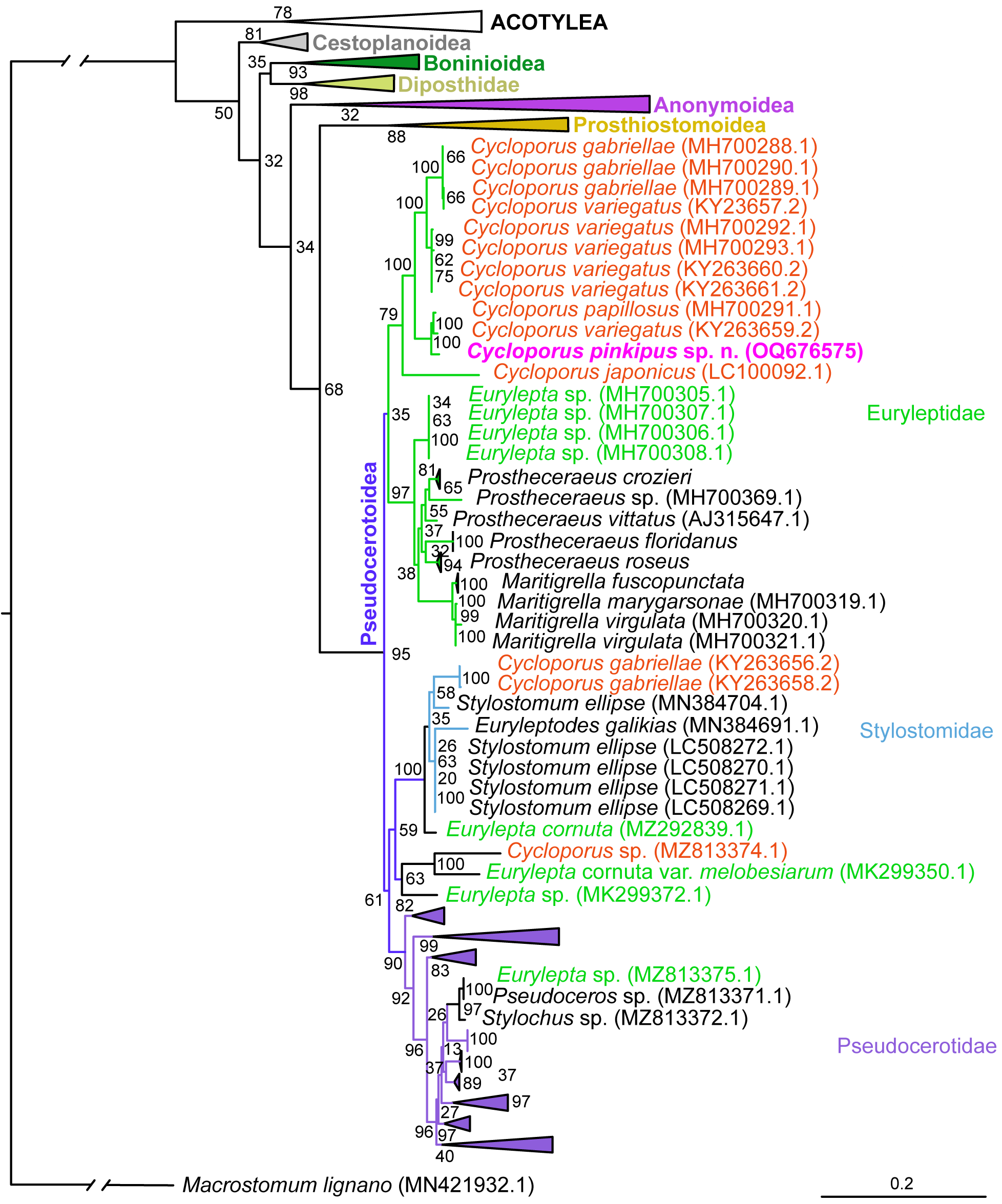
FIGURE 7. Phylogenetic maximum likelihood reconstruction using partial 28S sequences (accession numbers in brackets) of polyclads, rooted with Macrostomum lignano; branches other than Euryleptidae and Stylostomidae collapsed. Bootstrap nodal support of 200 non-parametric bootstrap replicates. Full tree in Suppl. Mat. 2. Cycloporus pinkipus sp. n. marked in pink. Additional representatives of Cycloporus written in red. Representatives of Eurylepta written in light green. Branches of Euryleptidae in light green. Branches of Stylostomidae in light blue. Branches of Pseudocerotidae in purple. Scale bar indicates the number of substitutions per site.
No known copyright restrictions apply. See Agosti, D., Egloff, W., 2009. Taxonomic information exchange and copyright: the Plazi approach. BMC Research Notes 2009, 2:53 for further explanation.
|
Kingdom |
|
|
Phylum |
|
|
Order |
|
|
Family |
|
|
Genus |