
Cycloporus pinkipus Egger & Dittmann, 2023
|
publication ID |
https://doi.org/10.11646/zootaxa.5319.2.5 |
|
publication LSID |
lsid:zoobank.org:pub:47B8DCDC-63C9-4DA1-8997-17DDD83B2BD3 |
|
DOI |
https://doi.org/10.5281/zenodo.8182479 |
|
persistent identifier |
https://treatment.plazi.org/id/9D46DD54-FFD0-D145-FF79-B100A8E8FAF9 |
|
treatment provided by |
Plazi (2023-07-25 09:46:59, last updated 2023-07-25 09:56:24) |
|
scientific name |
Cycloporus pinkipus Egger & Dittmann |
| status |
sp. n. |
Cycloporus pinkipus Egger & Dittmann sp. n.
( Figs. 1–6 View FIGURE 1 View FIGURE 2 View FIGURE 3 View FIGURE 4 View FIGURE 5 View FIGURE 6 )
Material examined. Cycloporus pinkipus sp. n. specimens #1 and #2 used for molecular analysis. Specimens #3 and #5 sagittally sectioned. Specimens #1, #2, #3, #4 and #5 used for live observations. Specimen #6 cross-sectioned.
Type material. Serial sections of holotype and paratypes submitted to the Natural History Museum Vienna, Austria. GenBank accession numbers of partial nuclear ribosomal subunits of specimen #2 are OQ676574 (18S) and OQ676575 (28S). The ZooBank registration number is urn:lsid:zoobank.org:act:6765D50D-C5C8-44D3-A032-29FDFAC6165F .
Holotype. One sagittally sectioned specimen (#3) stained with AZAN (NHMW-ZOO-EV-M-5880).
Paratype 1. One sagittally sectioned specimen (#5) stained with AZAN (NHMW-ZOO-EV-M-5881) .
Paratype 2. One cross-sectioned specimen (#6) stained with AZAN (NHMW-ZOO-EV-M-5882) .
Type locality. Port of Punat , Krk, Croatia (45°01’23’’N 14°37’41’’E) GoogleMaps .
Habitat. Animals were found in brown algae.
Etymology. The species epithet ‘pinkipus’ refers to the typical pink spots which characterises the dorsal colouration, and rhymes with ‘Cycloporus’.
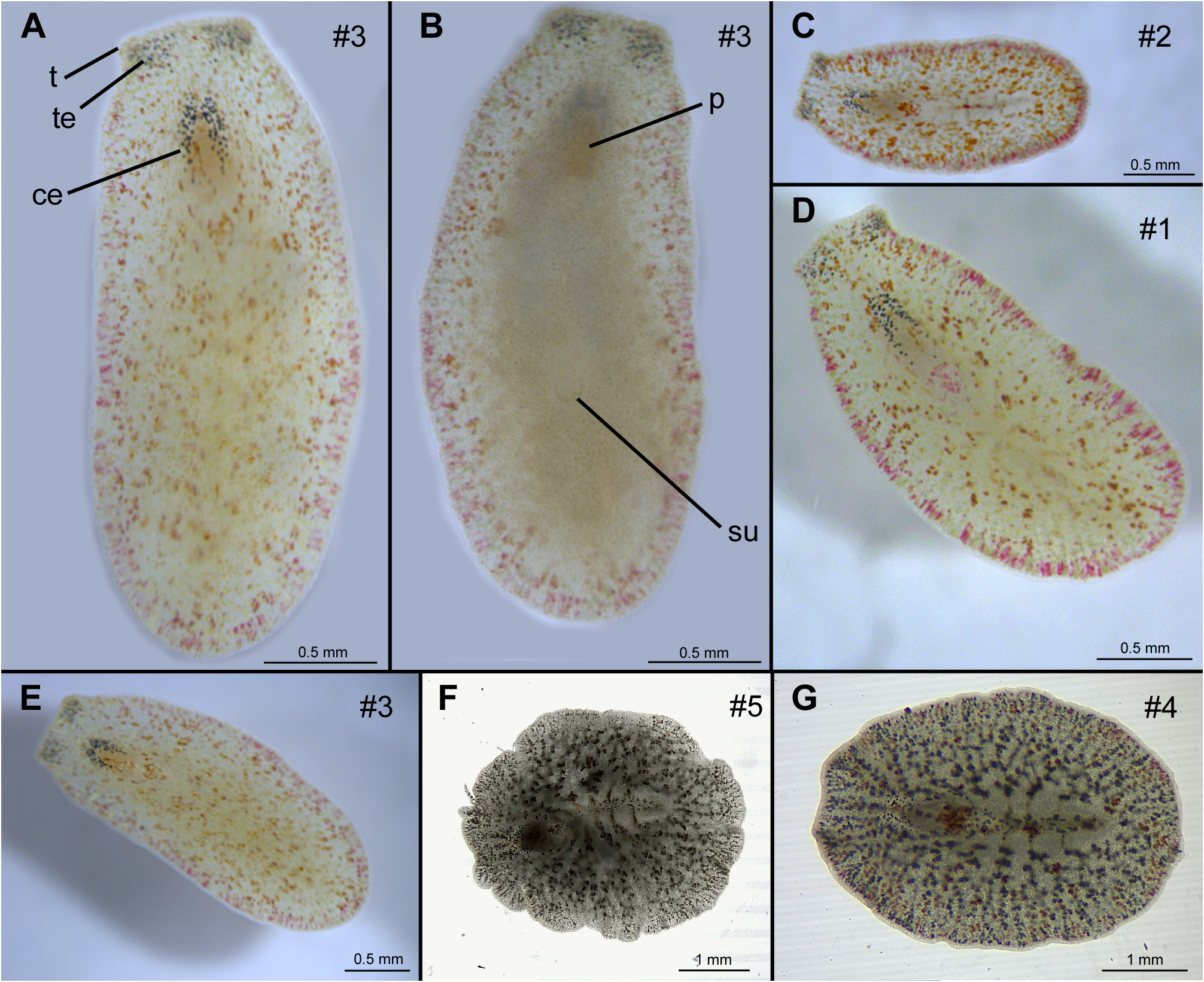
FIGURE 1. Live images of Cycloporus pinkipus sp. n. A–E. Incident light, F–G. Transmitted light. A., E. Dorsal view of the holotype (specimen #3). B. Ventral view of the holotype (specimen #3). C. Dorsal view of specimen #2. D. Dorsal view of specimen #1. F. Dorsal view of paratype 1 (specimen #5). G. Dorsal view of specimen #4. ce = cerebral eyes, p = pharynx, su = sucker, t = tentacle, te = tentacle eyes. Orientation: A–B anterior directed upwards, C–G anterior directed to the left.
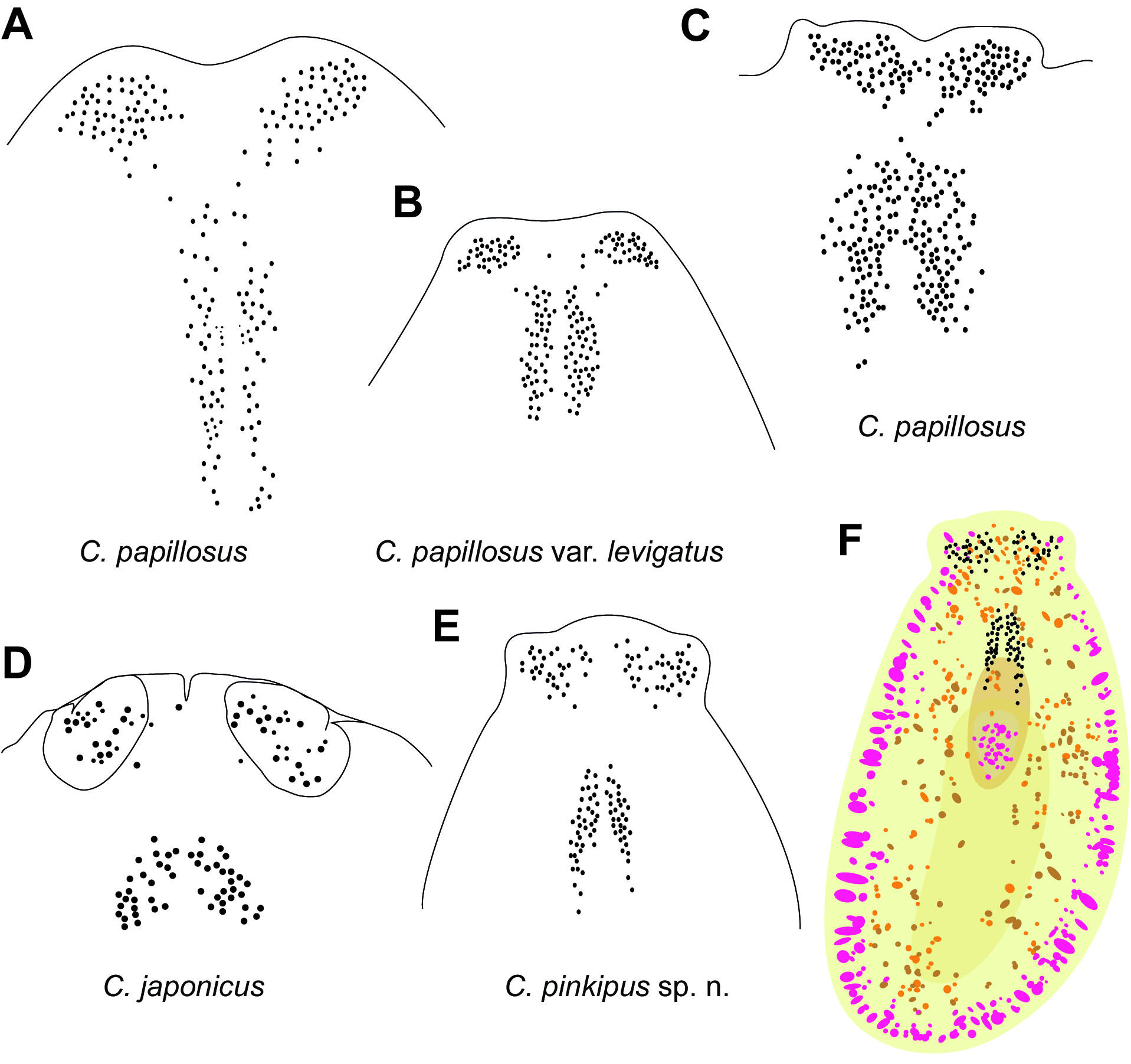
FIGURE 2. Schematic drawings of eye spot arrangement in similar Cycloporus species. A–E. Schematic drawings of eye spot clusters of different species and specimens after Lang (1884) (A–B.), after Marquina et al. (2015) (C.), and Kato (1937) (D.). F. Schematic drawing of the dorsal colouration of Cycloporus pinkipus sp. n. Anterior directed upwards. Not drawn to scale.

FIGURE 3. Sagittal sections of genital apparatus of Cycloporus pinkipus sp. n. (holotype; specimen #3). A. Male genital. B. Detailed view of male genital and mouth opening C. Detailed view of the ducts of prostatic vesicle and seminal vesicle. D. Female genital overview. E. Detailed view of female genital. ce = cerebral eyes, cg = cement glands, cp = cement pouch, fa = female atrium, fp = female pore, m = mouth opening, ma = male atrium, mp = male pore, p = pharynx, ps = penis sheath, pp = penis papilla, pv = prostatic vesicle, pvd = prostatic vesicle duct, st = stylet, sv = seminal vesicle, svd = seminal vesicle duct, vd = vas deferens. Orientation: anterior to the left.
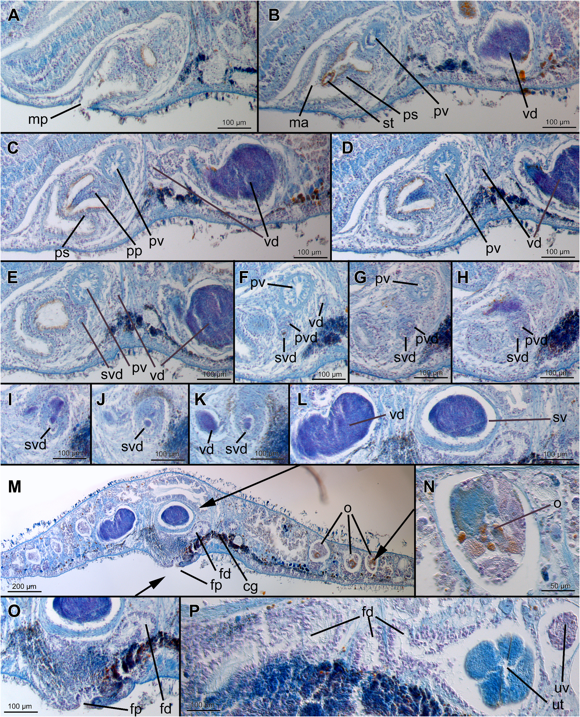
FIGURE 4. Sagittal sections of genital apparatus of Cycloporus pinkipus sp. n. (paratype 1; specimen #5). A–D. Penis papilla, stylet, prostatic vesicle and vas deferens. E–L. Junction between seminal vesical and vas deferens. M. Female genital. Arrows indicate the region of detailed views. N. Detailed view of ovary. O. Detailed view of female pore. P. Female duct, uterus and uterine vesicle. cg = cement glands, fd =female duct, fp = female pore, ma = male atrium, mp = male pore, o = ovary, pp = penis papilla, ps = penis sheath, pv = prostatic vesicle, pvd = prostatic vesicle duct, st = stylet, sv = seminal vesicle, svd = seminal vesicle duct, ut = uterus, uv = uterine vesicle, vd = vas deferens. Orientation: anterior to the left. Same scale bars in A–L., O–P.
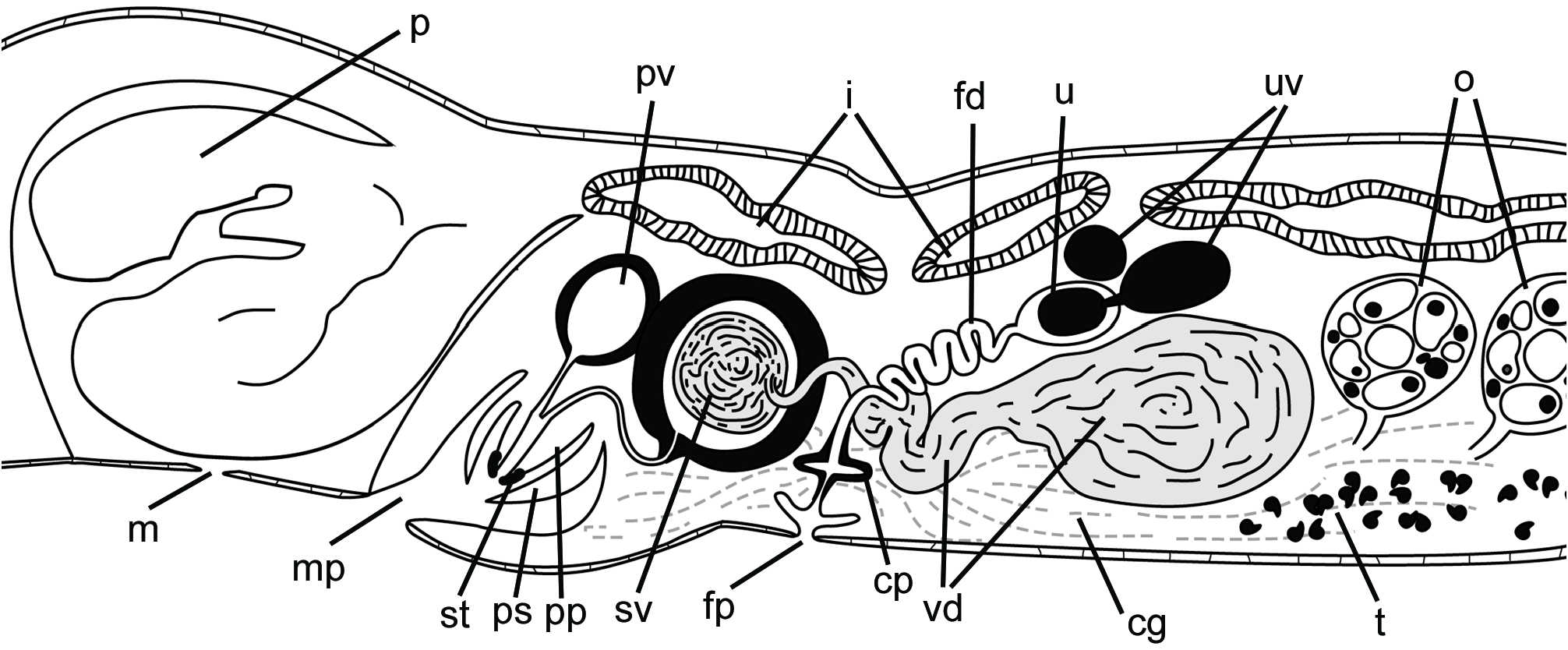
FIGURE 5. Sagittal reconstruction of the genital of Cycloporus pinkipus sp. n. cg = cement glands, cp = cement pouch, fd =female duct, fp = female pore, i = intestine, m = mouth opening, mp = male pore, o = ovary, p = pharynx, ps = penis sheath, pp = penis papilla, pv = prostatic vesicle, st = stylet, sv = seminal vesicle, t = testis, u = uterus, uv = uterine vesicle, vd = vas deferens. Orientation: anterior to the left.
| AZAN |
Akademia Nauk Azerbaijana-Bulgarian Academy of Science of Azerbaijan |
No known copyright restrictions apply. See Agosti, D., Egloff, W., 2009. Taxonomic information exchange and copyright: the Plazi approach. BMC Research Notes 2009, 2:53 for further explanation.
|
Kingdom |
|
|
Phylum |
|
|
Order |
|
|
Family |
|
|
Genus |