
Macrobiotus ovovittatus, Stec, 2024
|
publication ID |
https://doi.org/ 10.5852/ejt.2024.930.2481 |
|
publication LSID |
lsid:zoobank.org:pub:A09EB44B-286F-439A-A970-48F09416584A |
|
DOI |
https://doi.org/10.5281/zenodo.10904351 |
|
persistent identifier |
https://treatment.plazi.org/id/9C2931D3-8EBE-47AB-A3B0-991AB28F03A2 |
|
taxon LSID |
lsid:zoobank.org:act:9C2931D3-8EBE-47AB-A3B0-991AB28F03A2 |
|
treatment provided by |
Plazi |
|
scientific name |
Macrobiotus ovovittatus |
| status |
sp. nov. |
Macrobiotus ovovittatus sp. nov.
urn:lsid:zoobank.org:act:9C2931D3-8EBE-47AB-A3B0-991AB28F03A2
Figs 1–8 View Fig View Fig View Fig View Figure 4 View Fig View Fig View Fig View Fig , Tables 2–3 View Table 2 View Table 3
Etymology
The species name refers to the terminal discs of the egg processes which resemble crocheted napkins. From the Latin “egg” = “ovo” and “chaplet” = “vittatus”.
Material examined
32 animals, 57 eggs mounted on microscope slides in Hoyer’s medium, 15 animals and 15 eggs examined under SEM, 15 animals stained with orcein and two animals processed for DNA sequencing.
Type material
Holotype
GREENLAND • near Zackenberg Valley ; 74°29′0.766″ N, 20°32′18.308″ W; 77 m a.s.l.; Jul. 2021; M. Kolasa leg.; mixed sample of moss and lichen collected from the rock in arctic tundra; ISEA PAS, slide GL.001.01. GoogleMaps
Paratypes
GREENLAND • 46 animals; same collection data as for the holotype; ISEA PAS, slides GL.001.01 to GL.001.03, SEM stub TAR.015 GoogleMaps • 72 eggs; same collection data as for the holotype; ISEA PAS, slides GL.001.04 to GL.001.09, SEM stub TAR.015 GoogleMaps .
Description
Animals
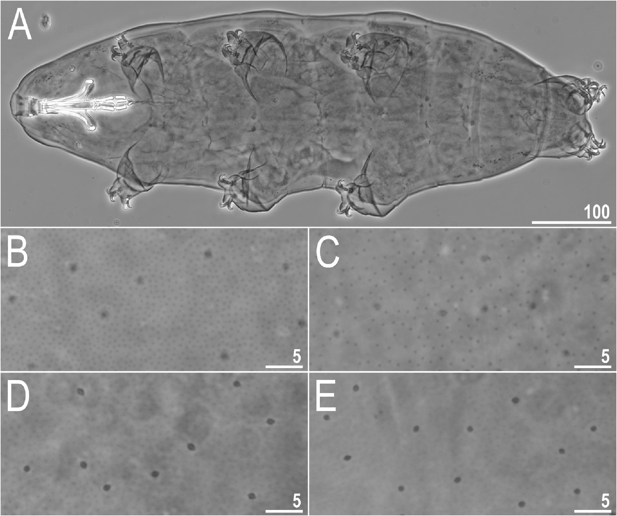
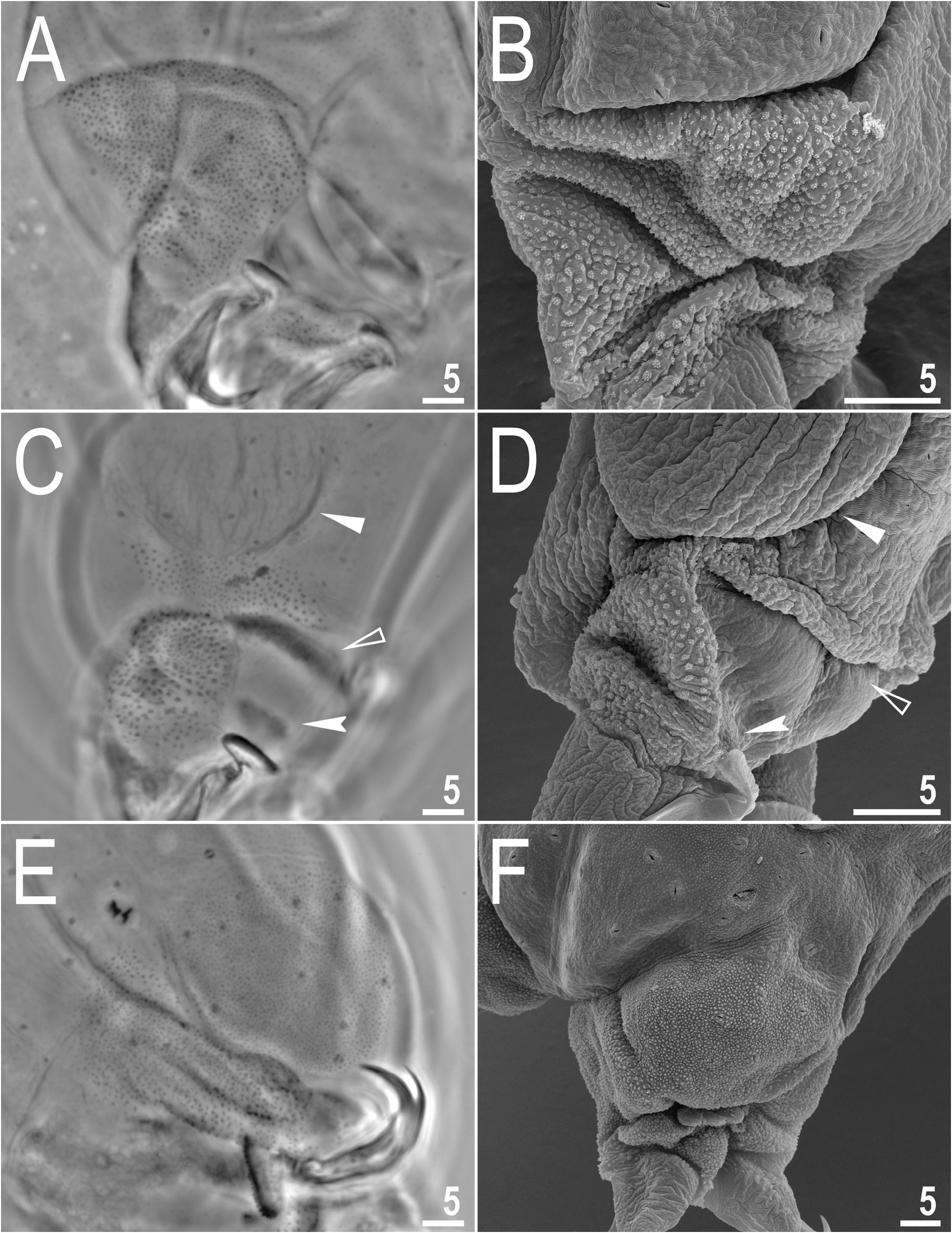
Body transparent in juveniles and white in adults, after fixation in Hoyer’s medium transparent ( Fig. 1A View Fig ). Eyes present. Round and oval pores (0.4–0.6 μm in diameter), scattered randomly throughout the cuticle (distributed more sparsely on the ventral side of the body) ( Figs 1B–E View Fig , 2A–B View Fig ), including the external and internal surface of all legs ( Fig. 3A–F View Fig ). Granulation is present on the entire body cuticle and clearly visible under PCM and SEM, with granulation on the ventral side of the body being less dense ( Figs 1B– E View Fig , 2A–B View Fig ). Moreover, evident dense granulation patches on the external and internal surface of all legs I–III are visible under PCM and SEM ( Fig. 3A–D View Fig ). This dense granulation is also present on the lateral and dorsal surfaces of legs IV ( Fig. 3E–F View Fig ). A pulvinus-shaped cuticular bulge is centrally present on the internal surface of all legs I–III ( Fig. 3C–D View Fig ). This structure is visible only if the legs are fully extended and well oriented.
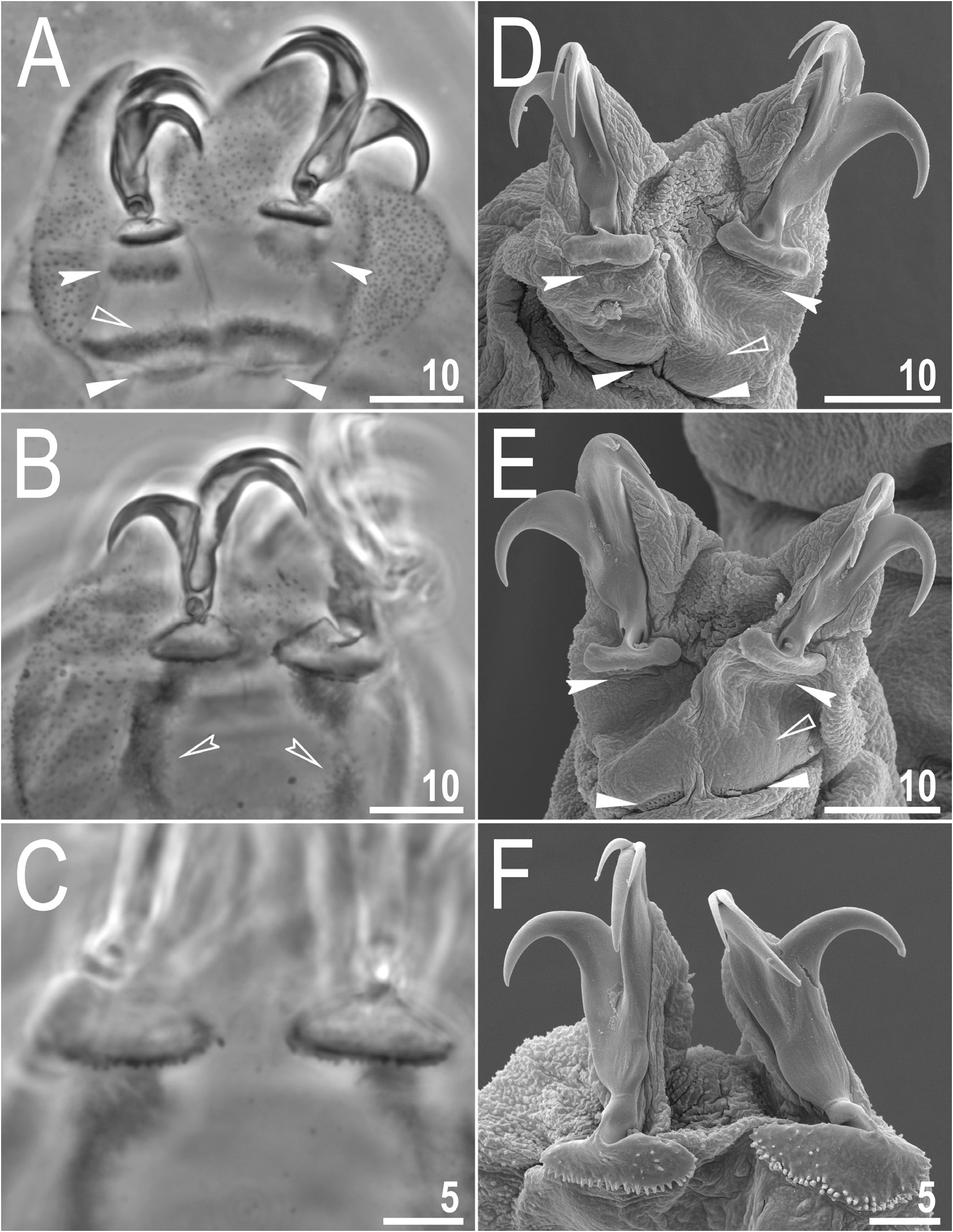
Claws small and slender, of the hufelandi type ( Fig. 4A–F View Figure 4 ). Primary branches with distinct accessory points, a long common tract, and an evident stalk connecting the claw to the lunula ( Fig. 4A–F View Figure 4 ). The lunulae on legs I–III are smooth ( Figs 4A, D, E View Figure 4 ), while there is a dentation in the lunulae on legs IV ( Fig. 4B, C, F View Figure 4 ). A single continuous cuticular bar and double muscle attachments are present above claws I–III ( Figs 3C–D View Fig , 4A, D, E View Figure 4 ). Shadowed extensions that extend from the lunulae of the claws on legs I–III are present and visible only under PCM ( Figs 3C View Fig , 4A View Figure 4 ). A horseshoe-shaped structure connects the anterior and posterior lunules on leg IV ( Fig. 4B View Figure 4 ).
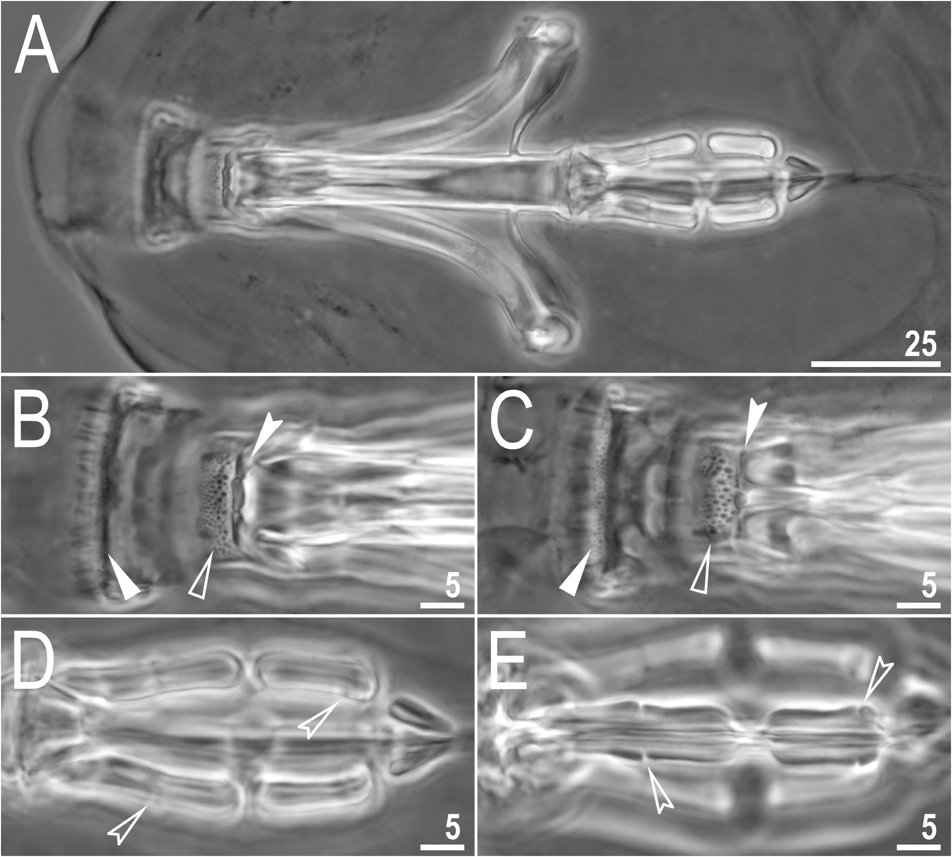
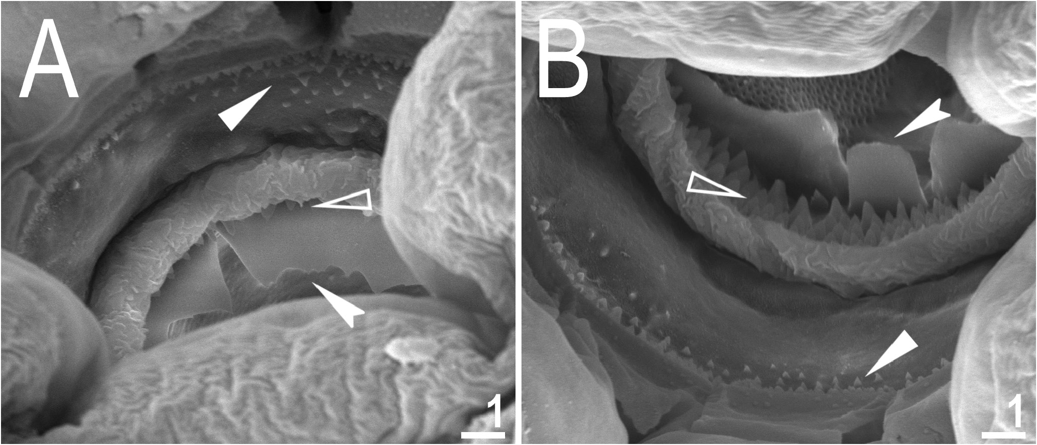
Mouth antero-ventral. Bucco-pharyngeal apparatus of Macrobiotus type, with ventral lamina and ten small peribuccal lamellae followed by six buccal sensory lobes ( Figs 5A View Fig , 6A–B View Fig ). Under PCM, the oral cavity armature is of hufelandi type – three bands of teeth are always visible ( Fig. 5B–C View Fig ). The first band of teeth is composed of numerous extremely small cones arranged in four to six rows located anteriorly in the oral cavity, at the bases of the peribuccal lamellae and just behind them ( Figs 5B–C View Fig , 6A–B View Fig ). The second band of teeth is located between the ring fold and the third band of teeth and comprises 4–6 rows of small cones, larger than those of the first band ( Figs 5B–C View Fig , 6A–B View Fig ). The teeth of the third band are located within the posterior portion of the oral cavity, between the second band of teeth and the opening of the buccal tube ( Figs 5B–C View Fig , 6A–B View Fig ). The third band of teeth is discontinuous and divided into the dorsal and ventral portions. Under PCM, the dorsal teeth are seen as three distinct transverse ridges, whereas the ventral teeth appear as two separate lateral transverse ridges between which a round median tooth is visible ( Fig. 5B–C View Fig ). Under SEM, both dorsal and ventral teeth are also clearly distinct ( Fig. 6A– B View Fig ). Under SEM, the margins of the dorsal teeth are clearly serrated ( Fig. 6A View Fig ) whereas the margins of the ventral teeth are less serrated ( Fig. 6B View Fig ). Pharyngeal bulb spherical, with triangular apophyses, two rod-shaped macroplacoids and a large triangular microplacoid ( Fig. 5A View Fig ). The macroplacoid length sequence being 2<1. The first and the second macroplacoid are constricted centrally and subterminally, respectively ( Fig. 5D–E View Fig ). The animals’ measurements and statistics are given in Table 2 View Table 2 .
Eggs
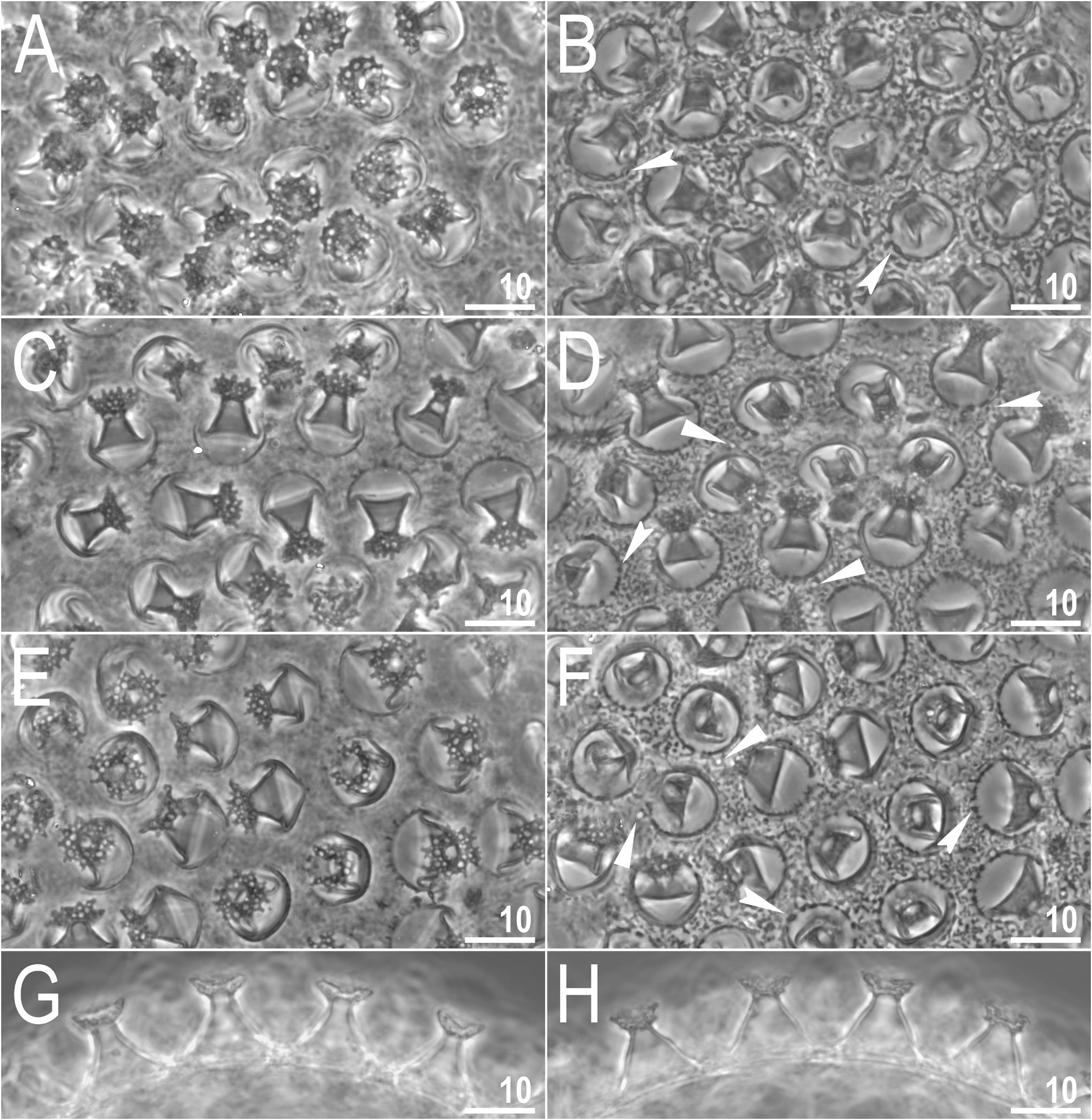
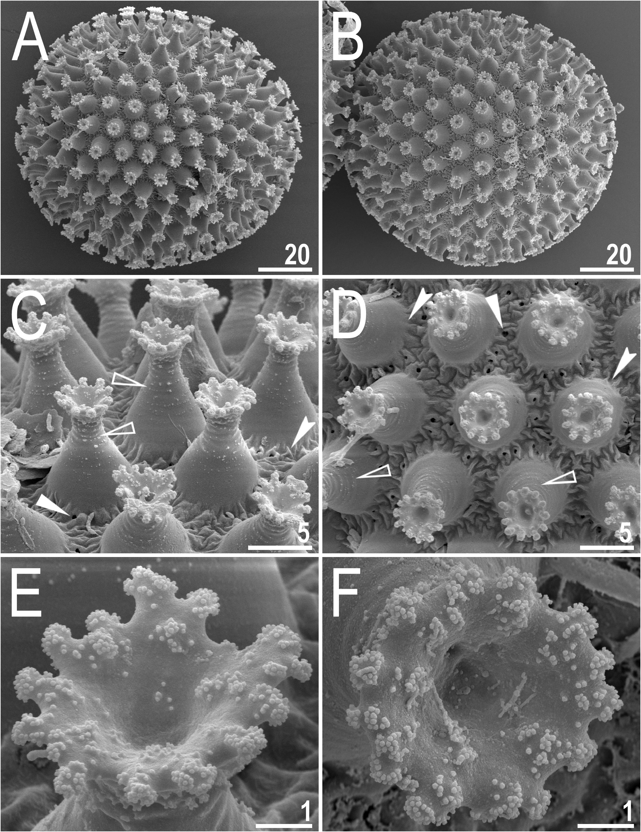
Laid freely, white, spherical and ornamented ( Figs 7A–H View Fig , 8A–F View Fig ). The surface between processes is of intermediate state between the maculatus and the persimilis type, that is, the surface is continuous/solid and clearly wrinkled with sparse, very small and irregularly spaced pores ( Figs 7B, D, F View Fig , 8C–D View Fig ). These pores are faintly visible under PCM but clearly visible under SEM (0.3–0.5 μm in diameter; Figs 7B, D, F View Fig , 8C–D View Fig ). Under PCM the wrinkles in egg surface are visible as dark dots/comas/bars making the impression of incomplete reticulation ( Fig. 7B, D, F View Fig ). The processes are in the shape of inverted goblets with concave conical trunks and well-defined terminal discs ( Figs 7A–H View Fig , 8A–F View Fig ). Faint annulations are visible on the trunk of the process, especially in its distal portion, which is also covered by fine granulation (characters visible only under SEM; Fig. 8C–D View Fig ). A crown of gently marked thickenings is visible around the bases of the processes as darker dots under PCM ( Fig. 7B, D, F View Fig ) and as thicker wrinkles at the processes bases under SEM ( Fig. 8C–D View Fig ). The terminal discs are cog-shaped, with a concave central area and 10–18 distinct teeth ( Figs 7A–H View Fig , 8A–F View Fig ). Terminal discs under PCM are covered by multiple light-refracting dots, and as such resemble crocheted napkins ( Fig. 7A, C, E View Fig ). These light refracting dots, when viewing the egg process laterally, give the impression that the terminal discs are rough and ragged (visible under PCM; Fig. 7 B, D, F–H View Fig ). However, the terminal discs under SEM are solid without any pores or light refracting dots, their teeth, are covered by small granules (visible only under SEM) that probably serve to improve the adhesive properties of the egg processes ( Fig. 8C–F View Fig ). The measurements and statistics of eggs are given in Table 3 View Table 3 .
Reproduction
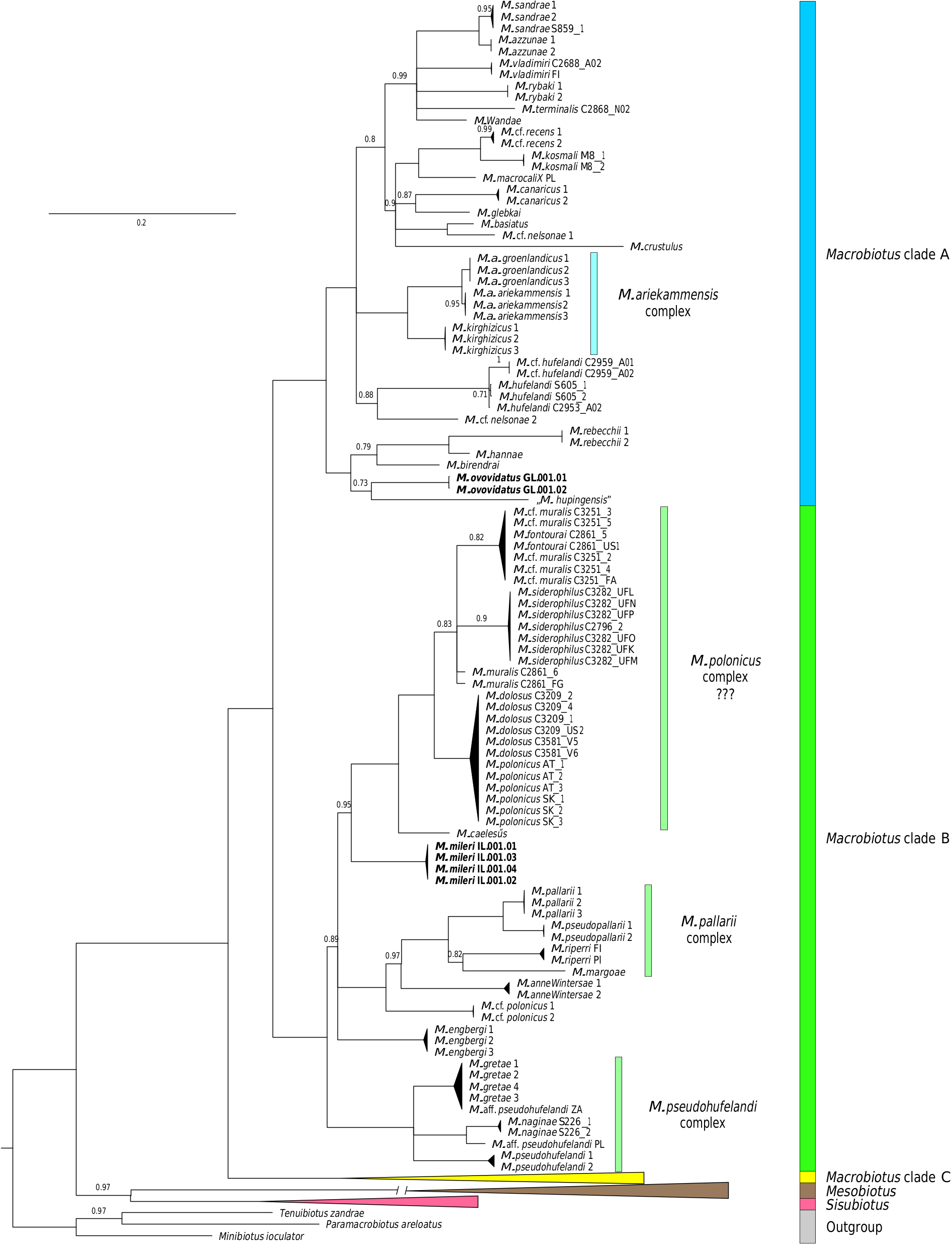
The reproduction mode of M. ovovittatus sp. nov. is unknown. Examination of orcein-stained specimens revealed no spermatozoa, but only developing oocytes. The same was true for the observation of freshly mounted individuals in Hoyer’s medium that did not reveal any sperm either but only developing oocytes. Therefore, this population could be parthenogenetic or hermaphroditic (considering its close relationship with hermaphroditic taxa in Fig. 21 View Fig ). In the second case, it might have been possible that there were no specimens in the development stage when the sperm could be detectable.
Differential diagnosis
By having (i) three bands of teeth in the oral cavity armature that are well visible under light microscope, (ii) entire body cuticle covered by granulation (sometimes visible only under SEM), (iii) eggs with inverted goblet shaped processes, the new species is the most similar to four other taxa of Macrobiotus , namely Macrobiotus joannae Pilato & Binda, 1983 reported from its type locality in Australia ( Pilato & Binda 1983), and several uncertain localities in central, eastern, and southeastern Russia ( Biserov 1990) and from Italy ( Bertolani et al. 2014), Macrobiotus hannae Nowak & Stec, 2018 known only from its type locality in Poland ( Nowak & Stec 2018), Macrobiotus punctillus Pilato, Binda & Azzaro, 1990 known only from its type locality in Chile ( Pilato et al. 1990) and Macrobiotus rebecchii Stec, 2022 known only from its type locality in Kyrgyzstan ( Stec 2022b). However, it can be easily distinguished from all of them by having a different morphology of the egg surface (the surface is continuous/solid and clearly wrinkled with sparse, very small and irregularly spaced pores in the new species vs chorion surface covered by evident reticulum in the other species), a different appearance of the terminal discs under PCM (the terminal discs are covered by multiple light-refracting dots, and as such resemble crocheted napkins vs terminal discs without light-refracting dots in other species).

| PAS |
PAS |
No known copyright restrictions apply. See Agosti, D., Egloff, W., 2009. Taxonomic information exchange and copyright: the Plazi approach. BMC Research Notes 2009, 2:53 for further explanation.
|
Kingdom |
|
|
Phylum |
|
|
Class |
|
|
Order |
|
|
Family |
|
|
Genus |