
Pheretima rubida, James, 2004
|
publication ID |
https://doi.org/10.5281/zenodo.4618925 |
|
DOI |
https://doi.org/10.5281/zenodo.4618784 |
|
persistent identifier |
https://treatment.plazi.org/id/9918E954-FFBB-E070-09F8-F8A45653F8F3 |
|
treatment provided by |
Carolina (2021-03-18 18:41:50, last updated by Plazi 2023-11-02 02:04:06) |
|
scientific name |
Pheretima rubida |
| status |
sp. nov. |
Pheretima rubida , new species
( Figs. 3I, J View Fig )
Material examined. – Holotype - adult ( NMA 003986 ), Philippines, Mindanao Island, Bukidnon Province, Mt. Kitanglad Range , 17 km S, 7 km E of Baungon, 8 10' N, 124 44' E, 1550 m. elevation, coll. D. Balete, no date. GoogleMaps
Etymology. – The species is named for its reddish pigmentation.
Description. – Red-brown pigment covering entire length, dorsal 2/3 of circumference, body 72 x 2.2 mm (vii), 2.7 (x), 3.1 mm (xxv), 110 segments; body cylindrical in cross-section with long taper towards tail. First dorsal pore 11/12, spermathecal pores paired in 7/8, 0.2 circumference apart, female pore single in xiv, openings of copulatory bursae paired in xviii, 0.12 circumference apart in 4 th setal line, 4 setae between openings. Setae regularly distributed around segmental equators; 34 setae on vii, 36 setae on xx; in vii ZZ: YZ = 3.0, in xx ZZ: YZ = 2.0, no ventral gaps. Clitellum annular xiv-xvi; posterior of vii, anterior of viii ventral side thickened to form a pad spanning spermathecal pores, pad thinner at midventral line ( Fig. 3I View Fig ).
Septa all thin, all present. Nephridia of intestinal segments pre-and post-septal at septum-body wall junctions, dense tufts of nephridia on anterior faces of 5/6, 6/7.
Large gizzard in viii, esophagus with vertical lamellae xixiv, intestinal origin xvi, simple caeca originating in xxvii, extending forward to xxv, ventral margin smooth; typhlosole xxvii-lxxii, simple fold 0.33 lumen diameter; 26 longitudinal blood vessels in intestinal wall xxvii-xl.
Hearts x-xiii esophageal, commissural vessels vi, vii, ix lateral; viii to gizzard; supra-esophageal vessel x-xiv, extraesophageal vessel enters esophageal wall x; efferent parietoesophageal vessels not seen.
Ovaries and funnels free in xiii, spermathecae paired, preseptal in vii with nephridia on ducts; each spermatheca with blocky sac-shaped ampulla, broad muscular duct shorter than ampulla, stalked diverticulum terminating in ovate receptacle, stalks with one or two kinks ( Fig. 3J View Fig ). Male sexual system holandric, testes and funnels enclosed in paired sacs in x, xi; all sacs separate; seminal vesicles xi, xii with dorsal lobe; vasa deferentia free from body wall en route to ental end of prostatic ducts; each prostate racemose, occupying xvixx, bilobed, lobes subdivided in three or four sections, stout muscular duct entering apex of copulatory bursa in xviii; coelomic surface of copulatory bursae muscular, lacking glandular or other projections; stout penis from roof of copulatory bursae, no pads flanking opening.
Remarks. – This species also keys to the P. sangirensis group in Sims & Easton (1972), but differs from the previously known species in having the combination of 4 setae between male openings, paired spermathecae in vii, intestinal origin in xvi, extensively pigmented body wall, esophageal lamellae in xiv, and a relatively large typhlosole and penes. Compared to P. paucisetosa , it differs in having pigmentation and a typhlosole, and lacking pads within the copulatory bursae.
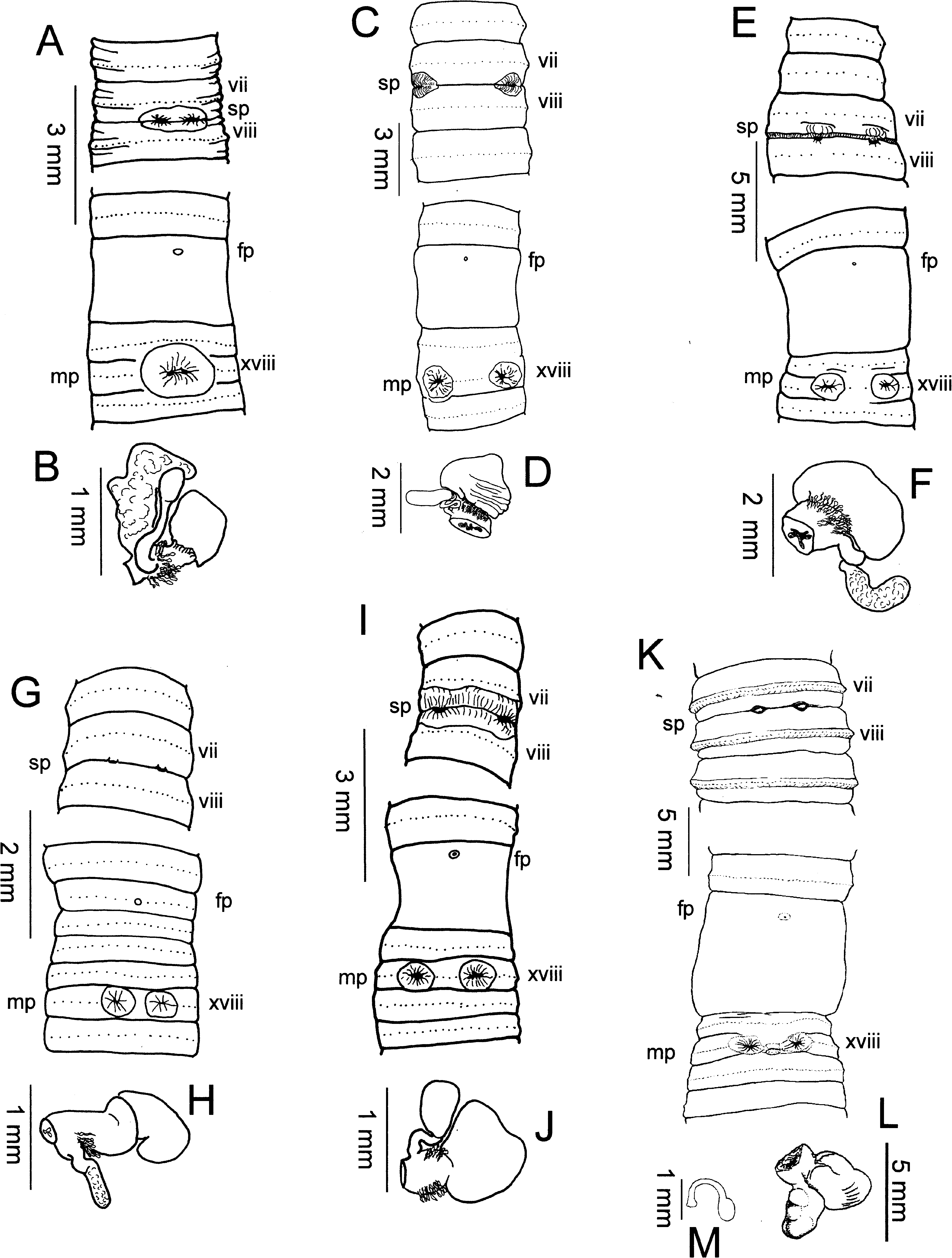
Fig. 3. Spermathecae, and ventral views showing spermathecal pores, female pores, and male pores of P. vicinipora (A, B), P. baungonensis (C, D), Pheretima paucisetosa (E, F), P. alba (G, H), P. rubida (I, J), and P. virgata (K, L, plus M, spermatophore). Symbols: fp, female pore(s); mp, male pores; sp, spermathecal pores.
No known copyright restrictions apply. See Agosti, D., Egloff, W., 2009. Taxonomic information exchange and copyright: the Plazi approach. BMC Research Notes 2009, 2:53 for further explanation.
|
Kingdom |
|
|
Phylum |
|
|
Class |
|
|
Order |
|
|
Family |
|
|
Genus |