
Parastenhelia Thompson & Scott, 1903
|
publication ID |
https://doi.org/ 10.11646/zootaxa.5051.1.13 |
|
publication LSID |
lsid:zoobank.org:pub:F94203E7-FCD1-4975-BAD3-0DF534806712 |
|
DOI |
https://doi.org/10.5281/zenodo.5572429 |
|
persistent identifier |
https://treatment.plazi.org/id/951887EA-FFCE-FF8D-FF51-D61EE7BCFCDD |
|
treatment provided by |
Plazi |
|
scientific name |
Parastenhelia Thompson & Scott, 1903 |
| status |
|
Parastenhelia Thompson & Scott, 1903
The description of two Sri Lankan species, P. hornelli and P. similis , by Thompson & Scott (1903) marked the start of the historical division of the genus into two groups, the spinosa -group, characterized by a distinctly elongate P1 exp- 2, and the hornelli -group, which unites species with subequal segments in the P1 exopod. Various species have been added since then to either group, almost solely on the basis of P1 morphology, without considering other characters that could be of taxonomic or phylogenetic significance.Attempts to delimit species boundaries within Parastenhelia have historically been made by two opposing factions, i.e. lumpers ( Lang 1948; Pesta 1959; Vervoort 1964; Wells & Rao 1987; Mielke 1974, 1990) versus splitters ( Sewell 1940; Gee 2006; Kuru & Karaytuğ 2015). Proponents of the first school of thought were primarily influenced by Lang (1948) who basically considered P. spinosa an amalgam of forms in which the middle segment of the exopod of P1 is at least four times as long as broad. Wells & Rao (1987) pointed out that P. spinosa , as revised by Lang, is one of the most variable species of harpacticoids, and that no clearcut distinctions can be made between the various forms that he included. Unfortunately, the adoption of his system as the best approach to the complex taxonomy of the genus has generated tremendous confusion.An extreme example is presented by Vervoort’s (1964) unwarranted recognition of extreme variability in specimens attributed to P. spinosa from the Caroline Islands. His grouping of two different forms – one with subequal exopodal segments in the P1 ( hornelli type), the other with a distinctly elongate P1 exp-2 ( spinosa type; note that his Fig. 70b depicts the exopod not the exopod as stated in the figure legend) – into P. spinosa defies any serious attempts to delimit species boundaries in the genus. As pointed out by Wells et al. (1982) the relative stability displayed by the proportions of the P1 exopodal segments in P. spinosa must invalidate Vervoort’s inclusion of a form which by his own admission approaches P. hornelli in several respects.
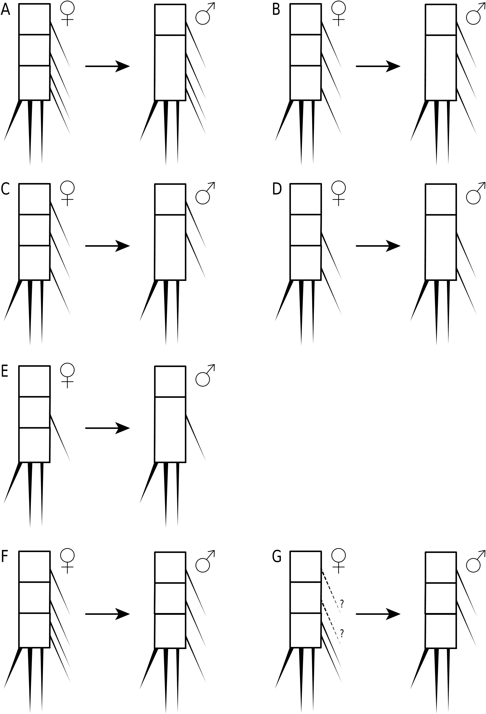
In his search of apomorphies that could define the genus Parastenhelia as a monophyletic lineage, Mielke (1990) highlighted the significance of the sexual dimorphism of the P3 endopod, involving the transformation of the outer spine on the distal segment into a fused apophysis and a reduction in the number of inner setae (to one in P. bulbosa and none in all other species) (Fig. 7). The absence of this character in P. reducta and P. megarostrum led him to claim that they do not belong to Parastenhelia . This view was corroborated by Gee (2006), who moved P. reducta as species incertae sedis to Karllangia (= Thalestrella ) and suggested that both P. megarostrum and P. pyriformis should be assigned to one or more new genera, a course of action that is formalized here (see below).
Various authors have pointed out that a revision of the genus is hampered by the taxonomic confusion surrounding its alleged type species P. spinosa ( Vervoort 1964; Wells & Rao 1987; Gee 2006; Kuru & Karaytuğ 2015). However, it has recently come to light that Lang’s (1948) fixation of P. spinosa as the type was invalid since it was not originally included (ICZN Art. 69.2) and that P. hornelli had already been fixed as the type species by subsequent designation ( Lang 1934: 24; Huys 2009: 96). This implies that any assessment of the monophyly of Parastenhelia must first centre on a review of P. hornelli (and allied species – hornelli group) since it provides the objective standard of reference for the application of the generic name it bears. Consequently, the genus is here restricted to a core group of four valid species which share a P1 exopod with three subequal segments in both sexes. Males of these species share a haplocer antennule with three segments distal to the geniculation, a 2-segmented P2 endopod (enp-2 and -3 forming a compound segment but total armature as in ♀) and a modified P3 endopod with spinous apophysis on enp-3 ( Table 1 View TABLE ; Figs 6 View FIGURE 6 –7).
Thompson & Scott (1903) described two Parastenhelia species from pearl oyster washings in the Gulf of Mannar off northwestern Sri Lanka: P. hornelli , which was represented by several females and males and was also found in general washings of dredged material in deep water off Galle (southwestern Sri Lanka), and P. similis of which only two females were recorded. The two species were considered morphologically similar with females being distinguished on the basis of proportional length differences in the antennulary segments (the whole appendage being distinctly longer and more slender in P. hornelli ), the morphology of the maxilliped, and the shape of the P5 exopod. Additional discrepancies are found in the P1 with the endopod being more slender, the inner seta of enp-1 distinctly longer, and the distal claws on enp-2 markedly longer (relative to the segment) in P. hornelli . Lang (1934) dismissed all these points and sank P. similis as a junior subjective synonym of P. hornelli . Since no variability in the antennule, P1 and P5 has so far been documented we regard these differences as sufficient justification to reinstate P. similis as a valid species.
Wells & Rao (1987) assessed the “ hornelli group” based on material from Mozambique, the Andaman Islands and New Zealand. They redescribed both sexes of P. hornelli from specimens of Middle and South Andaman, recognized two distinct size morphs among the female specimens (small form about 630 μm; large form about 885 μm; all ♂♂ 650 μm) and opted for the interim solution to place specimens previously identified as this species by Noodt (1955a) and Apostolov (1973), as well as the material identified as P. spinosa by Vervoort (1964), in P. hornelli . Careful inspection of Wells & Rao’s (1987) illustrations, however, shows that they were not dealing with P. hornelli but with P. similis . Evidence in support of this claim is found in the female morphology of the antennule (general facies and relative lengths of segments), maxilliped (palmar margin with conspicuous row of long spinules), P1 (length of endopod, inner seta of enp-1 and claws on enp-2), P5 exopod (exact shape and strong spinular ornamentation along inner and outer margins) and caudal rami (setae IV–V slightly swollen and irregularly shaped at base). The three differences between Thompson & Scott’s (1903) type material of P. similis and Wells & Rao’s (1987) specimens are (a) segmentation of female antennule (the minute segments 7–8 in Wells & Rao’s Fig 52e are shown as complelety fused in the original description), (b) absence of the small distal inner seta on P4 exp-3, and (c) absence of short inner seta on the endopodal lobe of P5, both of which are conceivable the result of imperfect observation. The records of P. hornelli from Odisha (formerly Orissa) in the Bay of Bengal ( Rao 1989), Lakshadweep ( Rao 1991) and Little Andaman ( Rao 1993) are accompanied by very brief, mostly uninformative descriptions and must be considered as unconfirmed. Other Indian records from Bhitarkanika in Odisha ( Chadha & Kar 1999), Kundugal channel ( Krishnaswamy 1953) and Porites Bay ( Krishnaswamy 1957) in Tamil Nadu, and the Andaman Islands ( Rao 1993; Jayabarathi 2016; Jayabarathi et al. 2012, 2015; Pongener et al. 2018) are equally indeterminable.
Noodt (1955a) provides a brief redescription, completely lacking in illustrations, of two females from the Sea of Marmara in northwestern Turkey which he attributes to P. hornelli . He states that both P1 and P5 agree exactly in form and armature with the illustrations of Thompson & Scott (1903) but it is not clear which of the Sri Lankan species he refers to. Since the shape of the exopod is different between P. hornelli and P. similis , Noodt’s (1955a) statement that it is widening proximally towards the baseoendopod (“Insbesondere ist der Exp. P5 durch seine Erweiterung zur Basis hin typisch”) may carry some significance since this feature is more expressed in P. hornelli ( Thompson & Scott 1903: Plate VII, Fig. 8). Noodt’s (1955a) report that the antennary exopod is 3-segmented must be an observational error since it is 2-segmented in the groundpattern in the Parastenheliidae ( Willen 2000) ; he also states that the proximal segment bears one seta but does not disclose how many elements are present on the remaining segments. Noodt (1955a) presents the armature pattern of P2–P4, noting variability in the presence/ absence of the inner seta on P2 exp-3. Although Wells & Rao (1987) pointed out that the pattern of P4 (exp: 1.1.223; enp: 1.1.221) is congruent with that given by Thompson & Scott (1903) (for both P. hornelli and P. similis ) it remains to be confirmed whether the presence of only two inner setae on P3–P4 exp- 3 in the Turkish specimens is genuine or reflects an oversight of the fine inner distal seta. Based on Noodt’s description no positive statement can be made as to the validity of his identification and pending re-examination of the material (and discovery of the male) his record must be considered suspect. The unillustrated records of P. hornelli from two beaches (Cunda Beach and Altınova Beach) in the Aegean Sea ( Karaytuğ & Sak 2006), in close proximity to the Sea of Marmara, possibly refer to the same species.
Although Vervoort (1964) voiced criticism of Lang’s (1948) broad taxonomic concept of P. spinosa , he indirectly expressed the opinion that both P. hornelli and P. ornatissima may also fall within the range of this species. Among the material from Ifalik Atoll, Vervoort describes a male and a female which he assigns to P. spinosa while admitting that they approach P. hornelli in the structure of the P1 exopod, where the three segments have about the same length. Vervoort (1964) dismisses the significance of this difference by accepting Lang’s (1948) view that it falls within the variability of P. spinosa – a concept that is mistakenly based on comparison of geographically separated and, most likely, non-conspecific populations. Since intrapopulation variability in the length of P1 exp-2 has never been recorded in any species so far, Vervoort’s (1964) interpretation is rejected here. His material displays a number of distinct differences with both P. hornelli and P. similis : (a) P3–P4 exp-3 without inner seta; (b) in addition to the common sexual dimorphism expressed in the endopods of P2–P3, the male P4 endopod also differs from that of the female in having only one inner seta on enp-3; this type of sexual dimorphism has previously been observed in Microthalestris littoralis f. penicillata sensu Willey (1935) and Parastenhelia bulbosa ( Gee 2006) , and as a further derived state in P. minuta and Penicillicaris penicillata sp. nov. where both inner setae are lost in the male ( Willey 1935; Pallares 1982; Fig. 6F–G View FIGURE 6 herein); and (c) male P5 exopod 2-segmented; this condition was recently described in P. aydini ( Kuru & Karaytuğ 2015) and in Johnwellsia bipartita gen. et sp. nov. (present account). Based on these differences it is obvious that Vervoort’s (1964) material from stations 589 and 590 cannot be attributed to P. spinosa or to either of Thompson & Scott’s (1903) species and must be assigned distinct specific rank (see below). Wells et al. ’s (1982) claim that Vervoort’s description and illustrations of the antenna are contradictory is irrelevant. His lateral habitus view (Fig. 68a) shows indeed the antennary exopod as 3-segmented under low magnification but the detailed drawing of the antenna (Fig. 68d) clearly reveals it as 2-segmented (as reiterated in the text: p. 186).
Apostolov (1973: 273, Fig. 9) reported two females and one male of P. hornelli on brown algae ( Cystoseira barbata ) near Tsarevo (formerly Michurin) along the Black Sea coast of Bulgaria. No text description was given, except for his statement that the male fits Thompson & Scott’s (1903) original description perfectly. Apostolov (1973) provides illustrations of the male P2 endopod (similar to that illustrated by Thompson & Scott), P3 endopod (previously unknown) and P5. The latter differs significantly in that it is only 1-segmented and the armature elements all are very short, consequently ruling out the possibility that the Bulgarian material belongs to either P. hornelli or P. similis . Similar to Noodt’s (1955a) specimens the antennary exopod is erroneously illustrated as 3-segmented causing Wells & Rao (1987) to suggest that both sets of specimens are conspecific. The female antennule and P5 exopod resemble their respective conditions as illustrated by Thompson & Scott (1903) for P. hornelli , the P4 also displays the same armature pattern (although confirmation of the exact number of inner setae on exp-3 is required in both), but the P1 endopod is comparatively longer and more slender. The only significant difference with Noodt’s (1955a) material is found in the absence of the inner seta on P2 enp-1 (a condition that is mirrored in the male). In summary, Apostolov’s (1973) Bulgarian specimens share several characters with P. hornelli from Sri Lanka but the 1-segmented condition of the male P5 exopod and deficiencies in the description of the antenna (and possibly P3–P4 exopods) prevent them from being assigned to the type species. Pending a re-examination of the Black Sea material, P. hornelli sensu Apostolov (1973) must be classified as indeterminable.
Apostolov & Pandourski (1999) reported one female and one male of P. hornelli from a littoral sandbank in Livingston Island (South Shetlands archipelago), north of the Antarctic Peninsula (see also Pesce & Pandourski 2002: 134). The authors do not provide a text description but state that their material confirms the variability previously observed in this species and cite the different number of setae and spines on P3 exp-3 and enp-3 as an example. Although they do not cite the work it is likely that Apostolov & Pandourski (1999) used Wells & Rao’s (1987) redescription of P. hornelli (= P. similis ) as a reference for comparision. Their illustration of the female P3 endopod (they do not figure the exopod) shows three well developed inner setae on the distal segment which contradicts the condition in P. similis ( Table 1 View TABLE ). The female P5 exopod approaches the shape (although more truncated) of Thompson & Scott’s (1903) P. hornelli while the length of the endopodal setae is different. The morphology of the male is said to be identical with P. hornelli but only the P2 endopod is illustrated; the proximal segment of this limb lacks the inner seta found in the type species. No information was given on the armature pattern of P2–P4 (except P3 endopod ♀) or the segmentation and setation of the male P6. Apostolov & Pandourski’s (1999) description is too fragmentary to allow any conclusions to be made about its identity and consequently it must be classified as an indeterminable record of P. hornelli .
Based on Wells & Rao’s (1987) report the only confirmed records of P. similis are from Sri Lanka ( Thompson & Scott 1903), Inhaca Island in Mozambique ( Wells 1967), northeastern New Zealand ( Wells et al. 1982) and the Andaman Islands ( Wells & Rao 1987). Authenticated records of P. hornelli remain restricted to the type populations from Galle and the Gulf of Mannar in Sri Lanka ( Thompson & Scott 1903). This is in stark contrast with the previously assumed circum-subtropical distribution ( Wells et al. 1982; Wells & Rao 1987) which included unconfirmed and (possibly) false records from the Aegean/Black Sea region ( Noodt 1955a; Apostolov 1973), Barbados ( Coull 1970a) and the U.S. Virgin Islands ( Coull 1971a; Hartzband & Hummon 1974) in the Atlantic, and the east coast of Peninsular Malaysia ( Zaleha et al. 2006) and the Caroline Islands ( Vervoort 1964) in the Pacific. The armature pattern of P2–P4 given by Zaleha et al. (2006) shows that they were dealing with a different species. In addition to the unconfirmed Indian records mentioned above, P. hornelli has also been reported from Gazi Bay in Kenya ( De Troch 2001 – as P. cfr. hornelli ) and along the Pacific seaboard of North America including the Nanaimo River delta ( Sibert 1981; Kask et al. 1982) and Fraser River estuary ( Sibert et al. 1982; Bravender et al. 1993; Johansen & Reis 1994) in British Columbia, Padilla Bay ( Simenstad et al. 1988) and Puget Sound ( Simenstad et al. 1991) in Washington State and the Santa Barbara Channel in southern California ( Spies et al. 1988). All these records are unillustrated and must be considered as unconfirmed and probably false. Parastenhelia oligochaeta has so far been recorded from the Andaman and Nicobar Islands (Chidyatapu, Mayabandar, Car Nicobar, Katchal and Great Nicobar Islands – Wells & Rao 1987) and from Bhitarkanika in Odisha, mainland India ( Chadha & Kar 1999).
Diagnosis. Parastenheliidae . Sexual dimorphism in antennule, P2–P3 endopods, P5–P6 and urosomal segmentation; occasionally P4 endopod and caudal ramus setae IV–V. Body subcylindrical, short; posterior margin of cephalothorax, somites bearing P2–P5 and remaining urosomites (except anal somite) with denticulodigitate hyaline frills. Rostrum defined at base, of moderate size, reaching at most to distal margin of second antennulary segment; linguiform. Anal operculum semicircular, naked or bordered with fine spinules. Caudal ramus wider than long, without conspicuous spinular ornamentation on dorsal surface; with at least six setae (seta I possibly absent), setae IV–V slightly swollen at base in ♀ in some species ( P. similis ).
Antennule elongate and 8- or 9-segmented in ♀, segment 1 not elongate, segments 7–8 (when 9-segmented) shortest, with aesthetascs on segments 4 and 8 or 9; haplocer in ♂ but segmentation and segmental homologies unconfirmed, with three segments distal to geniculation, segment 5 swollen and with aesthetasc. Antenna not sexually dimorphic; proximal endopodal segment largely separated from basis or forming allobasis, with pinnate seta on abexopodal margin; exopod 2-segmented, proximal segment with two setae, distal segment with 1–2 lateral and three apical elements; distal endopodal segment without penicillate elements. Mandible with 2–4 elements on basis; endopod with eight setae; exopod 1-segmented with three setae or represented by single seta arising from minute knob. Maxillulary coxal epipodite represented by one seta. Maxilla with three endites on syncoxa; endopod discrete, with 2–3 setae. Maxilliped with 2–3 setae on syncoxa; basis with 1–2 seta(e) on palmar margin; endopod represented by curved claw, accompanied by 1–2 accessory seta(e).
P1 inner basal spine not sexually dimorphic. P1 exopod 3-segmented; exp-2 about as long as others, typically with short inner seta; exp-3 small, with two pinnate spines, one geniculate seta and one naked seta. P1 endopod 2-segmented; enp-1 elongate, about 1.3 –1.5 times length of exopod, with long, pinnate, inner seta inserted in proximal third, segment margins without area of reduced chitinization; enp-2 very small, with one naked minute seta and two dentate claws. P2–P4 rami 3-segmented. P2 endopod ♂ 2-segmented; total number of spines/setae as in ♀. P3 endopod ♂ 3-segmented; without inner setae on enp-3 and outer spine modified into spinous apophysis. P4 endopod ♂ occasionally with one less inner seta on enp-3 ( P. willemvervoorti sp. nov.). Armature formula of P2–P4 as follows:
P 5 ♀ endopodal lobe with five setae, outermost shortest; inner margin without transverse striae. P 5 ♀ exopod elongate, with six elements. P 5 ♂ endopodal lobe with two elements, outer one shortest; exopod 1-, 2- or 3- segmented, with 6–7 elements in total. Vestigial P 6 ♀ represented by two minute setae. P 6 ♂ with three setae.
Type species. Parastenhelia hornelli Thompson & Scott, 1903 (by subsequent designation; Lang 1934: 24).
Other species. P. similis Thompson & Scott, 1903 ; P. oligochaeta Wells & Rao, 1987 ; P. willemvervoorti sp. nov.
Species inquirendae. Parastenhelia hornelli Thompson & Scott, 1903 sensu Noodt (1955a) , sensu Apostolov (1973), sensu Apostolov & Pandourski (1999) and sensu Zaleha et al. (2006).
No known copyright restrictions apply. See Agosti, D., Egloff, W., 2009. Taxonomic information exchange and copyright: the Plazi approach. BMC Research Notes 2009, 2:53 for further explanation.
|
Kingdom |
|
|
Phylum |
|
|
Class |
|
|
Order |
|
|
Family |